. 2013:4:1440.
doi: 10.1038/ncomms2443.

Doublecortin-like kinase enhances dendritic remodelling and negatively regulates synapse maturation

Affiliations

Doublecortin-like kinase enhances dendritic remodelling and negatively regulates synapse maturation

Euikyung Shin et al. Nat Commun. 2013.

Abstract

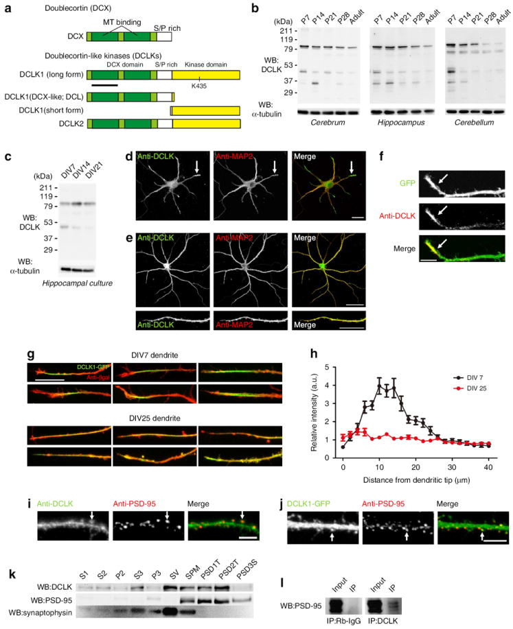
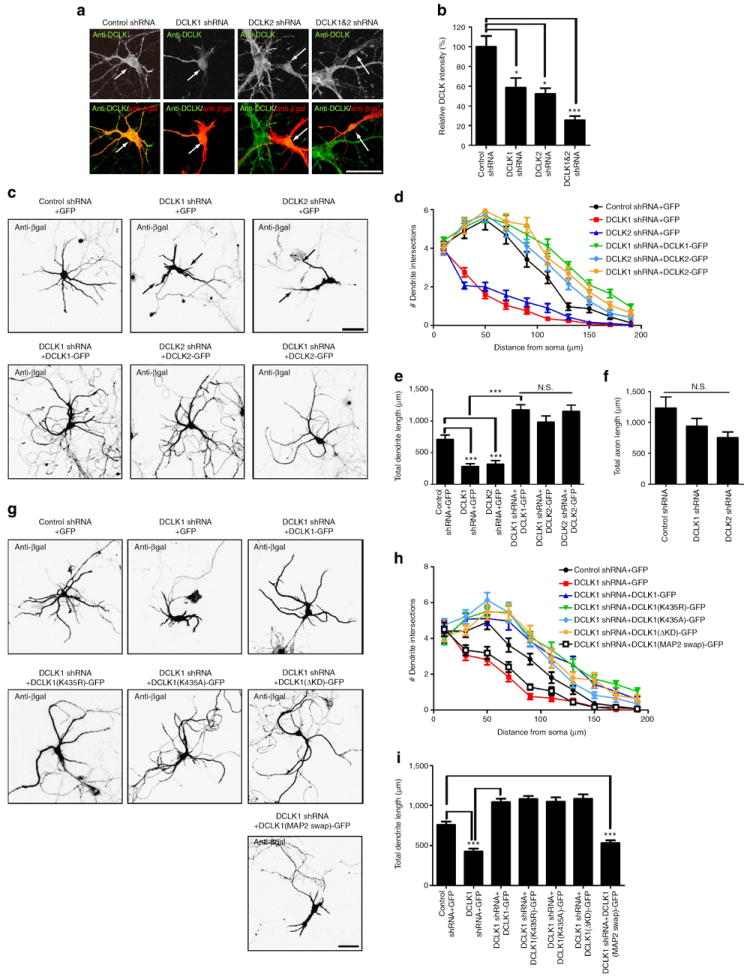
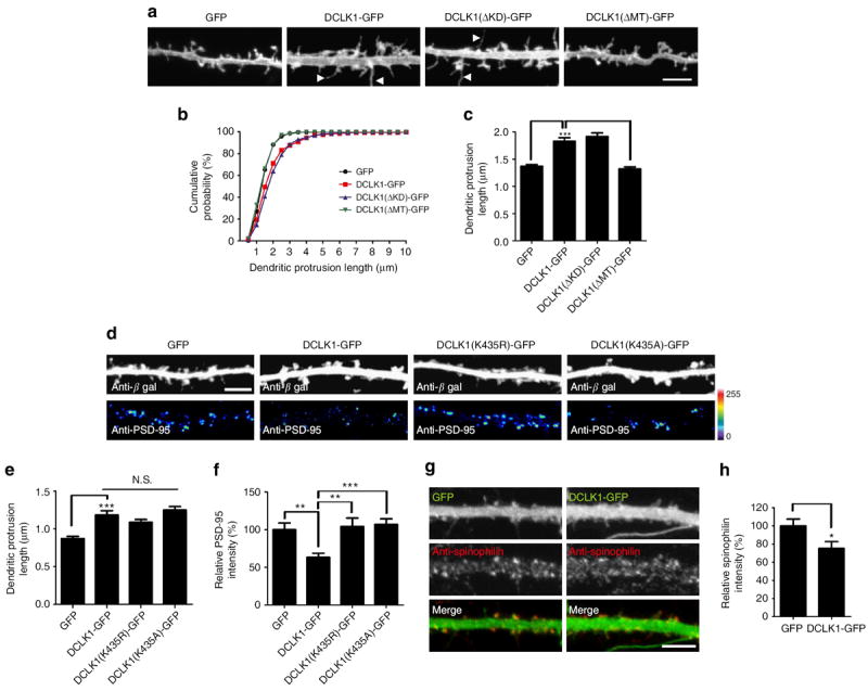
Dendritic morphogenesis and formation of synapses at appropriate dendritic locations are essential for the establishment of proper neuronal connectivity. Recent imaging studies provide evidence for stabilization of dynamic distal branches of dendrites by the addition of new synapses. However, molecules involved in both dendritic growth and suppression of synapse maturation remain to be identified. Here we report two distinct functions of doublecortin-like kinases, chimeric proteins containing both a microtubule-binding domain and a kinase domain in postmitotic neurons. First, doublecortin-like kinases localize to the distal dendrites and promote their growth by enhancing microtubule bundling. Second, doublecortin-like kinases suppress maturation of synapses through multiple pathways, including reduction of PSD-95 by the kinase domain and suppression of spine structural maturation by the microtubule-binding domain. Thus, doublecortin-like kinases are critical regulators of dendritic development by means of their specific targeting to the distal dendrites, and their local control of dendritic growth and synapse maturation.


Conflict of interest statement

Competing financial interests: The authors declare no competing financial interests.

Figures

Figure 1
Figure 1. Postnatal expression and subcellular localization of DCLKs
(a) Domain organizations of DCX, DCLK1 isoforms and DCLK2. A black line indicates the DCLK1 fragment used for antibody generation. S/P rich, serine/proline-rich domain. (b,c) Western blotting of DCLK protein. The immunoreactive bands of 85 kDa corresponding to the size of the full-length DCLK1 and DCLK2 proteins were detected in the extracts prepared from multiple brain regions (b) or dissociated hippocampal neurons at various days after plating (c). (d,e) Immunocytochemistry of dissociated hippocampal neurons at 7 DIV (d) and 24 DIV (e) using anti-DCLK and anti-MAP2 antibodies. DCLK proteins were preferentially concentrated in the distal part of dendrites at 7 DIV (arrows). (f) Immunocytochemistry of GFP-expressing hippocampal neurons with anti-DCLK antibody at 7 DIV. DCLK proteins were preferentially concentrated in the distal part of dendrites (arrows). (g) Transfection of DCLK1-GFP together with a lacZ reporter plasmid and subsequent immunocytochemistry with an anti-β-galactosidase antibody. (h) Relative intensity of DCLK1-GFP and anti-β-galactosidase immunoreactive signals at 7 DIV and 25 DIV. (DIV 7: n = 13 cells, DIV 25: n =15 cells.) (i,j) Presence of DCLK immunoreactivity and DCLK1-GFP fluorescence in PSD-95-positive spines (arrows). (k) Presence of DCLK immunoreactivity in PSD-1T and PSD-2T fractions, which contain purified PSDs (P2: crude synaptosomal pellet, S3: crude synaptic vesicle fraction, P3: lysed synaptosomal membrane fraction, SV: synaptic vesicle fraction, SPM: synaptic plasma membrane fraction, PSD-1T,PSD-2T: purified PSD fractions, PSD3S: PSD fraction after sarkosyl treatment). (l) Immunoprecipitation of PSD-95 by anti-DCLK antibody in extracts from the adult brain (rabbit IgG (Rb-IgG) as a control antibody of immunoprecipitation). All numeric data are given as mean ± s.e.m. Bar, 20 μm for d, 50 and 20 μm for e, 10 μm for f and g and 5 μm for i and j.
Figure 2
Figure 2. Regulation of dendritic growth by DCLKs
(a,b) Dissociated hippocampal neurons expressing GFP or DCLK1-GFP from 8 to 14 DIV. Note the more complex dendritic branching in neurons expressing DCLK1-GFP. (c,d) Sholl analysis and total dendritic lengths of neurons expressing GFP or DCLK1-GFP (number of cells analysed; GFP: 30 and DCLK1-GFP: 30; t-test: ***P<0.001). (e,f) Comparison of dendritic morphology before and after induction of DCLK1-GFP or GFP only, by Cre-dependent excision of loxp-stop-loxp sequences. The images before and 3 days after application of Cre-expressing recombinant adenoviruses were recorded (12 and 15 DIV). Prominent upregulation of dendritic growth was observed only in neurons expressing DCLK1-GFP (red dotted circles). (g) The total number of dendritic branches per neurons was significantly increased in neurons after induction of DCLK1-GFP expression. (number of cells analysed; GFP: 9 and DCLK1-GFP: 13; t-test: ***P<0.001.) (h) Overexpression of DCLK1-GFP suppressed pruning of dendrites and increased the rate of appearance of new dendritic segments (number of cells analysed; GFP: 9 and DCLK1-GFP: 13; t-test: *P<0.05). (i) The total length of dendrites per neurons was significantly increased in neurons overexpressing DCLK1-GFP. (Dendritic growth (%) = sum of dendritic length (after)/sum of dendritic length (before)) (Number of cells analysed; GFP: 9 and DCLK1-GFP: 13; t-test: **P<0.01). (j,k) Sholl analyses of neurons before and after induction of GFP (j) or DCLK1-GFP expression (k) (number of cells analysed; GFP: 9 and DCLK1-GFP: 13). (l) Anti-α-tubulin immunnostaining of growth-cone MTs in neurons transfected with plasmids expressing GFP or DCLK1-GFP. DCLK1-GFP expression induced tightly bundled MTs within growth cones. (m) Time-lapse imaging of both DCLK1-RFP and tubulin-GFP. DCLK1-positive segments show proximal to distal translocation in dendrites. Arrowheads indicate the distal end of the DCLK1-positive segment. All numeric data are given as mean ± s.e.m. Bar, 50 μm for a, b, e and f; 5 μm for l and m.
Figure 3
Figure 3. Dendritic growth was negatively regulated by DCLK shRNAs
(a,b) Expression of DCLK1 and DCLK2 shRNA plasmids in dissociated hippocampal neurons from 4 to 9 DIV (a). A marked decrease of DCLK immunoreactivity was observed (b) (number of cells analysed; control: 5, DCLK1 shRNA: 6, DCLK2 shRNA: 5, DCLK1 and DCLK2 shRNA: 5; one-way analysis of variance (ANOVA) followed by Tukey–Kramer multiple comparison tests: *P<0.05, ***P<0.001). (c) Neurons transfected with plasmids expressing DCLK1 and DCLK2 shRNAs showed severe impairment of dendritic growth (arrows), which could be rescued by expression of shRNA-resistant constructs for DCLK1 and 2. (d) Sholl analysis of dendritic complexity in neurons transfected with control, DCLK1 shRNA or DCLK2 shRNA plasmids, with or without rescue constructs (number of cells analysed; control: 31, DCLK1 shRNA: 29, DCLK2 shRNA: 30, DCLK1 shRNA plus DCLK1-GFP: 20, DCLK1 shRNA plus DCLK2-GFP: 19 and DCLK2 shRNA plus DCLK2-GFP: 20). (e) Total dendritic lengths of neurons expressing DCLK1 shRNAs or DCLK2 shRNAs, with or without rescue constructs (numbers of cells analysed were the same as in d; one-way ANOVA followed by Tukey–Kramer multiple comparison tests: ***P<0.001). NS, no statistical difference. (f) Total axonal lengths of neurons expressing DCLK1 shRNAs or DCLK2 shRNAs. Total axonal length measured at 5 DIV showed a tendency for reduction without statistical significance (number of cells analysed; control: 30, DCLK1 shRNA: 29 and DCLK2 shRNA: 28). (g) Neurons transfected with DCLK1 shRNA plasmid and expression vectors for various mutants of GFP-tagged DCLK1. Dendritic morphology was visualized by anti-β-galactosidase immunostaining. (h,i) Sholl analyses of dendritic complexity (h) and evaluation of total dendritic length (i) in neurons expressing DCLK1 shRNA plasmid together with expression vectors for various mutants of GFP-tagged DCLK1 (number of cells analysed; control shRNA + GFP: 32, DCLK1 shRNA + GFP: 30, sh + DCLK1-GFP: 27, sh + DCLK1(K435R)-GFP: 27, sh + DCLK1(K435A)-GFP: 28, sh + DCLK1(ΔKD)-GFP: 30 and sh + DCLK1(MAP2 swap)-GFP: 32; one-way ANOVA followed by Tukey–Kramer multiple comparison tests: ***P<0.001). All numeric data are given as mean ± s.e.m. Bar, 50 μm for a, c and g.
Figure 4
Figure 4. Regulation of postsynaptic functions by DCLK1
(a,b) Fluorescence images of dendrites expressing GFP, DCLK1-GFP, GFP-tagged DCX domain of DCLK1 (DCLK1(ΔKD)-GFP) or GFP-tagged kinase domain of DCLK1 (DCLK1(ΔMT)-GFP) (a). Neurons expressed the various forms of DCLKs from 18 to 20 DIV. Prominent reduction of PSD-95 immunoreactivity was observed in neurons overexpressing DCLK1-GFP or DCLK1(ΔMT)-GFP (b) (number of cells analysed; GFP: 19, DCLK1-GFP: 20, DCLK1(ΔKD)-GFP: 20 and DCLK1(ΔMT)-GFP: 20; one-way analysis of variance (ANOVA) followed by Tukey–Kramer multiple comparison tests: **P<0.01, ***P<0.001). (c–e) Anti-β-galactosidase staining of neurons expressing either GFP only or DCLK1-GFP (c). Density of dendritic protrusions was significantly reduced in DCLK1-GFP-expressing neurons (d) and the fraction of mushroom-type spines was also reduced (e) (number of cells analysed; GFP: 24, DCLK1-GFP: 26; t-test: *P<0.05, ***P<0.001). (f) Anti-β-galactosidase staining and anti-PSD-95 staining of neurons transfected with control shRNA plus GFP-expression plasmids, DCLK1 shRNA plus GFP-expression plasmid or DCLK1 shRNA plus shRNA-resistant DCLK1-GFP-expression plasmids. LacZ expression plasmids were included in all transfection experiments for the purpose of volume label. (g–j) Effects of DCLK1 knockdown on spines and PSDs. Neurons expressing DCLK1 shRNAs showed significant increase of spine volume (g) and the total intensity of PSD-95 clusters (h). These neurons also showed a slight, but significant, increase of dendritic protrusion density (i). Classification of spine types revealed increase in the fraction of mushroom-type spines (j). These effects of DCLK1 knockdown were rescued by expression of shRNA-resistant DCLK1-GFP (number of cells analysed; control: 20, DCLK1 shRNA: 18 and DCLK1 shRNA plus shRNA-resistant DCLK1: 20; one-way ANOVA followed by Tukey–Kramer multiple comparison tests or t-test: *P<0.05, **P<0.01, ***P<0.001) All numeric data are given as mean ± s.e.m. Bar, 5 μm for a; 10 μm for c and f.
Figure 5
Figure 5. Roles of DCLK1 domains on postsynaptic functions
(a) Morphology of dendrites labelled by DiI. Effects of overexpressing either GFP alone, wild-type DCLK1-GFP, the GFP-tagged DCX domain of DCLK1 (DCLK1(ΔKD)-GFP) or the GFP-tagged kinase domain of DCLK1 (DCLK1(ΔMT)-GFP) were evaluated. (b) Cumulative curves of spine/filopodia length in neurons expressing either GFP only, full-length DCLK1-GFP, DCLK1(ΔKD)-GFP or DCLK1(ΔMT)-GFP (GFP: n = 6 cells, 300 spines; DCLK1-GFP: n = 7cells, 350 spines; DCLK1(ΔKD)-GFP: n = 6 cells, 300 spines; and DCLK1(ΔMT)-GFP: n = 6 cells, 300 spines). (c) Length of dendritic protrusions in neurons expressing GFP only, full-length DCLK1-GFP, DCLK1(ΔKD)-GFP or DCLK1(ΔMT)-GFP (number of cells and spines analysed were the same as in b; one-way analysis of variance (ANOVA) followed by Tukey–Kramer multiple comparison tests: ***P<0.001). (d) Morphology of dendritic protrusions and PSD-95 immunoreactivity in neurons expressing GFP only, DCLK1-GFP and the GFP-tagged DCLK1 with kinase-dead point mutations (DCLK1 (K435R)-GFP and DCLK1 (K435A)-GFP). (e,f) Length of dendritic protrusions (e) and relative intensity of PSD-95 puncta (f) in neurons expressing GFP only, DCLK1-GFP, DCLK1 (K435R)-GFP and DCLK1 (K435A)-GFP (GFP: n = 30 cells, 1033 spines; DCLK1-GFP: n = 29 cells, 985 spines; DCLK1 (K435R)-GFP: n = 14 cells, 510 spines; and DCLK1 (K435A)-GFP: n = 20 cells, 657 spines; one-way ANOVA followed by Tukey–Kramer multiple comparison tests: **P<0.01, ***P<0.001). (g) Reduction of spinophilin content in dendrites by overexpression of DCLK1-GFP. (h) Quantification of spinophilin immunoreactivity in spines revealed suppression of spinophilin expression in neurons expressing DCLK1-GFP (GFP: n =19 and DCLK1-GFP: n =18; t-test: *P<0.05). All numeric data are given as mean ± s.e.m. Bar, 5 μm for a, d and g.
Figure 6
Figure 6. Regulation of AMPA receptor-mediated current by DCLK1
(a–e) mEPSC traces (a). Scale bar, 20 pA, 300 ms. The average and cumulative distribution of mEPSC amplitudes (b,d) and inter-event intervals (c,e) recorded from dissociated hippocampal neurons. Overexpression of DCLK1-GFP by infection of recombinant adenoviruses induced a decrease in mEPSC amplitudes and increase in inter-event intervals. (GFP: n = 6 cells and DCLK1-GFP: n = 8 cells; one-way analysis of variance (ANOVA): *P<0.05, **P<0.01.) (f) Traces of evoked EPSCs from neurons expressing either GFP only or DCLK1-GFP by using recombinant adenoviruses. Scale bar, 50 pA, 50 ms. (g) Mean and s.e.m of the AMPA/NMDA EPSC ratio in neurons overexpressing GFP alone or DCLK1-GFP. The graph shows significant suppression of the AMPA/NMDA EPSC ratio by DCLK1-GFP overexpression (GFP: n = 8 cells and DCLK1-GFP: n = 11 cells; one-way ANOVA: *P<0.05). All numeric data are given as mean ± s.e.m.
Figure 7
Figure 7. In vivo phenotypes of DCLK knockdown and knockout
(a,b) Golgi staining of the hippocampal CA1 area prepared from wild-type or DCLK1−/− mice (a) revealed an increase of mushroom-type spines in the latter (b) (WT: n = 3 and DCLK1-KO: n = 3 (littermates); t-test: *P<0.05) (c) The indistinguishable migratory properties of neuronal precursors in the somatosensory cortex with or without DCLK1 shRNA. (d–f) Cortical pyramidal neuron dendrites in vivo after in utero electroporation of DCLK1 shRNAs (d) showed reduction of dendritic complexity by Sholl analyses (e) and a smaller number of terminal dendritic branches (f) (control: n = 45 cells and DCLK1 shRNA: n = 45 cells; t-test: ***P<0.001). (g–i) GFP fluorescence of dendritic protrusions in pyramidal neurons after in utero electroporation of DCLK1 shRNAs (g) revealed an increase in spine width (h) and spine density (i) (control: n = 18 cells, 624 spines and DCLK1 shRNA: n = 17 cells, 501 spines; t-test: *P<0.05, ***P<0.001). (j,k) Manipulation of DCLK1 expression on the DCLK2−/− genetic background did not enhance impairment of dendritic growth. The complexity of dendrites (j) and the number of total terminal dendritic branches (k) were analysed in cortical pyramidal neurons expressing control or DCLK1 shRNAs on wild-type or DCLK2−/− background (WT+control shRNA: n = 22 cells; WT+DCLK1 shRNA: n = 17 cells; and DCLK2-KO+DCLK1 shRNA: n = 21 cells; one-way analysis of variance (ANOVA) followed by Tukey–Kramer multiple comparison tests: *P<0.05). NS, no statistical difference. (l,m) Expression of DCLK1 shRNAs on a DCLK2−/− genetic background further enhanced the increase in spine widths (WT+control shRNA: n = 22 cells, 768 spines; WT+DCLK1 shRNA: n = 17 cells, 622 spines; DCLK2-KO+DCLK1 shRNA: n = 20 cells, 802 spines; one-way ANOVA followed by Tukey–Kramer multiple comparison tests: **P<0.01,***P<0.001). All numeric data are given as mean ± s.e.m. Bar, 5 μm for a, g and l; 100 μm for c; and 50 μm for d.

References

    1. Allen KM, Walsh CA. Genes that regulate neuronal migration in the cerebral cortex. Epilepsy Res. 1999;36:143–154. - PubMed
    1. Gleeson JG, et al. Doublecortin, a brain-specific gene mutated in human X-linked lissencephaly and double cortex syndrome, encodes a putative signaling protein. Cell. 1998;92:63–72. - PubMed
    1. Taylor KR, Holzer AK, Bazan JF, Walsh CA, Gleeson JG. Patient mutations in doublecortin define a repeated tubulin-binding domain. J Biol Chem. 2000;275:34442–34450. - PubMed
    1. Francis F, et al. Doublecortin is a developmentally regulated, microtubule-associated protein expressed in migrating and differentiating neurons. Neuron. 1999;23:247–256. - PubMed
    1. Gleeson JG, Lin PT, Flanagan LA, Walsh CA. Doublecortin is a microtubule-associated protein and is expressed widely by migrating neurons. Neuron. 1999;23:257–271. - PubMed

Publication types

MeSH terms