

Crystallization and preliminary crystallographic analysis of two eukaryotic fructosyl peptide oxidases
- PMID: 23385752
- PMCID: PMC3564613
- DOI: 10.1107/S1744309112051445
Crystallization and preliminary crystallographic analysis of two eukaryotic fructosyl peptide oxidases
Abstract
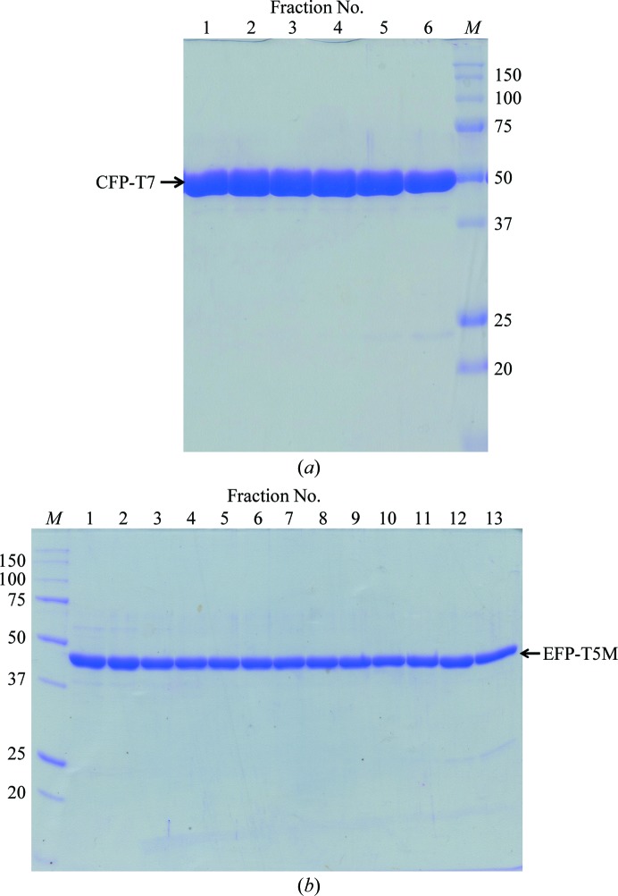
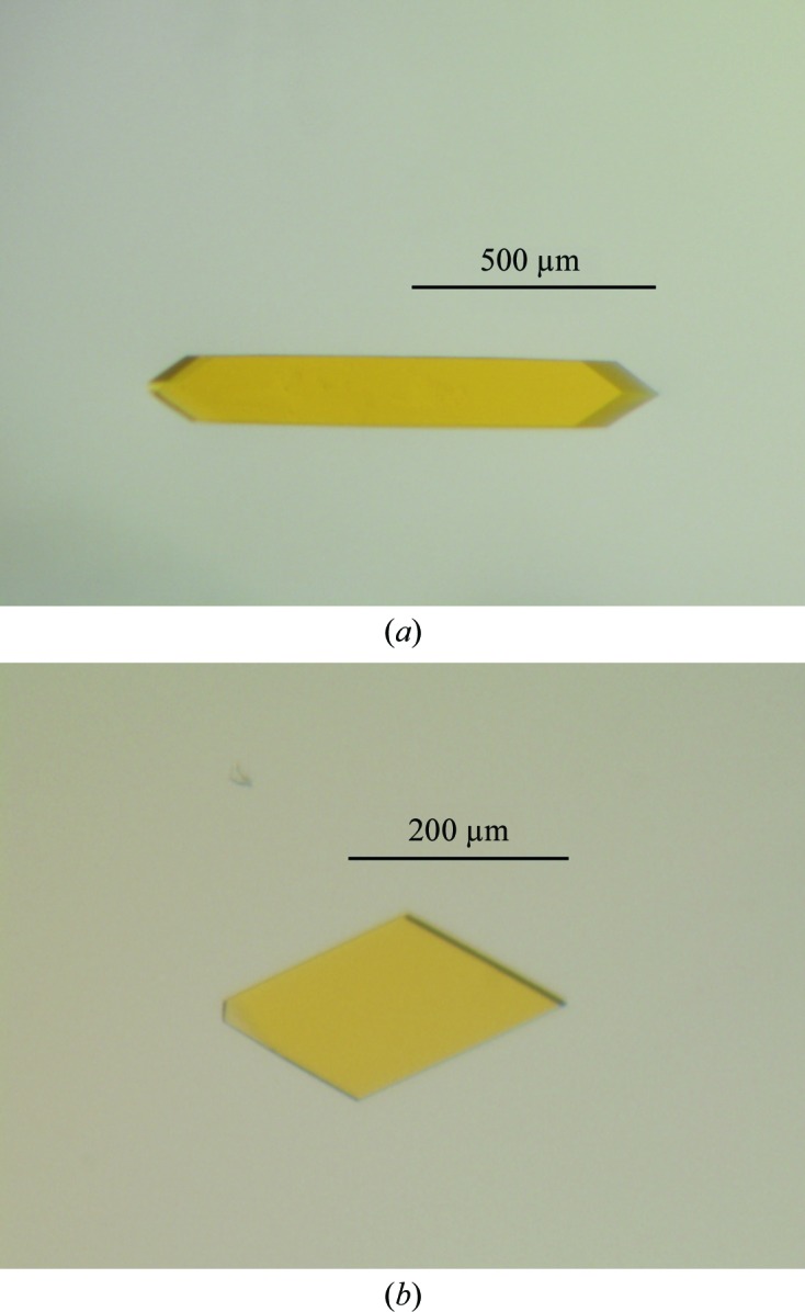
Fructosyl peptide oxidase (FPOX) catalyses the oxidation of α-glycated dipeptides such as N(α)-(1-deoxy-D-fructos-1-yl)-L-valyl-L-histidine (Fru-ValHis) and is used in the diagnosis of diabetes mellitus. Here, two thermostable mutants of FPOX, CFP-T7 and EFP-T5M, were crystallized by the sitting-drop vapour-diffusion method. The crystal of CFP-T7 belonged to the tetragonal space group P4(1)2(1)2, with unit-cell parameters a = b = 110.09, c = 220.48 Å, and that of EFP-T5M belonged to the monoclinic space group P2(1), with unit-cell parameters a = 43.00, b = 230.05, c = 47.27 Å, β = 116.99°. The crystals of CFP-T7 and EFP-T5M diffracted to 1.8 and 1.6 Å resolution, respectively.
Keywords: Coniochaeta sp.; Eupenicillium terrenum; diagnosis of diabetes; fructosyl peptide oxidase; haemoglobin A1c.
Figures
Similar articles
-
Expression, purification, crystallization and preliminary X-ray diffraction analysis of EtFPOX from Eupenicillium terrenum sp.Acta Crystallogr Sect F Struct Biol Cryst Commun. 2013 Jun;69(Pt 6):666-8. doi: 10.1107/S1744309113012128. Epub 2013 May 25. Acta Crystallogr Sect F Struct Biol Cryst Commun. 2013. PMID: 23722849 Free PMC article.
-
Engineering an efficient mutant of Eupenicillium terrenum fructosyl peptide oxidase for the specific determination of hemoglobin A1c.Appl Microbiol Biotechnol. 2019 Feb;103(4):1725-1735. doi: 10.1007/s00253-018-9529-9. Epub 2019 Jan 3. Appl Microbiol Biotechnol. 2019. PMID: 30607487
-
Crystallization and preliminary crystallographic analysis of bacterial fructosyl amino acid oxidase.Acta Crystallogr Sect F Struct Biol Cryst Commun. 2005 Feb 1;61(Pt 2):196-8. doi: 10.1107/S1744309104034372. Epub 2005 Jan 20. Acta Crystallogr Sect F Struct Biol Cryst Commun. 2005. PMID: 16510992 Free PMC article.
-
An amperometric biosensor for specific detection of glycated hemoglobin based on recombinant engineered fructosyl peptide oxidase.Int J Biol Macromol. 2020 Jan 1;142:855-865. doi: 10.1016/j.ijbiomac.2019.10.025. Epub 2019 Oct 14. Int J Biol Macromol. 2020. PMID: 31622711
-
A comprehensive review on fructosyl peptide oxidase as an important enzyme for present hemoglobin A1c assays.Biotechnol Appl Biochem. 2025 Feb;72(1):268-281. doi: 10.1002/bab.2647. Epub 2024 Aug 4. Biotechnol Appl Biochem. 2025. PMID: 39099239 Review.
References
-
- Bunn, H. F., Gabbay, K. H. & Gallop, P. M. (1978). Science, 200, 21–27. - PubMed
-
- Halwachs-Baumann, G., Katzensteiner, S., Schnedl, W., Pürstner, P., Pieber, T. & Wilders-Truschnig, M. (1997). Clin. Chem. 43, 511–517. - PubMed
-
- Hirokawa, K., Gomi, K. & Kajiyama, N. (2003). Biochem. Biophys. Res. Commun. 311, 104–111. - PubMed
-
- Hirokawa, K., Ichiyanagi, A. & Kajiyama, N. (2008). Appl. Microbiol. Biotechnol. 78, 775–781. - PubMed
MeSH terms
Substances
LinkOut - more resources
Full Text Sources
Other Literature Sources
Miscellaneous
