. 2013 Mar;26(1):8-18.
doi: 10.1007/s13577-012-0057-0. Epub 2013 Feb 6.

Pathogenesis of Hand-Foot Syndrome induced by PEG-modified liposomal Doxorubicin

Affiliations

Pathogenesis of Hand-Foot Syndrome induced by PEG-modified liposomal Doxorubicin

Noriyuki Yokomichi et al. Hum Cell. 2013 Mar.

Abstract

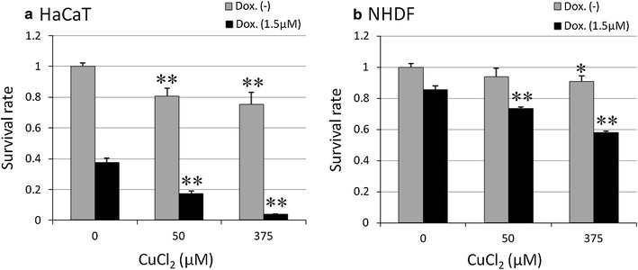
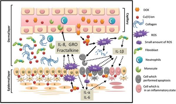
PEGL-DOX is an excellent treatment for recurrent ovarian cancer that rarely causes side-effects like cardiotoxicity or hair loss, but frequently results in Hand-Foot Syndrome (HFS). In severe cases, it can become necessary to reduce the PEGL-DOX concentration or the duration of the drug therapy, sometimes making it difficult to continue treatment. In this study, we prepared an animal model to compare the effects of DOX versus PEGL-DOX, and we noticed that only treatment with PEGL-DOX resulted in HFS, which led us to conclude that extravasation due to long-term circulation was one of the causes of HFS. In addition, we were able to show that the primary factor leading to the skin-specific outbreaks in the extremities was the appearance of reactive oxygen species (ROS) due to interactions between DOX and the metallic Cu(II) ions abundant in skin tissue. ROS directly disturb the surrounding tissue and simultaneously induce keratinocyte-specific apoptosis. Keratinocytes express the thermoreceptor TRPM2, which is thought to be able to detect ROS and stimulate the release of chemokines (IL-8, GRO, Fractalkine), which induce directed chemotaxis in neutrophils and other blood cells. Those cells and the keratinocytes then undergo apoptosis and simultaneously release IL-1β, IL-1α, and IL-6, which brings about an inflammatory state. In the future, we plan to develop preventative as well as therapeutic treatments by trapping the ROS.


Figures

Fig. 1
Fig. 1
Survival rate of human skin cells following DOX treatments. HaCaT and NHDF cells were cultured with DOX for 24 h before counting. The values are presented as mean ± SD (n = 3). Significantly different from control: *p < 0.05, **p < 0.01
Fig. 2
Fig. 2
Rat paws (SD, female, 7 weeks) after intravenous injection of DOX or PEGL-DOX. a Appearance immediately after 10 mg/kg injection. b Appearance after multiple doses. Doses were administered once every 3 days, and photos were taken on the 10th day. Blue circles indicate particularly inflamed areas
Fig. 3
Fig. 3
Tissue staining in rat (SD, female, 7 weeks) paw skin after 10 mg/kg PEGL-DOX injection. a H&E staining. Epidermal layer was thinned with respect to control (epidermal layer is shown in blue between the pink stratum corneum and lighter blue dermal layer). b Picrosirius red staining. Color of stain in order of decreasing strength and thickness of fibers: red, yellow, green. PEGL-DOX group displayed disarranged and broken collagen fibers. c TUNEL staining. Red marks nuclei, green marks apoptosis. Only basal cells show signs of apoptosis. (ac) scale bar 50 μm
Fig. 4
Fig. 4
Rat Cytokine Antibody Array. Skin tissue from HFS-affected areas after multiple 10 mg/kg PEGL-DOX injections. Results were normalized to controls. Graph shows increased production of chemokines with respect to control. The values are presented as mean ± S.D
Fig. 5
Fig. 5
Comparison of photographs after intravenous injection of 36 mg/kg 1.5 ml PEGL-FS or FS in hairless rats (female, 7 weeks). a Photos cropped from whole-body visualization under long-wavelength UV lamp immediately after, 3 and 7 h after injection. PEGL-FS remained fluorescing in the paws 7 h post-injection. b Tissue sections cut 1 h and 7 h after injection. PEGL-FS diffused faster from the dermis through the epidermis to the stratum corneum, lingered in the dermis longer, and showed slightly higher fluorescence than FS. Scale bar (b) 100 μm
Fig. 6
Fig. 6
Changes in chemokine and cytokine production in HaCaT and NHDF cells following addition of DOX or DOX + Cu(II) ions to culture medium. a Production of chemokines IL-8, GRO, and Fractalkine (ng/ml). HaCaT cells exhibited DOX- and Cu(II)-dependent production of IL-8 and GRO. The values are presented as mean ± SD (n = 3). Significantly different from control: *p < 0.05, p < 0.01. b Production of cytokines IL-1α, IL-1β, and IL-6 (ng/ml). HaCaT cells exhibited DOX/Cu(II)-dependent increased production of all cytokines shown, and NHDF cells exhibited substantially increased production of IL-1β only
Fig. 7
Fig. 7
Survival rate of human cells in the presence or absence of 1.5 μM DOX and varying concentrations of Cu(II) ions. a HaCaT cells. Survival rate declined with increasing Cu(II) ion concentration in the presence of DOX. b NHDF cells showed relatively little response to DOX and Cu(II) ions. The values are presented as mean ± SD (n = 3). Significantly different from control: *p < 0.05, **p < 0.01
Fig. 8
Fig. 8
Rescue of DOX/Cu(II)-induced decline in HaCaT cell survival rate by SOD. The values are presented as mean ± SD (n = 3). Significantly different from DOX, DOX + CuCl2, and DOX + CuCl2 + SOD, respectively: *p < 0.05, *p < 0.01
Fig. 9
Fig. 9
Illustration of HFS mechanism. As known, doxorubicin would produce ROS in the presence of metal ions. The illustration is shown in the case of the presence of Cu(II) ions because doxorubicin would react with Cu(II) ions to produce ROS. Doxorubicin could induce the apoptosis of skin cells by ROS which stimulates keratinocytes (epidermal layer cells) and blood cells by the productions of chemokines and cytokines by themselves. Additionally, ROS cause collagen breakdown

References

    1. Zuehle RL. Erythematous eruption of the palms and soles associated with mitotane therapy. Dermatologia. 1974;148:90–92. doi: 10.1159/000251603. - DOI - PubMed
    1. Green AE, Peter GR. Pegylated liposomal doxorubicin in ovarian cancer. Int J Nanomed. 2006;1:229–239. doi: 10.2217/17435889.1.2.229. - DOI - PMC - PubMed
    1. Ministry of Health, Labour and Welfare, Tokyo: The total of the year monthly report of statistics change of the population. 2010.
    1. Sadzuka Y, Tsuruda T, Sonobe T. The characterization and cytotoxicity of mixed PEG-DSG modified liposomes. Yakugaku Zasshi. 2005;125:149–157. doi: 10.1248/yakushi.125.149. - DOI - PubMed
    1. Ihaka R, Gentlemen R. A language for data analysis and graphics. J Comp Graph Stat. 1996;5:299–314.

LinkOut - more resources