Review
. 2013 Mar;22 Suppl 2(Suppl 2):S276-95.
doi: 10.1007/s00586-013-2676-x. Epub 2013 Feb 6.

Junctional spinal disorders in operated adult spinal deformities: present understanding and future perspectives

Affiliations
Review

Junctional spinal disorders in operated adult spinal deformities: present understanding and future perspectives

V Arlet et al. Eur Spine J. 2013 Mar.

Abstract

Introduction: Junctional spinal disorders have become one of the greatest challenges in spinal deformity surgery. They can occur at any age but are mostly seen in adult deformity surgery and are most often observed as the patient gets older.

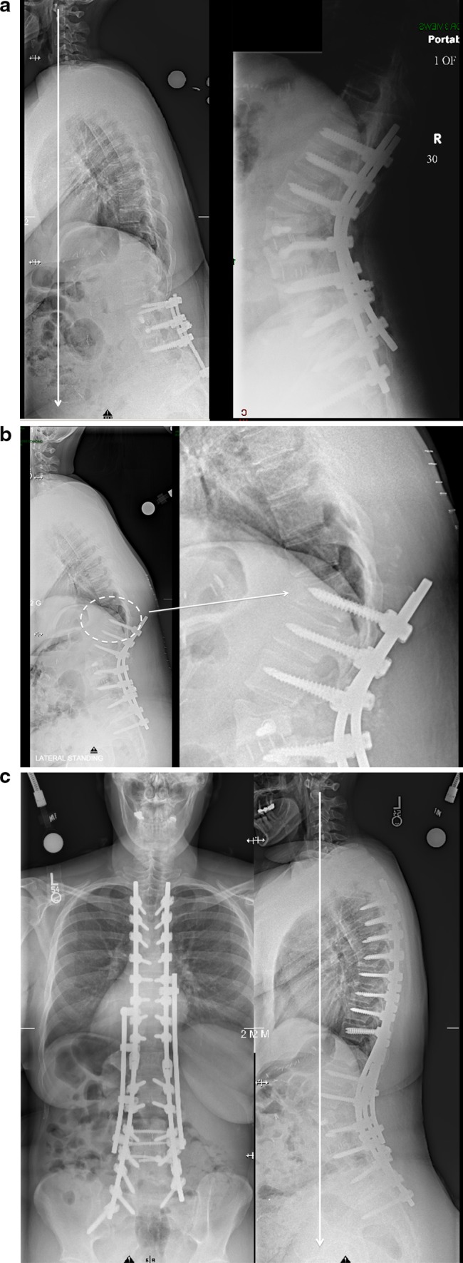
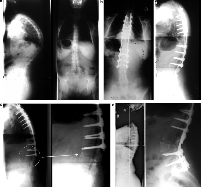
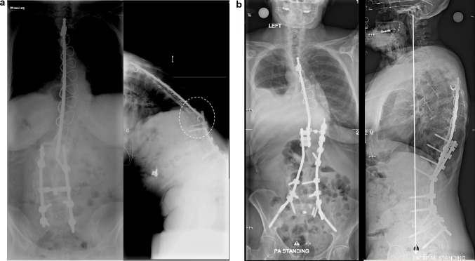
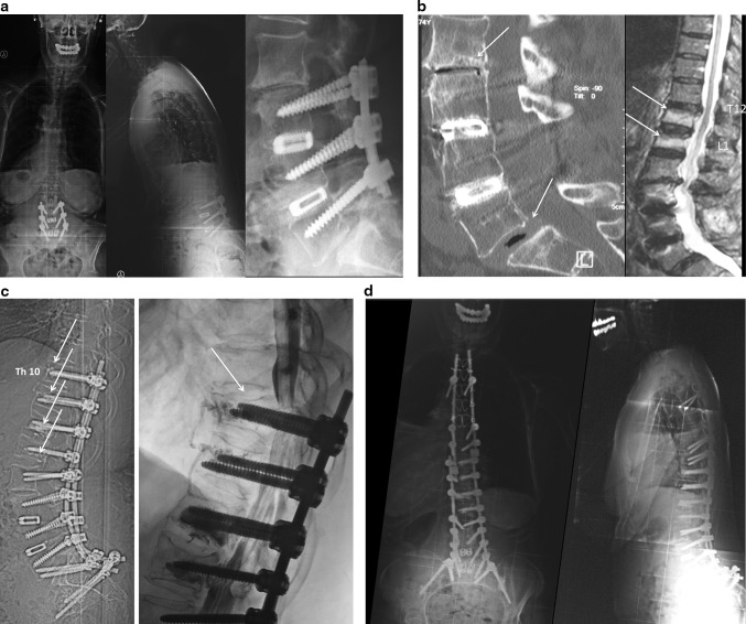
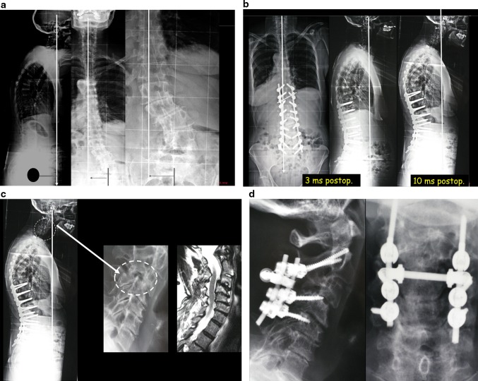
Definitions: Different forms can be individualized according to their types and location: one can observe simple segmental degeneration above or below instrumentation with or without spinal stenosis. Or the situation may be more complex with proximal junctional kyphosis, distal junctional kyphosis and intercalary junctional kyphosis where the junctional kyphosis occurs between two instrumented segments of the spine. Junctional scoliosis may also be observed as a new curve that did not exist after the index surgery.

Pathophysiology: Many different factors have been described being associated or the cause of junctional problems: old age, increased BMI, osteoporosis, etc. The role of pre-existing and postoperative sagittal imbalance plays a definitive role in their pathogenesis. As well the weakened posterior elements and or fatty degeneration of the posterior muscles are key factors in the occurrence of these problems. Multiple different radiologic parameters to describe and achieve perfect sagittal balance have been described knowing that the pelvic incidence of the patients is the key element that governs lumbar lordosis of the patient and hence the sagittal balance. Away from the spine one has to integrate the issues of the knees and the hips in the presentation of these junctional problems whether they are the cause or one of the consequences of the sagittal malalignment. Likewise the non-instrumented part of the spine (thoracic and or cervical spine) will also play a role in the pathogenesis or prevention of these junctional problems if they are stiff and or autofused along with their respective deformity.

Treatment: To prevent the occurrence of such junctional problems some basic surgical rules must be observed, but still lots remain unknown such as how much restoration of lordosis is really necessary, how to create a smoother transition between the instrumented and non-instrumented spine, which metal and where to use it, which implants to use as our widely used pedicle screw system may be one of the causes of these problems. Clinically these junctional problems can be asymptomatic and require only observation, or require revision surgery. Revision will require in most cases decompression of the neural elements, extension of the instrumentation and spinal osteotomies.

Conclusion: Definitively the issue of junctional spinal disorder after deformity surgery will require further extensive research to minimize this problem especially in our aging population.


Figures

Fig. 1
Fig. 1
Junctional scoliosis with degeneration below a previous fusion. a 56-year-old patient with previous scoliosis surgery 25 years before complaining of severe leg pain, back pain and inability to stand up straight. b Below the fusion that extended to L4, one can see severe facet arthropathy; on the lateral x-rays there is loss of lumbar lordosis. The myelogram confirms the spinal stenosis (white arrow). c Surgery with extension of the instrumentation to the pelvis, TLIF and Smith Petersen osteotomies to restore the lordosis. Good sagittal and coronal balance
Fig. 2
Fig. 2
Proximal junctional kyphosis (PJK). a Flat back syndrome after multiple low back surgeries in a 45-year-old obese patient: treatment of the sagittal imbalance with a pedicle subtraction osteotomy at L2. Observe the rods left slightly longer proximally as though PJK was an anticipated risk. b 2 weeks postoperatively, patient feels a crack in her back: X-rays shows a proximal junctional kyphosis with fracture through the pedicle of T11 (dashed circle left and magnifiedpicture right). c Treatment with extension of the fusion proximally with end to end connectors and a four rods construct because of the increased BMI of the patient
Fig. 3
Fig. 3
Distal junctional kyphosis (DJK). a 32-year-old female with a history of ankylosing spondylitis and progressive thoracic kyphosis. b Treatment with multiple transdiscal osteotomies and one PSO at the apex of the thoracic curve: good restoration of sagittal profile. c 6 months postoperatively: progressive loss of reduction with distal junctional kyphosis (dashed circle). The instrumentation should have been extended down to L3 that was the stable sagittal vertebra. d Treatment with extension of the instrumentation to the sacrum and a new PSO at L3: good restoration of the sagittal balance
Fig. 4
Fig. 4
Intercalary junctional kyphosis (IJK). a 62-year-old patient with a history of scoliosis surgery 30 years ago and lumbosacral fusion 10 years ago: Progressive flat back deformity and kyphosis in the intercalary segment that was not instrumented (Dashed circle). b Correction with first stage: anterior 5/1 fusion with wedge allograft, second stage PSO at L2 and connection to the previous Harrington rod (the left iliac screw broke at 1-year follow-up and was asymptomatic)
Fig. 5
Fig. 5
PJK related to natural history of progressive kyphosis as observed in natural aging: a degenerative scoliosis in 63-year-old obese female (fusion T10 pelvis). b Two years after decompression laminectomy instrumentation fusion T10 pelvis, one can observe a thoracic Cobb of 60°, patient pelvis is not retroverted, the plumb line is still behind the femoral heads. c Six years later the patient is now 69, there has been an increase in the PJK: Cobb is measured 80°. The patient remains asymptomatic, yet there is some imbalance with the plumb line falling in front of the femoral heads. The increased thoracic kyphosis parallels the ones observed in natural aging
Fig. 6
Fig. 6
PJK with failure of posterior ligament complex. a 62-year-old female, overweight, four previous lumbar spine surgeries: seen for intractable and leg pain and inability to stand straight. Notice the failure of the posterior ligament system (white arrows). b MRI and CT demonstrates the failure of the PLC with facet incompetency, spinal stenosis and end plate failure on both sides at L2/L1. There is also atrophy of the posterior musculature. c Treatment with posterior vertebral column resection, and instrumentation T3 to the sacro-pelvis [note the new PJK at T3 with cut out of the proximal screw in the disc space (dashed circle and white arrow), hopefully only mildly symptomatic]
Fig. 7
Fig. 7
PJK and fracture of the UIV (wedge compression fracture). a 60-year-old female with a history of multiple previous spine surgeries, osteoporotic compression fractures and sagittal decompensation. b Treatment with pedicle subtraction osteotomy at L2 and PSF sacrum to T10 (left). 2 weeks after surgery fracture of the UIV with wedge compression fracture (dashed circle and triangle) (middle and right picture). c Treatment with extension of the instrumentation to the upper thoracic spine: note the new PJK at the cervicothoracic junction (white arrow) (patient had some lower cervical pain)
Fig. 8
Fig. 8
Proximal and distal junctional disease, creation of a smooth transition. a 77-year-old patient, Parkinson, with previous circumferential surgery L3–L5 severe back and leg pain and inability to stand straight. b CT confirms the proximal and distal junctional disease with gas in the disc space at L2–L3 and L5–S1 (arrows left picture). MRI shows proximal junctional kyphosis with end plate fracture at T12 and L1 at distance from the L3–L5 fusion (arrows right picture). c Treatment with posterior fusion T10 pelvis with cement augmentation of the pedicle screws at T10, T11, T12 and L1 (arrows) (left picture). A few months later PJK with VCF of T10 and spinal stenosis (arrow on right). d Revision with extension to the upper thoracic spine using mostly sublaminar wires to decrease the stiffness of the instrumentation
Fig. 9
Fig. 9
PJK and fracture dislocation of the spine. a 56-year-old patient with a history of chronic steroid use and rheumatoid arthritis. Had previously undergone two surgeries: one for flat back deformity, and the previous one for PJK at the level of the TL junction that required extension to the upper thoracic spine. b Two months after her instrumentation: develops weakness in her legs CT scan shows a dislocated spine at the upper part of the instrumentation (dashed circle and arrow). c Surgery will be reduction of the dislocation extension of the fusion to the cervical spine: neurologic recuperation
Fig. 10
Fig. 10
Fracture through fusion mass below instrumentation. a 72-year-old female, 3 previous spine surgeries over 10 years. Patient has a L3 S1 fusion and instability/stenosis at L3/L2 and L2/L1 as well as lack of lordosis. b Posterior fusion with instrumentation down to L4 (L5 and S1 were not instrumented because the fusion was solid): 2 months after the surgery: acute pain: CT shows the fracture through the inferior aspect of L4 through the fusion mass (white arrows). c Revision and extension to S1
Fig. 11
Fig. 11
Creation of a smooth transition with vertebroplasties and hook. a 75-year-old patient with multiple previous surgeries intractable back and leg pain and inability to stand straight. b First stage is anterior femoral wedge allograft at 5/1, second stage 2 days later with posterior fixation T10 pelvis. c Close up view of the smooth transition creation: The T11 screws are inserted in a down going fashion and embedded in the cement above and around. Above in T10 only a supralaminar hook is inserted. At last above the instrumentation at T9 a prophylactic vertebroplasty is carried out
Fig. 12
Fig. 12
PJK happening at a distance in the cervical spine. a 71-year-old female patient with degenerative scoliosis and sagittal and coronal imbalance. b After correction of her sagittal and coronal imbalance with S1 T10 fusion: good restoration of her balance. However, the 3 months lateral X-rays show the beginning of subluxation C3/C4 and at 10 months postop one can observe sagittal decompensation and cervical kyphosis observed with significant pain. c Close up view: C2–C3 is spontaneously fused, the segment C4–C7 severely osteoarthritic and stiff, the thoracic spine was also stiff. d Treatment with posterior cervical fusion C2–C5

References

    1. Jimbo S, Kobayashi T, Aono K, Atsuta Y, Matsuno T. Epidemiology of degenerative lumbar scoliosis: a community-based cohort study. Spine. 2012;37(20):1763–1770. doi: 10.1097/BRS.0b013e3182575eaa. - DOI - PubMed
    1. Schwab F, Dubey A, Gamez L, El Fegoun AB, Hwang K, Pagala M, Farcy JP. Adult scoliosis: prevalence, SF-36, and nutritional parameters in an elderly volunteer population. Spine. 2005;30(9):1082–1085. doi: 10.1097/01.brs.0000160842.43482.cd. - DOI - PubMed
    1. Aebi M. The adult scoliosis. Eur Spine J. 2005;10:925–948. doi: 10.1007/s00586-005-1053-9. - DOI - PubMed
    1. Denis F, Sun EC, Winter RB. Incidence and risk factors for proximal and distal junctional kyphosis following surgical treatment for Scheuermann kyphosis: minimum five-year follow-up. Spine. 2009;34(20):E729–E734. doi: 10.1097/BRS.0b013e3181ae2ab2. - DOI - PubMed
    1. Hostin R, McCarthy I, O’Brien M, Bess S, Line B, Boachie-Adjei O, Burton D, Gupta M, Ames C, Deviren V, Kebaish K, Shaffrey C, Wood K, Hart R, International Spine Study Group (2012) Incidence, mode, and location of acute proximal junctional failures following surgical treatment for adult spinal deformity. Spine (Phila Pa 1976) [Epub ahead of print] - PubMed