

Axon diversity of lamina I local-circuit neurons in the lumbar spinal cord
- PMID: 23386329
- PMCID: PMC3738926
- DOI: 10.1002/cne.23311
Axon diversity of lamina I local-circuit neurons in the lumbar spinal cord
Abstract
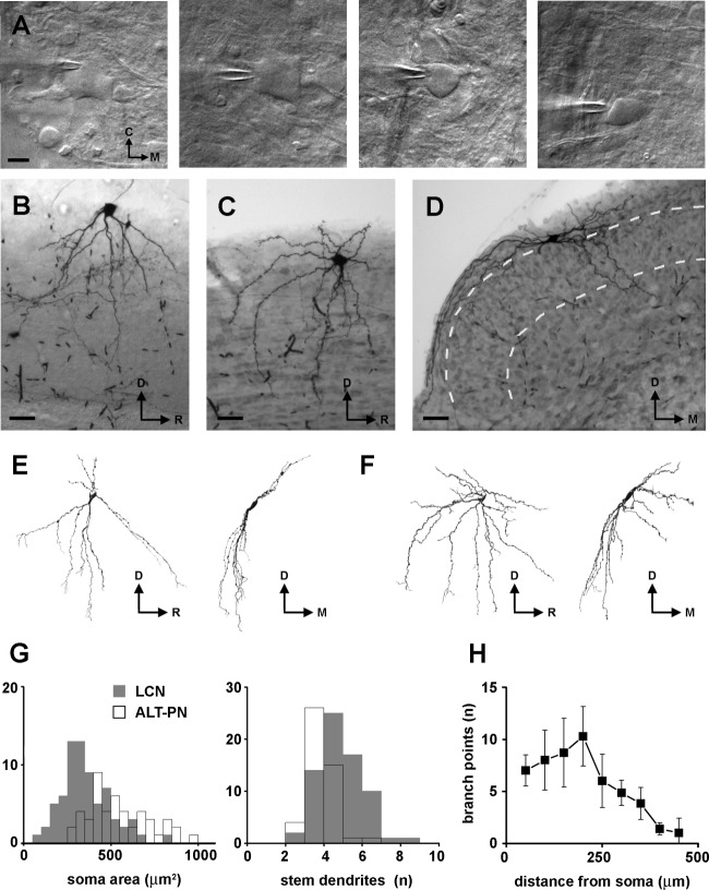
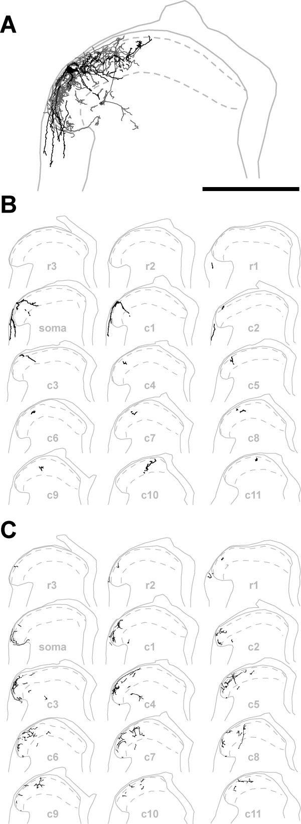
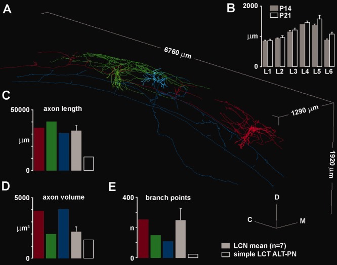
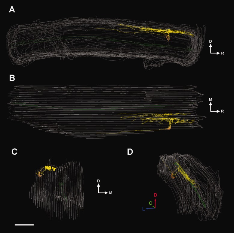
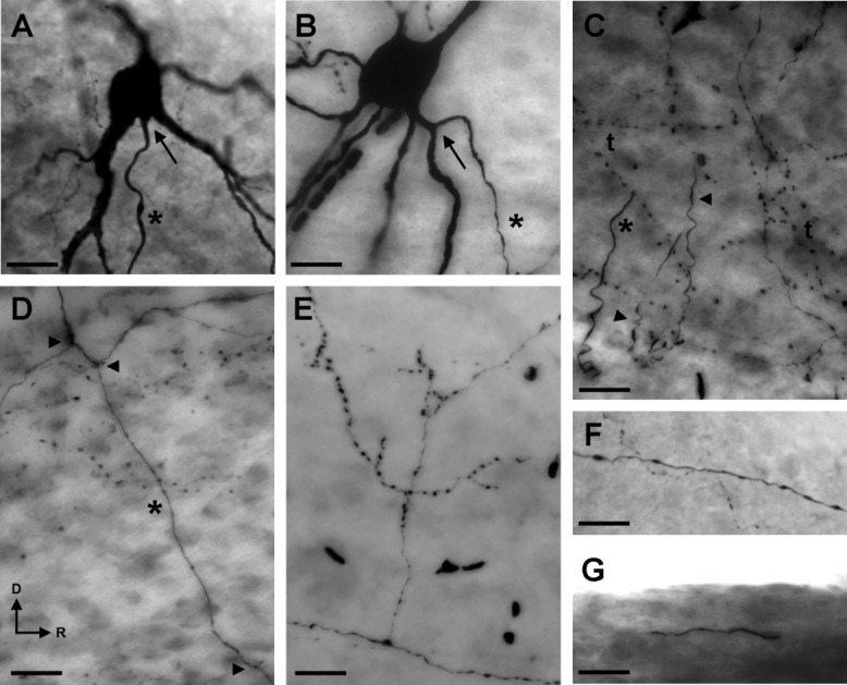
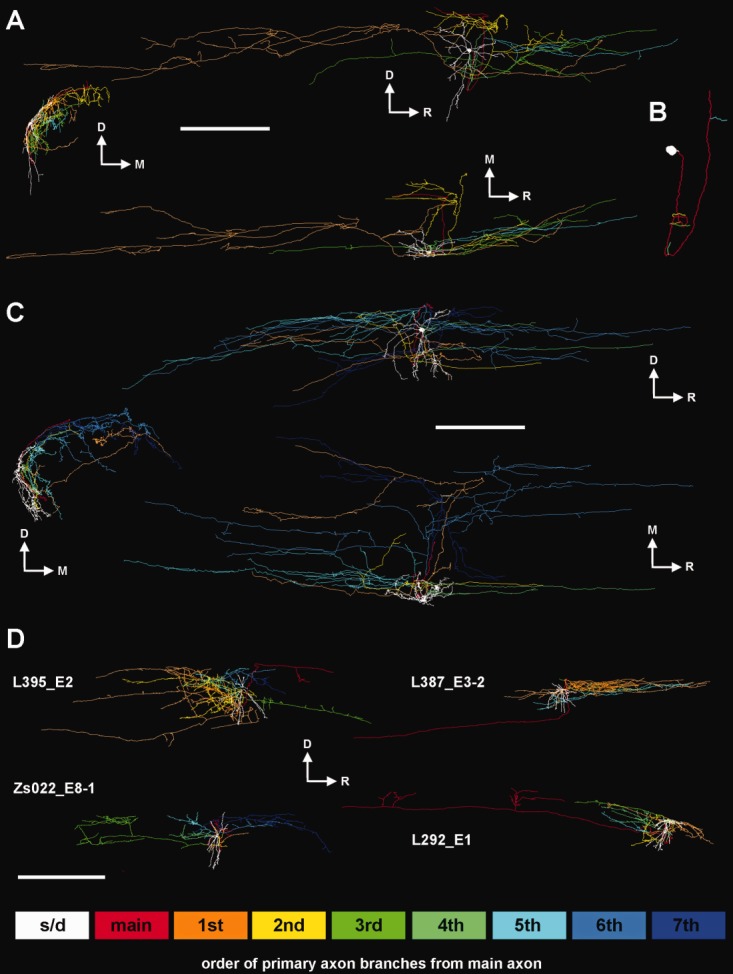
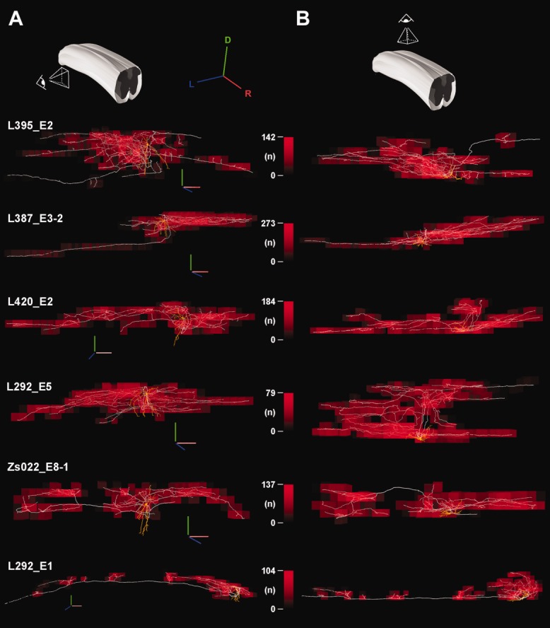
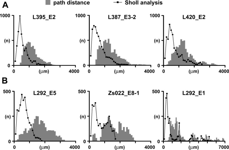
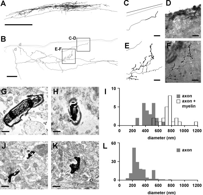
Spinal lamina I is a key area for relaying and integrating information from nociceptive primary afferents with various other sources of inputs. Although lamina I projection neurons have been intensively studied, much less attention has been given to local-circuit neurons (LCNs), which form the majority of the lamina I neuronal population. In this work the infrared light-emitting diode oblique illumination technique was used to visualize and label LCNs, allowing reconstruction and analysis of their dendritic and extensive axonal trees. We show that the majority of lamina I neurons with locally branching axons fall into the multipolar (with ventrally protruding dendrites) and flattened (dendrites limited to lamina I) somatodendritic categories. Analysis of their axons revealed that the initial myelinated part gives rise to several unmyelinated small-diameter branches that have a high number of densely packed, large varicosities and an extensive rostrocaudal (two or three segments), mediolateral, and dorsoventral (reaching laminae III-IV) distribution. The extent of the axon and the occasional presence of long, solitary branches suggest that LCNs may also form short and long propriospinal connections. We also found that the distribution of axon varicosities and terminal field locations show substantial heterogeneity and that a substantial portion of LCNs is inhibitory. Our observations indicate that LCNs of lamina I form intersegmental as well as interlaminar connections and may govern large numbers of neurons, providing anatomical substrate for rostrocaudal "processing units" in the dorsal horn.
Copyright © 2013 Wiley Periodicals, Inc., A Wiley Company.
Figures


Similar articles
-
Neurons in the lateral part of the lumbar spinal cord show distinct novel axon trajectories and are excited by short propriospinal ascending inputs.Brain Struct Funct. 2016 May;221(4):2343-60. doi: 10.1007/s00429-015-1046-3. Epub 2015 Apr 26. Brain Struct Funct. 2016. PMID: 25912439
-
Diverse firing properties and Aβ-, Aδ-, and C-afferent inputs of small local circuit neurons in spinal lamina I.Pain. 2016 Feb;157(2):475-487. doi: 10.1097/j.pain.0000000000000394. Pain. 2016. PMID: 26797505
-
Projection neurons in lamina III of the rat spinal cord are selectively innervated by local dynorphin-containing excitatory neurons.J Neurosci. 2012 Aug 22;32(34):11854-63. doi: 10.1523/JNEUROSCI.2707-12.2012. J Neurosci. 2012. PMID: 22915126 Free PMC article.
-
Spinal lamina I neurons that express neurokinin 1 receptors: morphological analysis.Neuroscience. 2000;97(2):335-45. doi: 10.1016/s0306-4522(00)00035-x. Neuroscience. 2000. PMID: 10799765
-
Novel aspects of signal processing in lamina I.Neuropharmacology. 2024 Apr 1;247:109858. doi: 10.1016/j.neuropharm.2024.109858. Epub 2024 Jan 28. Neuropharmacology. 2024. PMID: 38286189 Review.
Cited by
-
History of Spinal Cord "Pain" Pathways Including the Pathways Not Taken.Front Pain Res (Lausanne). 2022 Jun 9;3:910954. doi: 10.3389/fpain.2022.910954. eCollection 2022. Front Pain Res (Lausanne). 2022. PMID: 35756909 Free PMC article. Review.
-
Pacemaker Neurons and the Development of Nociception.Neuroscientist. 2014 Jun;20(3):197-202. doi: 10.1177/1073858414521499. Epub 2014 Feb 7. Neuroscientist. 2014. PMID: 24510073 Free PMC article. Review.
-
Projection Neuron Axon Collaterals in the Dorsal Horn: Placing a New Player in Spinal Cord Pain Processing.Front Physiol. 2020 Dec 21;11:560802. doi: 10.3389/fphys.2020.560802. eCollection 2020. Front Physiol. 2020. PMID: 33408637 Free PMC article. Review.
-
Concurrent Oncolysis and Neurolesion Repair by Dual Gene-Engineered hNSCs in an Experimental Model of Intraspinal Cord Glioblastoma.Cells. 2024 Sep 11;13(18):1522. doi: 10.3390/cells13181522. Cells. 2024. PMID: 39329707 Free PMC article.
-
Contralateral Afferent Input to Lumbar Lamina I Neurons as a Neural Substrate for Mirror-Image Pain.J Neurosci. 2023 May 3;43(18):3245-3258. doi: 10.1523/JNEUROSCI.1897-22.2023. Epub 2023 Mar 22. J Neurosci. 2023. PMID: 36948583 Free PMC article.
References
-
- Alle H, Geiger JR. Analog signalling in mammalian cortical axons. Curr Opin Neurobiol. 2008;18:314–320. - PubMed
-
- Beal JA, Penny JE, Bicknell HR. Structural diversity of marginal (lamina I) neurons in the adult monkey (Macaca mulatta) lumbosacral spinal cord: a golgi study. J Comp Neurol. 1981;202:237–254. - PubMed
-
- Bennett GJ, Abdelmoumene M, Hayashi H, Hoffert MJ, Dubner R. Spinal cord layer I neurons with axon collaterals that generate local arbors. Brain Res. 1981;209:421–426. - PubMed
-
- Berbel P, Innocenti GM. The development of the corpus callosum in cats: a light- and electron-microscopic study. J Comp Neurol. 1988;276:132–156. - PubMed
-
- Bice TN, Beal JA. Quantitative and neurogenic analysis of the total population and subpopulations of neurons defined by axon projection in the superficial dorsal horn of the rat lumbar spinal cord. J Comp Neurol. 1997a;388:550–564. - PubMed
Publication types
MeSH terms
Substances
Grants and funding
LinkOut - more resources
Full Text Sources
Other Literature Sources
Miscellaneous
