. 2013 Mar 15;288(11):7572-7579.
doi: 10.1074/jbc.M112.414904. Epub 2013 Feb 5.

Toll-like receptor 2 mediates peripheral nerve injury-induced NADPH oxidase 2 expression in spinal cord microglia

Affiliations

Toll-like receptor 2 mediates peripheral nerve injury-induced NADPH oxidase 2 expression in spinal cord microglia

Hyoungsub Lim et al. J Biol Chem. .

Abstract

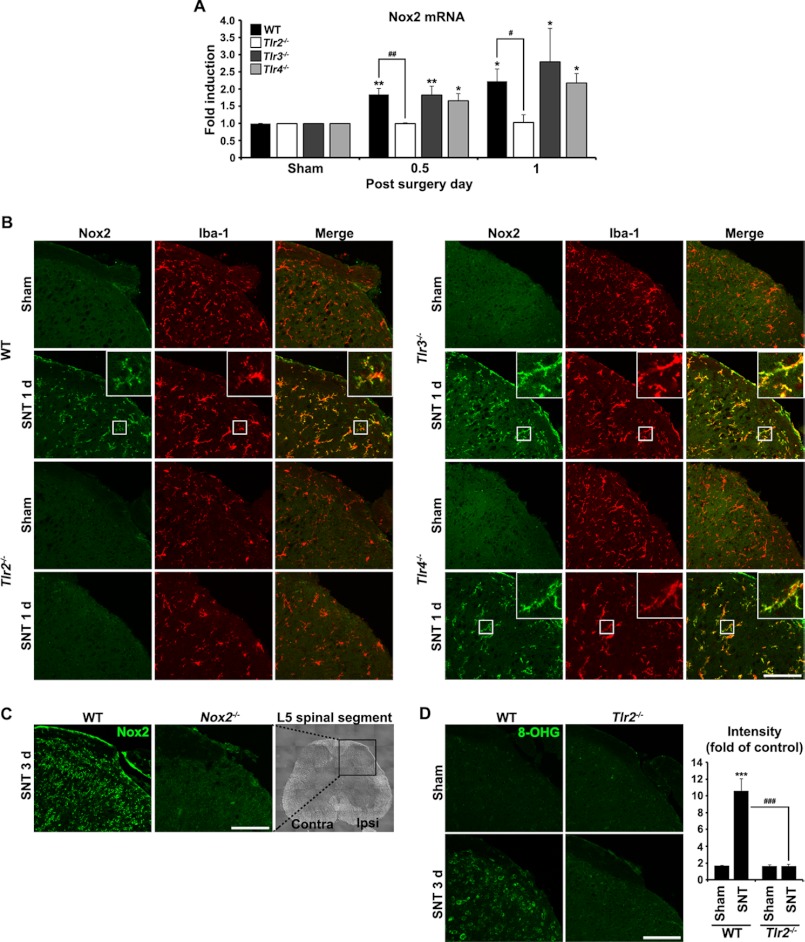
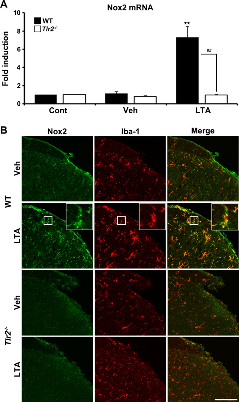
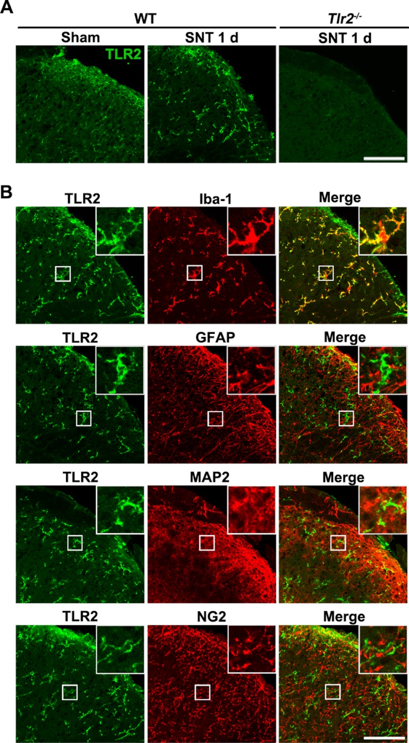
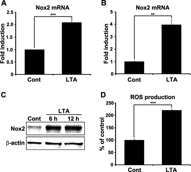
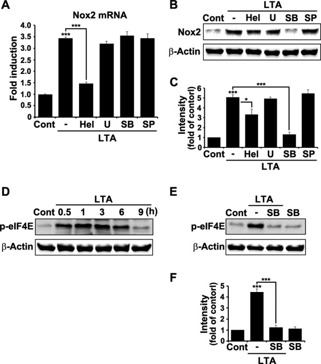
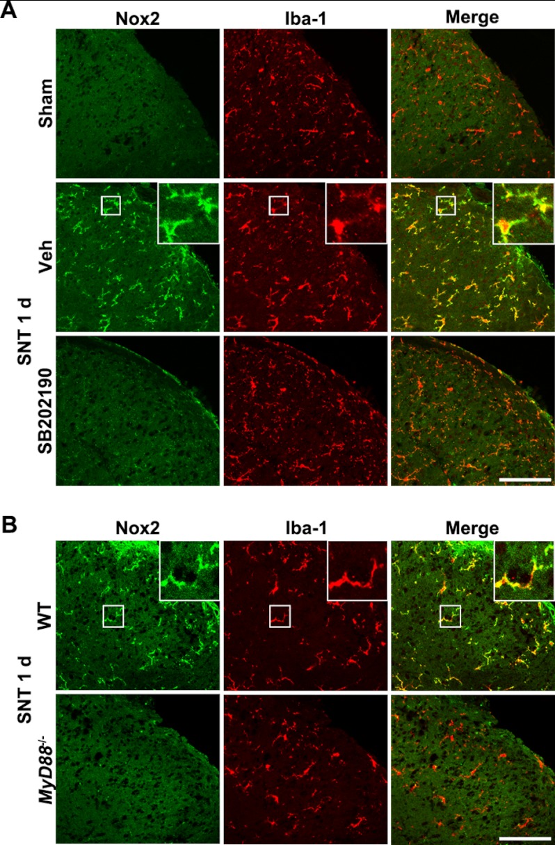
We have previously reported that NADPH oxidase 2 (Nox2) is up-regulated in spinal cord microglia after spinal nerve injury, demonstrating that it is critical for microglia activation and subsequent pain hypersensitivity. However, the mechanisms and molecules involved in Nox2 induction have not been elucidated. Previous studies have shown that Toll-like receptors (TLRs) are involved in nerve injury-induced spinal cord microglia activation. In this study, we investigated the role of TLR in Nox2 expression in spinal cord microglia after peripheral nerve injury. Studies using TLR knock-out mice have shown that nerve injury-induced microglial Nox2 up-regulation is abrogated in TLR2 but not in TLR3 or -4 knock-out mice. Intrathecal injection of lipoteichoic acid, a TLR2 agonist, induced Nox2 expression in spinal cord microglia both at the mRNA and protein levels. Similarly, lipoteichoic acid stimulation induced Nox2 expression and reactive oxygen species production in primary spinal cord glial cells in vitro. Studies on intracellular signaling pathways indicate that NF-κB and p38 MAP kinase activation is required for TLR2-induced Nox2 expression in glial cells. Conclusively, our data show that TLR2 mediates nerve injury-induced Nox2 gene expression in spinal cord microglia via NF-κB and p38 activation and thereby may contribute to spinal cord microglia activation.


Figures

FIGURE 1.
FIGURE 1.
L5 SNT-induced Nox2 up-regulation is abrogated in TLR2 knock-out mice. A, Nox2 mRNA expression in the L5 spinal cord segment after L5 SNT was measured by real-time RT-PCR. Total RNA was isolated from the L5 spinal cord tissues of sham-operated control mice and SNT-injured mice (each group, n = 3) at 12 h and 1 dpi (each time point, n = 3). The Nox2 transcript level is presented as the fold induction compared with the levels measured in the control mice of each group. Data are represented as mean ± S.E. (Student's t test; *, p < 0.05; **, p < 0.01, versus sham control in each mice group; #, p < 0.05; ##, p < 0.01, versus TLR2 knock-out mice). B, the L5 spinal cord sections were prepared from WT, TLR2, TLR3, and TLR4 knock-out mice with or without L5 SNT (1 dpi) and then used for immunostaining with Iba-1 and Nox2 antibodies. Representative spinal cord sections are shown (each group, n = 3; scale bar, 100 μm). C, to confirm the specificity of Nox2 immunoreactivity, spinal cord sections from WT and Nox2 knock-out mice at 3 dpi were immunostained with anti-Nox2 antibody (scale bar, 100 μm). D, spinal cord sections were immunostained with antibody against 8-OHG, a nucleotide oxidation marker, at 3 dpi. Compared with TLR2 knock-out mice, 8-OHG-IR in WT mice was dramatically increased following L5 SNT. The fluorescent intensity of 8-OHG-IR signals was measured and presented as the fold induction compared with the corresponding level measured in sham-operated mice. Data are represented as mean ± S.E. (Student's t test, ***, p < 0.001, versus sham control in WT mice, ###, p < 0.001, versus TLR2 knock-out mice, each group, n = 3; scale bar, 100 μm).
FIGURE 2.
FIGURE 2.
LTA stimulation induces Nox2 expression in spinal cord microglia in vivo. A, At 12 h after intrathecal injection of 5 μl of LTA (50 μg/ml in PBS, each group, n = 3) or vehicle (Veh, each group, n = 3), total RNA was isolated from L5 spinal cord tissues and used to determine Nox2 mRNA expression by real-time RT-PCR. Data are expressed as mean ± S.E. (Student's t test, **, p < 0.01, versus vehicle control in WT mice group, ##, p < 0.01, versus TLR2 knock-out mice). B, Nox2 protein expression in the ipsilateral L5 spinal cord dorsal horn at 1 day (1 d) after 5 μl of LTA (50 μg/ml in PBS) or vehicle intrathecal injection was evaluated by immunohistochemistry. Spinal cord sections were immunostained with Nox2 and Iba-1 antibodies. Nox2-IR-signals were increased in Iba-1-IR microglia in WT mice after LTA administration, but not in TLR2 knock-out microglia. Vehicle-injected mice served as the control and representative spinal cord sections are shown (each group, n = 3; scale bar, 100 μm). Cont, control.
FIGURE 3.
FIGURE 3.
TLR2 is up-regulated in spinal cord microglia by L5 SNT. A, spinal cord sections from sham-operated or L5 SNT-injured WT mice were immunostained with TLR2 antibody. TLR2-IR signals were increased in the ipsilateral spinal cord dorsal horn upon L5 SNT. To confirm the TLR2 antibody specificity, spinal cord sections from TLR2 knock-out mice at 1 dpi were immunostained with anti-TLR2 antibody (scale bar, 100 μm). B, spinal cord sections from L5 SNT-injured mice (1 dpi) were double-immunostained with antibodies against TLR2 and a cell type-specific marker for microglia (Iba-1), astrocytes (glial fibrillary acidic protein; GFAP), neurons (MAP2), and oligodendrocyte precursor cells (NG2) (scale bar, 100 μm). TLR2-IR signals were detected only in Iba-1-IR microglia.
FIGURE 4.
FIGURE 4.
TLR2 stimulation induces Nox2 expression in spinal cord glial cells. Primary spinal cord mixed glial cells (A) and primary microglia (B) were stimulated with LTA (2 μg/ml) for 3 h. Total RNA was prepared from each sample and used for real-time RT-PCR to measure Nox2 mRNA expression. Three independent experiments were performed using primary cells cultured from different donors. The Nox2 transcript level is presented as the fold induction, and data are expressed as mean ± S.E. (Student's t test, **, p < 0.01; ***, p < 0.001). C, Nox2 protein expression was determined by Western blot assay in primary spinal cord glial cells at 6 and 12 h after LTA (2 μg/ml) treatment. β-Actin was used as a loading control. D, intracellular ROS production in spinal cord glial cells was measured using cell permeable fluorescent dye, CM-H2DCFDA (10 μm), after TLR2 stimulation. At 12 h after LTA (2 μg/ml) treatment, intracellular ROS generation was increased in spinal cord glial cells. Data are represented as mean ± S.E. (Student's t test, ***, p < 0.001). Cont, control.
FIGURE 5.
FIGURE 5.
NF-κB and p38 MAP kinase activation is required for Nox2 expression after TLR2 stimulation. A, primary mouse brain glial cells were stimulated with LTA (2 μg/ml) for 3 h in the presence or absence of the following NF-κB or MAPK inhibitors: helenalin (Hel; NF-κB inhibitor, 5 μm), U0126 (U; ERK inhibitor, 5 μm), SB202190 (SB; p38 inhibitor, 5 μm), and SP600125 (SP; JNK inhibitor, 5 μm). The inhibitors were pretreated for 1 h prior to the LTA stimulation. Total RNA was isolated from each sample and used to analyze Nox2 mRNA transcript level by real-time RT-PCR. Data are expressed as mean ± S.E. (Student's t test; ***, p < 0.001). B, brain glial cells were stimulated with LTA (2 μg/ml) for 6 h with or without NF-κB or MAPK inhibitors. Cell lysates were prepared from each sample and used for Western blot analysis to measure Nox2 expression. Three independent experiments were performed, and a representative Western blot image is shown. C, the band intensity of the Nox2 protein was measured and presented as the fold increase compared with the control, which was normalized to the intensity of β-actin (Student's t test; *, p < 0.05; ***, p < 0.001). D, brain glial cells were stimulated with LTA (2 μg/ml) for various time points. Cell lysates were used to measure phosphorylated eIF4E (p-eIF4E) by Western blot analysis. E, glial cells were stimulated with LTA (2 μg/ml) for 1 h in the presence or absence of SB202190. p38 inhibitor was pretreated for 1 h before TLR2 stimulation. Three experiments were independently performed, and a representative Western blot image is shown. F, the band intensity of the p-eIF4E was measured and normalized to the corresponding intensity of β-actin. Data are represented as fold increase compared with the control (Cont; Student's t test, ***, p < 0.001).
FIGURE 6.
FIGURE 6.
p38 activation and MyD88 are required for L5 SNT-induced Nox2 expression in spinal cord microglia in vivo. A, spinal cord sections were prepared from sham-operated or L5 SNT-injured WT mice (1 dpi) and double-immunostained with Nox2 and Iba-1 antibodies. SB202190 (10 μg) or vehicle (Veh) was intrathecally injected prior to L5 SNT. Representative images are shown (each group, n = 3; scale bar, 100 μm). B, the L5 spinal cord sections were prepared from WT and MyD88 knock-out mice upon L5 SNT (1 dpi) and then immunostained with Iba-1 and Nox2 antibodies. Representative spinal cord sections are shown (each group, n = 3; scale bar, 100 μm). 1 d, 1 day.

References

    1. Watkins L. R., Maier S. F. (2003) Glia: a novel drug discovery target for clinical pain. Nat. Rev. Drug Discov. 2, 973–985 - PubMed
    1. Milligan E. D., Mehmert K. K., Hinde J. L., Harvey L. O., Martin D., Tracey K. J., Maier S. F., Watkins L. R. (2000) Thermal hyperalgesia and mechanical allodynia produced by intrathecal administration of the human immunodeficiency virus-1 (HIV-1) envelope glycoprotein, gp120. Brain Res. 861, 105–116 - PubMed
    1. Raghavendra V., Tanga F., DeLeo J. A. (2003) Inhibition of microglial activation attenuates the development but not existing hypersensitivity in a rat model of neuropathy. J. Pharmacol. Exp. Ther. 306, 624–630 - PubMed
    1. Coull J. A., Beggs S., Boudreau D., Boivin D., Tsuda M., Inoue K., Gravel C., Salter M. W., De Koninck Y. (2005) BDNF from microglia causes the shift in neuronal anion gradient underlying neuropathic pain. Nature 438, 1017–1021 - PubMed
    1. Kawasaki Y., Zhang L., Cheng J. K., Ji R. R. (2008) Cytokine mechanisms of central sensitization: distinct and overlapping role of interleukin-1beta, interleukin-6, and tumor necrosis factor-α in regulating synaptic and neuronal activity in the superficial spinal cord. J. Neurosci. 28, 5189–5194 - PMC - PubMed

Publication types

MeSH terms

LinkOut - more resources