. 2013 Feb 19;110(8):2846-51.
doi: 10.1073/pnas.1300741110. Epub 2013 Feb 5.

Kissing and nanotunneling mediate intermitochondrial communication in the heart

Affiliations

Kissing and nanotunneling mediate intermitochondrial communication in the heart

Xiaohu Huang et al. Proc Natl Acad Sci U S A. .

Abstract

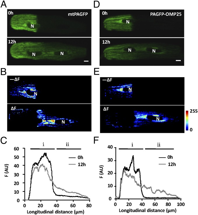
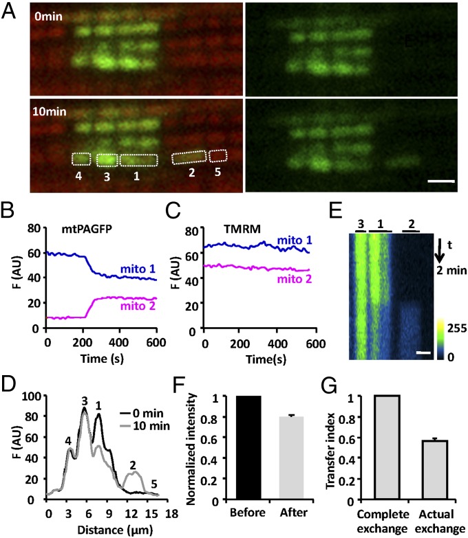
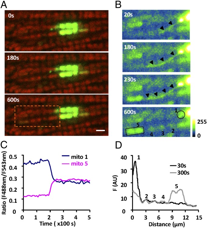
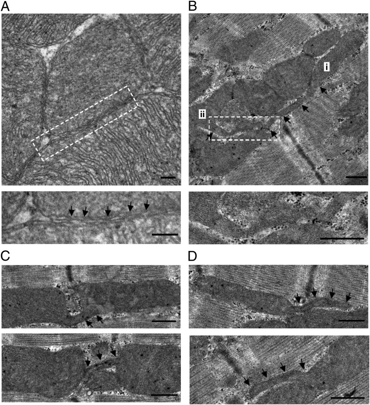
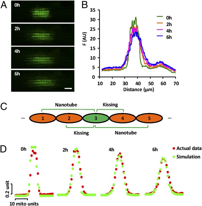
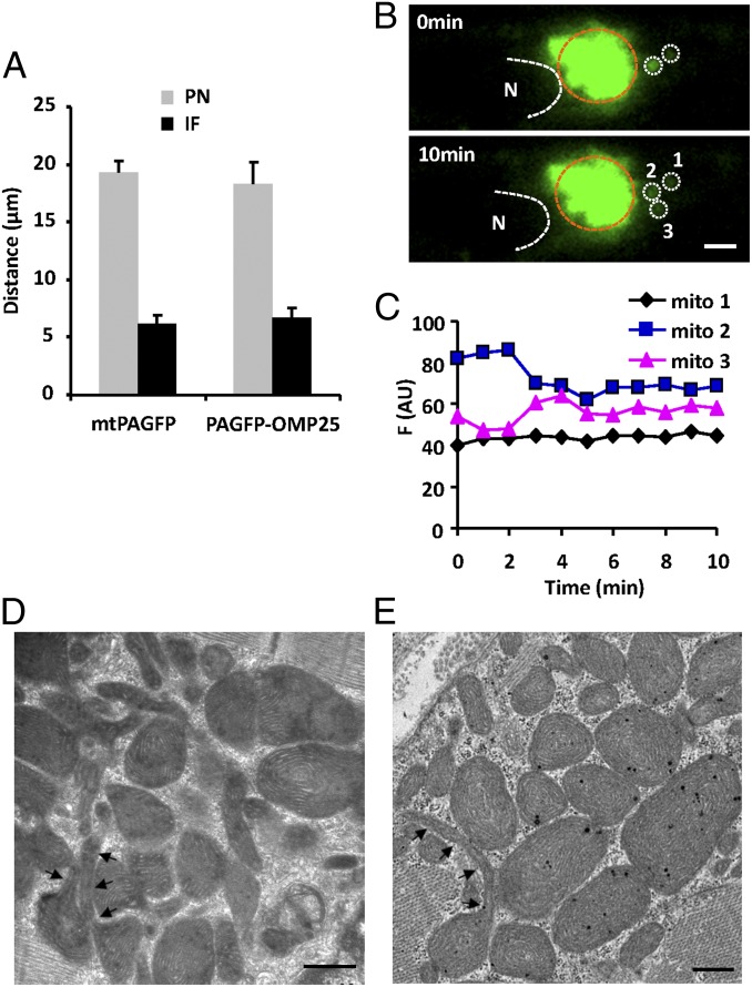
Mitochondria in many types of cells are dynamically interconnected through constant fusion and fission, allowing for exchange of mitochondrial contents and repair of damaged mitochondria. However, constrained by the myofibril lattice, the ∼6,000 mitochondria in the adult mammalian cardiomyocyte display little motility, and it is unclear how, if at all, they communicate with each other. By means of target-expressing photoactivatable green fluorescent protein (PAGFP) in the mitochondrial matrix or on the outer mitochondrial membrane, we demonstrated that the local PAGFP signal propagated over the entire population of mitochondria in cardiomyocytes on a time scale of ∼10 h. Two elemental steps of intermitochondrial communications were manifested as either a sudden PAGFP transfer between a pair of adjacent mitochondria (i.e., "kissing") or a dynamic nanotubular tunnel (i.e., "nanotunneling") between nonadjacent mitochondria. The average content transfer index (fractional exchange) was around 0.5; the rate of kissing was 1‰ s(-1) per mitochondrial pair, and that of nanotunneling was about 14 times smaller. Electron microscopy revealed extensive intimate contacts between adjacent mitochondria and elongated nanotubular protrusions, providing a structural basis for the kissing and nanotunneling, respectively. We propose that, through kissing and nanotunneling, the otherwise static mitochondria in a cardiomyocyte form one dynamically continuous network to share content and transfer signals.


Conflict of interest statement

The authors declare no conflict of interest.

Figures

Fig. 1.
Fig. 1.
Intermitochondrial communication in adult rat cardiomyocytes. (A and D) Confocal images of cardiomyocytes transfected with adeno-mtPAFGP (A) or adeno-PAGFP-OMP25 (D), immediately after photoactivation (Upper) or 12 h later (Lower). (Scale bars: 5 μm.) (B and E) Regional loss (−△F) and gain of fluorescence (△F) at 12 h after photoactivation. N, nucleus. (C and F) Distribution of PAGFP fluorescence along the cell length at indicated time points. i, photoactivated area; ii, nonactivated area.
Fig. 2.
Fig. 2.
Kissing between a pair of adjacent mitochondria. (A) Confocal images of interfibrillar mitochondria in cardiomyocytes expressing mtPAGFP and stained with TMRM, at indicated time points after photoactivation. (Scale bar: 2 μm.) (B) Time courses of mtPAGFP fluorescence in adjacent mitochondria, mito-1 and mito-2. (C) As in B, for TMRM fluorescence. (D) Spatial profiles showing the mtPAGFP fluorescence intensity along the mitochondrial bundle immediately or 10 min after photoactivation. (E) Space–time visualization of a mitochondrial kissing event. Time runs from top to bottom and mitochondrial location is shown horizontally. (F) Normalized mtPAGFP fluorescence intensity in donor mitochondria before and after kissing. (G) Mitochondrial transfer index.
Fig. 3.
Fig. 3.
Nanotunneling between two distant mitochondria. (A) Confocal images show the overlapped mtPAGFP (green) and TMRM signals (red) at indicated time points after photoactivation. (Scale bar: 2 μm.) (B) Enlarged views of the area outlined by the rectangle in A with enhanced mtPAGFP signal. Note the emergence and disappearance of a tubular mitochondrial structure (arrowheads) that mediated the remote transfer of mtPAGFP between two well-separated mitochondria without affecting the intermediate ones. (C) Time courses of the ratio of the mtPAGFP/TMRM fluorescence (F488/F543) for mito-1 and mito-5 marked in A. (D) Distribution of mtPAGFP fluorescence in mitochondria as indicated in B at 30 and 300 s after photoactivation.
Fig. 4.
Fig. 4.
Ultrastructure of cardiac mitochondria. (A) TEM data showing intimate kissing junctions between adjacent mitochondria. In the enlarged detail, the outer membranes of the two closely apposed mitochondria establish close contacts at repeated sites (arrows). (B) A nanotubular structure bridging long-distance mitochondria shown by i and ii. (C) Nanotubules connecting adjacent mitochondrial pairs. Note that the tubular structure in the upper panel traverses the gap at the Z-line to connect the mitochondrial pair. (D) Nanotubules extending (traced by arrows) from mitochondria in the interfibrillar area. (Scale bar: 500 nm.)
Fig. 5.
Fig. 5.
Model simulation of mitochondrial communication. (A) Confocal images of the mtPAFGP signal at indicated time points after photoactivation of a 10.6 μm × 5.9 μm rectangular region. (Scale bar: 5 μm.) (B) Spatial profiles of mtPAFGP distribution corresponding to A. (C) A model of dynamic content transfer in a linear mitochondrial bundle (Materials and Methods). (D) Monte Carlo simulations at 0 h (after 50 kissing and 2 nanotunneling events), 2, 4, and 6 h (with 1,048 kissing and 52 nanotunneling events), overlaid with experimentally measured fluorescence distributions.
Fig. 6.
Fig. 6.
Perinuclear mitochondrial communication. (A) Average travel distance of mtPAFGP and PAGFP-OMP25 signals 2 h after photoactivation, in mitochondria in the perinuclear (PN) and interfibrillar (IF) areas. (B) Confocal images showing mitochondrial kissing in the PN area. Region surrounded by brown dashed line indicates photoactivated area. (Scale bar: 2 μm.) (C) Fluorescence intensity changes in donor (mito 2) and acceptor mitochondria (mito 3) marked in B. Note the lack of change in mito 1 next to the kissing pair. (D and E) Electron micrographs of perinuclear mitochondria. The arrowheads track nanotubular structures of mitochondria. (Scale bars: 500 nm.)

References

    1. Chan DC. Mitochondria: Dynamic organelles in disease, aging, and development. Cell. 2006;125(7):1241–1252. - PubMed
    1. Westermann B. Mitochondrial fusion and fission in cell life and death. Nat Rev Mol Cell Biol. 2010;11(12):872–884. - PubMed
    1. Chen H, et al. Mitochondrial fusion is required for mtDNA stability in skeletal muscle and tolerance of mtDNA mutations. Cell. 2010;141(2):280–289. - PMC - PubMed
    1. Chen H, et al. Mitofusins Mfn1 and Mfn2 coordinately regulate mitochondrial fusion and are essential for embryonic development. J Cell Biol. 2003;160(2):189–200. - PMC - PubMed
    1. Chen H, Chan DC. Mitochondrial dynamics—fusion, fission, movement, and mitophagy—in neurodegenerative diseases. Hum Mol Genet. 2009;18(R2):R169–R176. - PMC - PubMed

Publication types

MeSH terms

Substances