. 2013 Jan 31:7:3.
doi: 10.3389/fnins.2013.00003. eCollection 2013.

Rapid Glucocorticoid-Induced Activation of TRP and CB1 Receptors Causes Biphasic Modulation of Glutamate Release in Gastric-Related Hypothalamic Preautonomic Neurons

Affiliations

Rapid Glucocorticoid-Induced Activation of TRP and CB1 Receptors Causes Biphasic Modulation of Glutamate Release in Gastric-Related Hypothalamic Preautonomic Neurons

Carie R Boychuk et al. Front Neurosci. .

Abstract

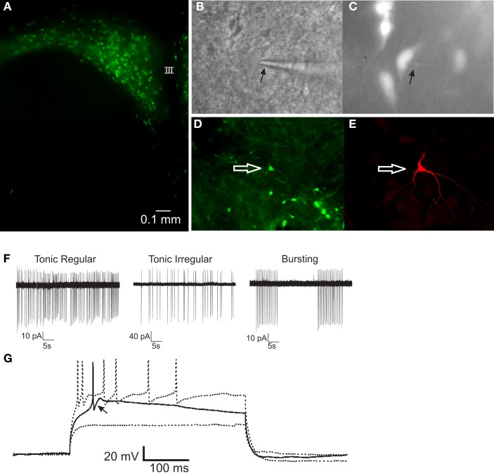
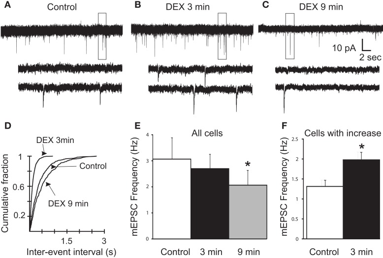
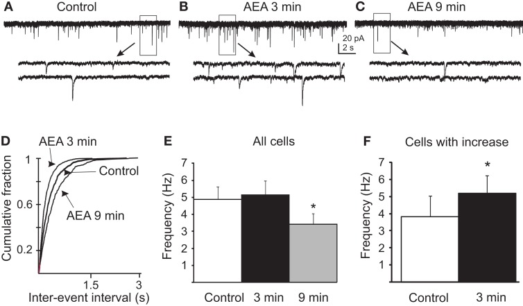
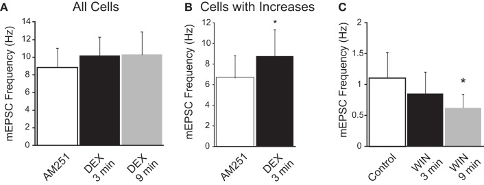
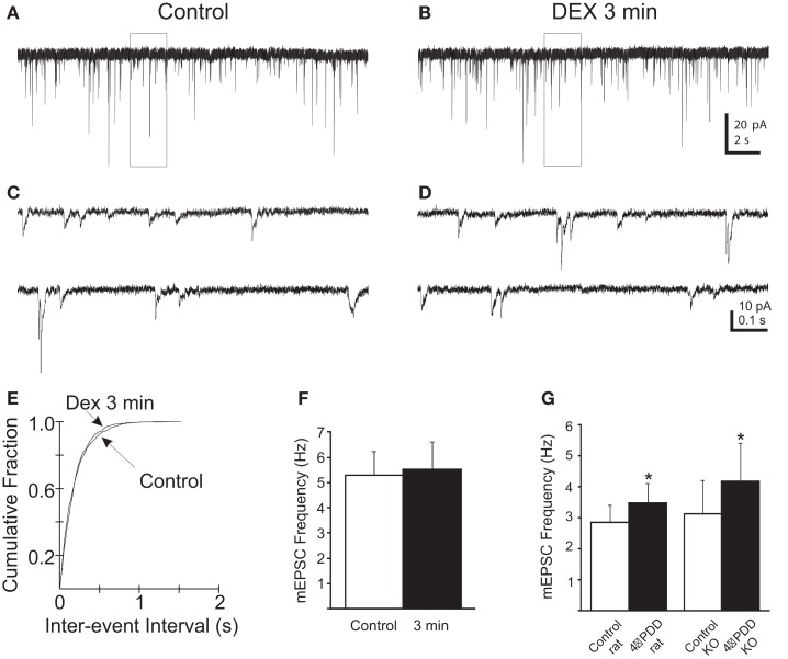
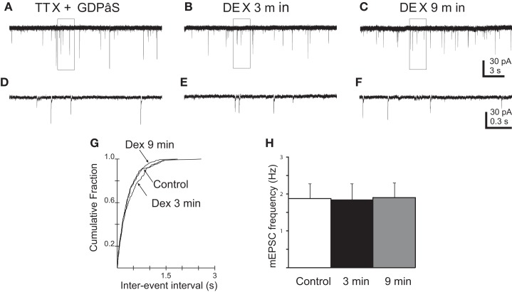
Glucocorticoids rapidly regulate synaptic input to neuroendocrine cells in the hypothalamic paraventricular nucleus (PVN) by inducing the retrograde release of endogenous messengers. Here we investigated the rapid effects of dexamethasone (DEX) on excitatory synaptic input to feeding-related, preautonomic PVN neurons using whole-cell patch-clamp recordings. In ∼50% of identified gastric-related preautonomic PVN neurons, DEX elicited a biphasic synaptic response characterized by an initial rapid and transient increase in the frequency of miniature excitatory postsynaptic currents (mEPSCs), followed by a decrease in mEPSC frequency within 9 min; remaining cells displayed only a decrease in mEPSC frequency. The late-phase decrease in mEPSC frequency was mimicked by the cannabinoid receptor agonists anandamide (AEA) and WIN 55,212-2, and it was blocked by the CB1 receptor antagonist AM251. The biphasic DEX effect was mimicked by AEA. The early increase in mEPSCs was mimicked by activation of transient receptor potential vanilloid type 1 (TRPV1) receptors with capsaicin and by activation of TRPV4 receptors with 4-α-PDD. The increase was reduced, but not blocked, by selective TRPV1 antagonists and in TRPV1 knockout mice; it was blocked completely by the broad-spectrum TRPV antagonist ruthenium red and by combined application of selective TRPV1 and TRPV4 antagonists. The DEX effects were prevented entirely by intracellular infusion of the G-protein inhibitor, GDPβS. Thus, DEX biphasically modulates synaptic glutamate onto a subset of gastric-related PVN neurons, which is likely mediated by induction of a retrograde messenger. The effect includes a TRPV1/4 receptor-mediated transient increase and subsequent CB1 receptor-mediated suppression of glutamate release. Multiphasic modulation of glutamate input to PVN neurons represents a previously unappreciated complexity of control of autonomic output by glucocorticoids and endogenous cannabinoids.

Keywords: cannabinoid; paraventricular nucleus; vanilloid.


Figures

Figure 1
Figure 1
PRV-152-infected gastric-related preautonomic neuron recorded in the PVN. (A) Fluorescence micrograph of a PVN slice (300 μm) showing EGFP-labeled, gastric-related preautonomic neurons approximately 90 h after PRV-152 inoculation of the stomach. (B) Example of a PVN neuron that was recorded in the slice preparation. Arrow indicates to recording pipette. (C) The same neuron as in (B) illuminated with epifluorescence indicating it contained GFP. (D) Image of a slice from which a GFP-labeled PVN neuron was recorded. The arrow indicates the recorded cell. (E) Fluorescence image of the recorded cell [same as in (D)] filled with biocytin and visualized with an Avidin-Texas Red conjugate. (F) Loose-patch recordings of action potential activity demonstrating typical firing patterns in three PRV-152 labeled, gastric-related PVN neurons. (G) Current-clamp recording from a gastric-related preautonomic neuron that generated a low-threshold spike (arrow) in response to depolarizing current pulses (70–90 pA) from a membrane potential of −90 mV [same neuron as shown in (D,E)]. III, third ventricle.
Figure 2
Figure 2
Rapid biphasic changes in mEPSC frequency in PVN gastric-related preautonomic neurons elicited by dexamethasone (DEX). (A–C) Sequential recordings of mEPSCs observed in an EGFP-labeled PVN neuron at a holding potential of −60 mV in control conditions (A) and after 3 min (B) and 9 min (C) of bath application of 10 μM DEX. Bottom traces show the boxed areas in the upper traces on an expanded time scale. (D) Cumulative probability plots of inter-mEPSC interval distribution from the same cell showed a significant increase at 3 min (leftward shift) and a significant decrease at 9 min (rightward shift) in mEPSC frequency (K–S test, p < 0.02). (E) Mean group changes in mEPSC frequency caused by 10 μM DEX at 3 and 9 min (n = 11; ANOVA, Tukey’s; *p < 0.05 from control). (F) Mean group frequency changes in the subset of neurons in which the K–S test detected an increase in mEPSC frequency (n = 6; *p < 0.05, t-test).
Figure 3
Figure 3
Anandamide biphasically altered mEPSC frequency in gastric-related preautonomic neurons. (A–C) Sequential recordings of mEPSCs observed in EGFP-labeled PVN neurons at a holding potential of −60 mV in control conditions (A) and after 3 min (B) and 9 min (C) of bath application of 10 μM AEA. Bottom traces show the boxed areas in the upper traces on an expanded time scale. (D) Cumulative probability plots of inter-mEPSC interval distribution from the same cell showed a significant increase at 3 min (leftward shift) and a significant decrease at 9 min (rightward shift) in mEPSC frequency (p < 0.02; K–S test). (E) Pooled data from all tested cells showing changes in frequency of mEPSCs caused by 10 μM AEA at 3 and 9 min (n = 9; *p < 0.05 from control and 3 min; ANOVA, Tukey’s). (F) Pooled data from the subset of neurons that responded to AEA with an increase in mEPSC frequency (45%) showing changes in the frequency of mEPSCs at 3 min (n = 5; * p < 0.05, t-test).
Figure 4
Figure 4
CB1 receptor dependence of dexamethasone (DEX)-induced suppression of mEPSC frequency. (A) In the presence of the CB1 receptor antagonist (AM251; 10 μM), DEX did not elicit a significant decrease in mEPSC frequency (n = 6; ANOVA, Tukey’s). (B) In AM251, DEX increased mEPSC frequency in a subset of neurons. The graph illustrates the mean frequency change in cells in which an increase in mEPSC frequency was detected (n = 4; *p < 0.05, t-test). (C) The synthetic cannabinoid agonist, WIN 55,212-2 (WIN; 10 μM) decreased the frequency of mEPSCs (n = 8; *p < 0.05 from control; ANOVA, Tukey’s); an increase in mEPSC frequency was not observed at either the early or late time point in the presence of WIN.
Figure 5
Figure 5
Rapid increase in mEPSC frequency in PVN gastric-related preautonomic neurons elicited by dexamethasone (DEX) at 3 min depends on TRPV1 and TRPV4 receptors. (A,B) Sequential recordings of mEPSCs observed in an EGFP-labeled PVN neuron at a holding potential of −60 mV in the presence of AM251 and the selective TRPV1 (5′-iRFT) and TRPV4 (RN1734) antagonists during control conditions (A) and after 3 min (B) of bath application of 10 μM DEX. (C,D) Expanded traces from the boxed areas in the traces shown in (A,B). (E) Cumulative probability plots of inter-mEPSC interval distribution from the same cell showed no significant increase at 3 min in mEPSC frequency (p < 0.02; K–S test). (F) No change in group means in mEPSC frequency after by 10 μM DEX at 3 and 9 min (n = 5; p > 0.05; ANOVA, Tukey’s). (G) 4-α-PDD (1 μM), a selective TRPV4 receptor agonist, increased the mEPSC frequency in gastric-related preautonomic PVN neurons from rat (n = 4) and TRPV1 knockout mice (n = 6; p < 0.05; t-test).
Figure 6
Figure 6
Blockade of G-protein activity prevented DEX-induced changes in mEPSC frequency in PVN gastric-related preautonomic neurons. (A–C) Sequential recordings of mEPSCs observed in an EGFP-labeled PVN neuron at a holding potential of −60 mV loaded with the G-protein inhibitor, GDPβS, during control conditions (A), after 3 (B) and 9 min (C) of bath application of 10 μM DEX. (D–F) Expanded traces from the boxed areas in the traces shown in (A–C). (G) Cumulative probability plots of inter-mEPSC interval distribution from the same cell showed no significant increase at any time in mEPSC frequency (p > 0.05; K–S test). (H) No change in group means in mEPSC frequency after by 10 μM DEX at 3 and 9 min (n = 5; p > 0.05; ANOVA, Tukey’s).

References

    1. Cano G., Card J. P., Rinaman L., Sved A. F. (2000). Connections of Barrington’s nucleus to the sympathetic nervous system in rats. J. Auton. Nerv. Syst. 79, 117–12810.1016/S0165-1838(99)00101-0 - DOI - PubMed
    1. Cano G., Sved A. F., Rinaman L., Rabin B. S., Card J. P. (2001). Characterization of the central nervous system innervation of the rat spleen using viral transneuronal tracing. J. Comp. Neurol. 439, 1–1810.1002/cne.1331 - DOI - PubMed
    1. Carreno F. R., Ji L. L., Cunningham J. T. (2009). Altered central TRPV4 expression and lipid raft association related to inappropriate vasopressin secretion in cirrhotic rats. Am. J. Physiol. Regul. Integr. Comp. Physiol. 296, R454–R46610.1152/ajpregu.90460.2008 - DOI - PMC - PubMed
    1. Caterina M. J. (2007). Transient receptor potential ion channels as participants in thermosensation and thermoregulation. Am. J. Physiol. Regul. Integr. Comp. Physiol. 292, R64–R7610.1152/ajpregu.00446.2006 - DOI - PubMed
    1. Ch’ng T. H., Spear P. G., Struyf F., Enquist L. W. (2007). Glycoprotein D-independent spread of pseudorabies virus infection in cultured peripheral nervous system neurons in a compartmented system. J. Virol. 81, 10742–1075710.1128/JVI.00981-07 - DOI - PMC - PubMed

LinkOut - more resources