. 2013;4(2):104-16.
doi: 10.7150/jca.5002. Epub 2013 Jan 10.

In Vitro Assessment of the Inflammatory Breast Cancer Cell Line SUM 149: Discovery of 2 Single Nucleotide Polymorphisms in the RNase L Gene

Affiliations

In Vitro Assessment of the Inflammatory Breast Cancer Cell Line SUM 149: Discovery of 2 Single Nucleotide Polymorphisms in the RNase L Gene

Brandon T Nokes et al. J Cancer. 2013.

Abstract

Background: Inflammatory breast cancer (IBC) is a rare, highly aggressive form of breast cancer. The mechanism of IBC carcinogenesis remains unknown. We sought to evaluate potential genetic risk factors for IBC and whether or not the IBC cell lines SUM149 and SUM190 demonstrated evidence of viral infection.

Methods: We performed single nucleotide polymorphism (SNP) genotyping for 2 variants of the ribonuclease (RNase) L gene that have been correlated with the risk of prostate cancer due to a possible viral etiology. We evaluated dose-response to treatment with interferon-alpha (IFN-α); and assayed for evidence of the putative human mammary tumor virus (HMTV, which has been implicated in IBC) in SUM149 cells. A bioinformatic analysis was performed to evaluate expression of RNase L in IBC and non-IBC.

Results: 2 of 2 IBC cell lines were homozygous for RNase L common missense variants 462 and 541; whereas 2 of 10 non-IBC cell lines were homozygous positive for the 462 variant (p= 0.09) and 0 of 10 non-IBC cell lines were homozygous positive for the 541 variant (p = 0.015). Our real-time polymerase chain reaction (RT-PCR) and Southern blot analysis for sequences of HMTV revealed no evidence of the putative viral genome.

Conclusion: We discovered 2 SNPs in the RNase L gene that were homozygously present in IBC cell lines. The 462 variant was absent in non-IBC lines. Our discovery of these SNPs present in IBC cell lines suggests a possible biomarker for risk of IBC. We found no evidence of HMTV in SUM149 cells. A query of a panel of human IBC and non-IBC samples showed no difference in RNase L expression. Further studies of the RNase L 462 and 541 variants in IBC tissues are warranted to validate our in vitro findings.

Keywords: HMTV; MMTV; RNase L.; SUM149; inflammatory breast cancer; interferon-alpha.


Conflict of interest statement

Competing Interests: The authors have declared that no competing interest exists.

Figures

Figure 1
Figure 1
Schematic representation of the innate IFN-RNase L pathway as influenced by viral infection. Viral infection triggers the release of Type I interferons, which act on the infected cell in an autocrine fashion. IFN signaling initiated the Oligoadenylate Synthase (OAS)/RNase L pathway for nucleic acid degradation (Adapted from Silverman 2007) [50].
Figure 2
Figure 2
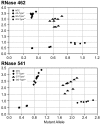
Single nucleotide polymorphism (SNP) allele discrimination scatter plot forribonuclease (RNase) L variants (A) 462 (rs486907) and (B) 541 (rs627928). The Y-axis indicates detection of the 462Q(A) and 541D (B) allele by a FAM reporter (diamonds), and the X-axis indicates detection of the 462R (A) and 541E (B) allele by a VIC reporter (circles). Heterozygous genotypes displayed amplified copies of both alleles and were found in an intermediate position between homozygous mutants and homozygous wildtype cell lines (triangles). The No Template Control (NTC) wells are denoted as squares. *Non-IBC cell lines, **Corresponds to IBC cell lines.
Figure 3
Figure 3
Plot of expression of gene RNASEL on Affymetrix expression microarrays of IBC and non-IBC tumor samples. This plot was generated on the NCBI GEO website from data set GSE5847 originally published by Boersma et al. 2008 [34]. Log expression value (red) and rank order of expression value (blue) are shown for each sample.
Figure 4
Figure 4
Cell proliferation as a function of Interferon (IFN- α) dosing. Increasing doses of IFN-α decreased cell proliferation significantly in a dose and time-dependent manner in SUM149 cells. Cells were treated with 0, 500, 1000, 2500, and 5000 U/ml of IFN-α for 24 and 48 hours. At each time point, the IFN-α was removed, the cells rinsed with PBS, and cell proliferation was evaluated (* P < 0.05, ** P < 0.01, (*** P < 0.001). The reduction in cell proliferation induced by treatment with IFN-α is an IBC-specific response. The 2 non-IBC cell lines (A) MDA-MB-231 and (B) BT474 did not demonstrate a significant decrease in cell proliferation with IFN-α treatment (* P < 0.05, ** P < 0.01, *** P < 0.001).
Figure 5
Figure 5
IFN- α response abrogated with antibody block. (A) SUM149 cells were treated for 24 hours with increased doses of IFN-α; cell proliferation decreased in a dose-dependent manner. (B) IFN-α complexed to IFN-α specific antibody abrogated the dose-dependent decrease in cell proliferation. For IFN-α block experiments, each treatment was pre-incubated for 15 minutes with 1 μg/ml IFN-α specific antibody (* P < 0.05, ** P < 0.01, *** P < 0.001).
Figure 6
Figure 6
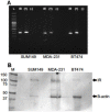
(A) Reverse transcription polymerase chain reaction (RT-PCR) and (B) Western blot for human IFN-α Receptor. (A) RT-PCR showed that the IFN- α receptor was basally expressed in untreated cultured SUM149, MDA-MB-231, and BT474 cells. L: DNA ladder, IR: IFN-α receptor, (+): GAPDH controls, (-): no template negative controls. (B) Western blot analysis indicating human IFN-α receptor protein expressed in the membrane fraction of SUM149, MDA-MB-231, and BT474 cell lines. M: Molecular weight marker, IR: Interferon-alpha receptor type I. β-actin served as controls for SDS-PAGE protein loading and for complete immunoblot transfer.
Figure 7
Figure 7
PCR products from SUM149 genomic DNA. Agarose gel electrophoresis of PCR amplicons from genomic DNA (A) and RT-PCR amplicons from RNA (B) using 4 primer sets (see materials and methods for sequences) for the HMTV env/LTR and late LTR regions. Expected product sizes if HMTV sequences were present are shown at the bottom of each gel. A. PCR analysis resulted in nonspecific amplicons from chromosomes 7 and 17. The white box highlights bands of interest that were cloned and sequenced. B. RT-PCR using several primer pairs over an annealing temperature gradient produced nonspecific amplicons, as compared with the positive controls (+) GAPDH. Molecular weight marker was 47kDa.
Figure 8
Figure 8
Southern blot analysis for HMTV sequences in SUM149 genome. Southern blot analysis indicated the env region of HMTV was not present in the SUM149 genome. (A) Ethidium bromide visualization of BamHI-digested and gel electrophoresed SUM149 genomic DNA and a synthetic 172 base-pair HMTV fragment. (B) Southern blot probed with biotinylated HMTV env probe. L: DNA ladder, S: 10 μg digested SUM149 genomic DNA, (+): HMTV controls.

References

    1. Gonzalez-Angulo AM, Hennessy BT, Broglio K. et al. Trends for inflammatory breast cancer: is survival improving? Oncologist. 2007;12:904–12. - PubMed
    1. Bristol IJ, Woodward WA, Strom EA. et al. Locoregional treatment outcomes after multimodality management of inflammatory breast cancer. Int J Radiat Oncol Biol Phys. 2008;72:474–84. - PMC - PubMed
    1. Charafe-Jauffret E, Tarpin C, Viens P, Bertucci F. Defining the molecular biology of inflammatory breast cancer. Semin Oncol. 2008;35:41–50. - PubMed
    1. Hance KW, Anderson WF, Devesa SS, Young HA, Levine PH. Trends in inflammatory breast carcinoma incidence and survival: the surveillance, epidemiology, and end results program at the National Cancer Institute. J Natl Cancer Inst. 2005;97:966–75. - PMC - PubMed
    1. Dirix LY, Van Dam P, Prove A, Vermeulen PB. Inflammatory breast cancer: current understanding. Curr Opin Oncol. 2006;18:563–71. - PubMed

LinkOut - more resources