

Mesoporous silica nanoparticle nanocarriers: biofunctionality and biocompatibility
- PMID: 23387478
- PMCID: PMC3686852
- DOI: 10.1021/ar3000986
Mesoporous silica nanoparticle nanocarriers: biofunctionality and biocompatibility
Abstract
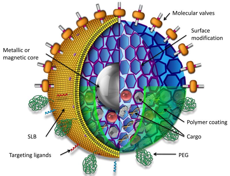
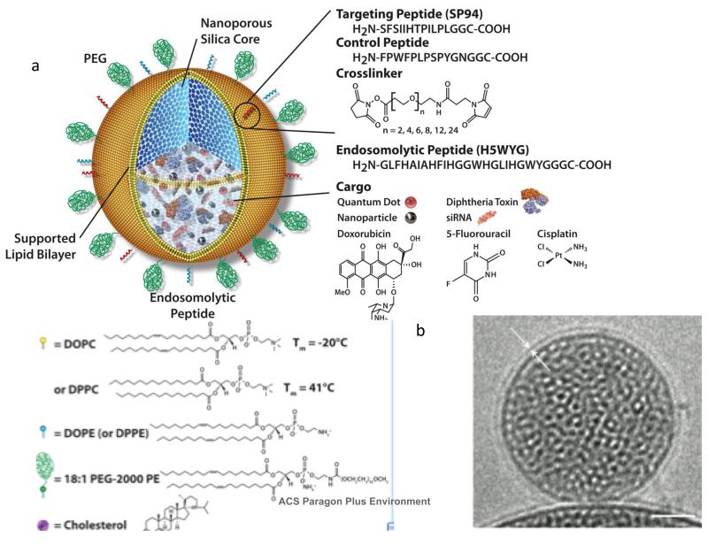
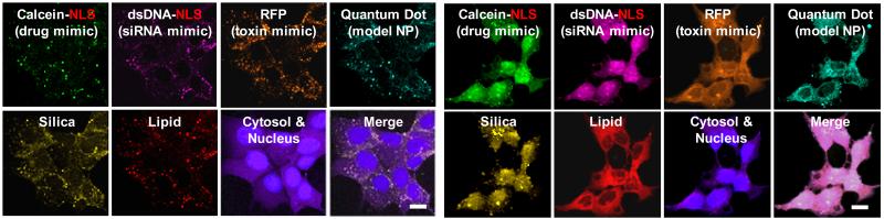
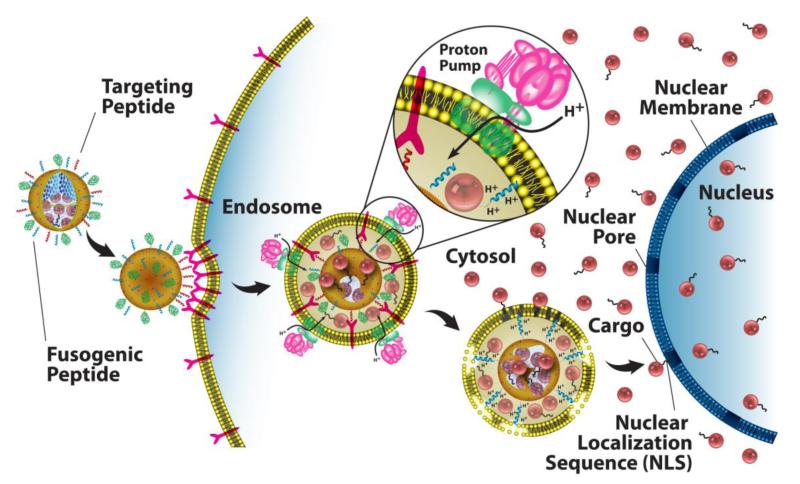
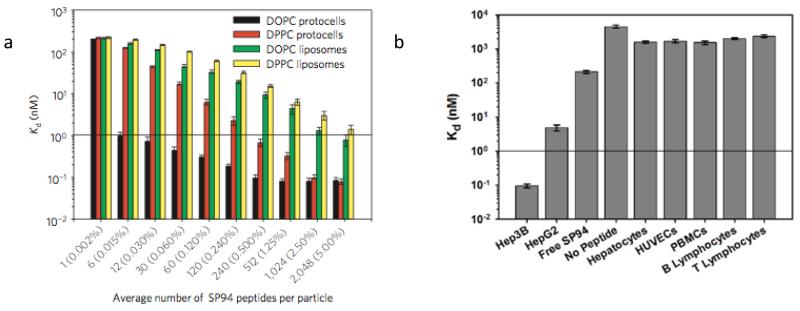
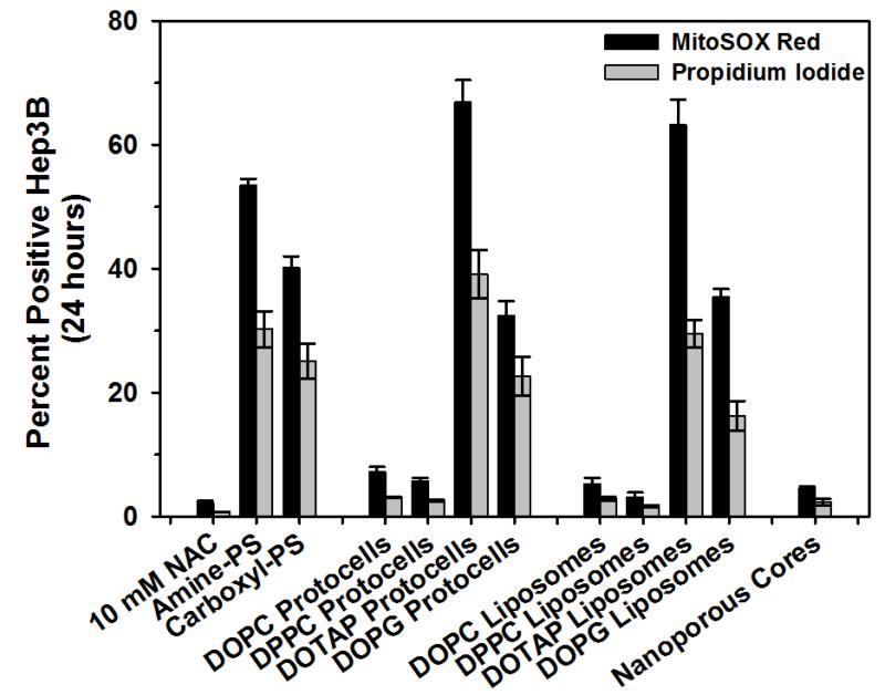
The study of ordered mesoporous silica materials has exploded since their discovery by Mobil researchers 20 years ago. The ability to make uniformly sized, porous, and dispersible nanoparticles using colloidal chemistry and evaporation-induced self-assembly has led to many applications of mesoporous silica nanoparticles (MSNPs) as "nanocarriers" for delivery of drugs and other cargos to cells. The exceptionally high surface area of MSNPs, often exceeding 1000 m²/g, and the ability to independently modify pore size and surface chemistry, enables the loading of diverse cargos and cargo combinations at levels exceeding those of other common drug delivery carriers such as liposomes or polymer conjugates. This is because noncovalent electrostatic, hydrogen-bonding, and van der Waals interactions of the cargo with the MSNP internal surface cause preferential adsorption of cargo to the MSNP, allowing loading capacities to surpass the solubility limit of a solution or that achievable by osmotic gradient loading. The ability to independently modify the MSNP surface and interior makes possible engineered biofunctionality and biocompatibility. In this Account, we detail our recent efforts to develop MSNPs as biocompatible nanocarriers (Figure 1 ) that simultaneously display multiple functions including (1) high visibility/contrast in multiple imaging modalities, (2) dispersibility, (3) binding specificity to a particular target tissue or cell type, (4) ability to load and deliver large concentrations of diverse cargos, and (5) triggered or controlled release of cargo. Toward function 1, we chemically conjugated fluorescent dyes or incorporated magnetic nanoparticles to enable in vivo optical or magnetic resonance imaging. For function 2, we have made MSNPs with polymer coatings, charged groups, or supported lipid bilayers, which decrease aggregation and improve stability in saline solutions. For functions 3 and 4, we have enhanced passive bioaccumulation via the enhanced permeability and retention effect by modifying the MSNP surfaces with positively charged polymers. We have also chemically attached ligands to MSNPs that selectively bind to receptors overexpressed in cancer cells. We have used encapsulation of MSNPs within reconfigurable supported lipid bilayers to develop new classes of responsive nanocarriers that actively interact with the target cell. Toward function 4, we exploit the high surface area and tailorable surface chemistry of MSNPs to retain hydrophobic drugs. Finally, for function 5, we have engineered dynamic behaviors by incorporating molecular machines within or at the entrances of MSNP pores and by using ligands, polymers, or lipid bilayers. These provide a means to seal-in and retain cargo and to direct MSNP interactions with and internalization by target cells. Application of MSNPs as nanocarriers requires biocompatibility and low toxicity. Here the intrinsic porosity of the MSNP surface reduces the extent of hydrogen bonding or electrostatic interactions with cell membranes as does surface coating with polymers or lipid bilayers. Furthermore, the high surface area and low extent of condensation of the MSNP siloxane framework promote a high rate of dissolution into soluble silicic acid species, which are found to be nontoxic. Potential toxicity is further mitigated by the high drug capacity of MSNPs, which greatly reduces needed dosages compared with other nanocarriers. We anticipate that future generations of MSNPs incorporating molecular machines and encapsulated by membrane-like lipid bilayers will achieve a new level of controlled cellular interactions.
Figures
Similar articles
-
Stimuli-responsive mesoporous silica nanoparticles: A custom-tailored next generation approach in cargo delivery.Mater Sci Eng C Mater Biol Appl. 2021 May;124:112084. doi: 10.1016/j.msec.2021.112084. Epub 2021 Mar 31. Mater Sci Eng C Mater Biol Appl. 2021. PMID: 33947574 Review.
-
Enhanced efficacy and drug delivery with lipid coated mesoporous silica nanoparticles in cancer therapy.Eur J Pharm Biopharm. 2021 Aug;165:31-40. doi: 10.1016/j.ejpb.2021.04.020. Epub 2021 May 4. Eur J Pharm Biopharm. 2021. PMID: 33962002
-
Enhancing Cellular Uptake and Doxorubicin Delivery of Mesoporous Silica Nanoparticles via Surface Functionalization: Effects of Serum.ACS Appl Mater Interfaces. 2015 Dec 9;7(48):26880-91. doi: 10.1021/acsami.5b09483. Epub 2015 Nov 20. ACS Appl Mater Interfaces. 2015. PMID: 26562468
-
Synthesis of lactoferrin mesoporous silica nanoparticles for pemetrexed/ellagic acid synergistic breast cancer therapy.Colloids Surf B Biointerfaces. 2020 Apr;188:110824. doi: 10.1016/j.colsurfb.2020.110824. Epub 2020 Jan 24. Colloids Surf B Biointerfaces. 2020. PMID: 32023511
-
Functionalized mesoporous silica nanoparticles in anticancer therapeutics.Semin Cancer Biol. 2021 Feb;69:365-375. doi: 10.1016/j.semcancer.2019.08.022. Epub 2019 Aug 20. Semin Cancer Biol. 2021. PMID: 31442571 Review.
Cited by
-
Comprehensive Survey on Nanobiomaterials for Bone Tissue Engineering Applications.Nanomaterials (Basel). 2020 Oct 13;10(10):2019. doi: 10.3390/nano10102019. Nanomaterials (Basel). 2020. PMID: 33066127 Free PMC article. Review.
-
Epigallocatechin-3-gallate/mineralization precursors co-delivery hollow mesoporous nanosystem for synergistic manipulation of dentin exposure.Bioact Mater. 2022 Nov 29;23:394-408. doi: 10.1016/j.bioactmat.2022.11.018. eCollection 2023 May. Bioact Mater. 2022. PMID: 36474660 Free PMC article.
-
Emerging vaccine delivery systems for COVID-19: Functionalised silica nanoparticles offer a potentially safe and effective alternative delivery system for DNA/RNA vaccines and may be useful in the hunt for a COVID-19 vaccine.Drug Discov Today. 2020 Sep;25(9):1556-1558. doi: 10.1016/j.drudis.2020.06.020. Epub 2020 Jun 24. Drug Discov Today. 2020. PMID: 32592866 Free PMC article. No abstract available.
-
Positive impact of IGF-1-coupled nanoparticles on the differentiation potential of human chondrocytes cultured on collagen scaffolds.Int J Nanomedicine. 2015 Feb 4;10:1131-43. doi: 10.2147/IJN.S72872. eCollection 2015. Int J Nanomedicine. 2015. PMID: 25709437 Free PMC article.
-
Antisense vicR-Loaded Dendritic Mesoporous Silica Nanoparticles Regulate the Biofilm Organization and Cariogenicity of Streptococcus mutans.Int J Nanomedicine. 2022 Mar 21;17:1255-1272. doi: 10.2147/IJN.S334785. eCollection 2022. Int J Nanomedicine. 2022. PMID: 35340824 Free PMC article.
References
-
- Kresge CT, Leonowicz ME, Roth WJ, Vartuli JC, Beck JS. Ordered mesoporous molecular sieves synthesized by a liquid-crystal template mechanism. Nature. 1992;359:710–712.
-
- Brinker CJ, Lu YF, Sellinger A, Fan HY. Evaporation-induced self-assembly: Nanostructures made easy. Advanced Materials. 1999;11:579–585.
-
- Beck JS, Vartuli JC, Roth WJ, Leonowicz ME, Kresge CT, Schmitt KD, Chu CTW, Olson DH, Sheppard EW. A new family of mesoporous molecular sieves prepared with liquid crystal templates. Journal of the American Chemical Society. 1992;114:10834–10843.
-
- Grün M, Lauer I, Unger KK. The synthesis of micrometer- and submicrometer-size spheres of ordered mesoporous oxide MCM-41. Advanced Materials. 1997;9:254–257.
-
- Huh S, Wiench JW, Yoo J-C, Pruski M, Lin VSY. Organic Functionalization and Morphology Control of Mesoporous Silicas via a Co-Condensation Synthesis Method. Chemistry of Materials. 2003;15:4247–4256.
Publication types
MeSH terms
Substances
Grants and funding
LinkOut - more resources
Full Text Sources
Other Literature Sources
