

Gene signature distinguishes patients with chronic ulcerative colitis harboring remote neoplastic lesions
- PMID: 23388545
- PMCID: PMC3836269
- DOI: 10.1097/MIB.0b013e3182802bac
Gene signature distinguishes patients with chronic ulcerative colitis harboring remote neoplastic lesions
Abstract
Background: Individuals with ulcerative colitis (UC) are at increased risk for colorectal cancer. The standard method of surveillance for neoplasia in UC by colonoscopy is invasive and can miss flat lesions. We sought to identify a gene expression signature in nondysplastic mucosa without active inflammation that could serve as a marker for remote neoplastic lesions.
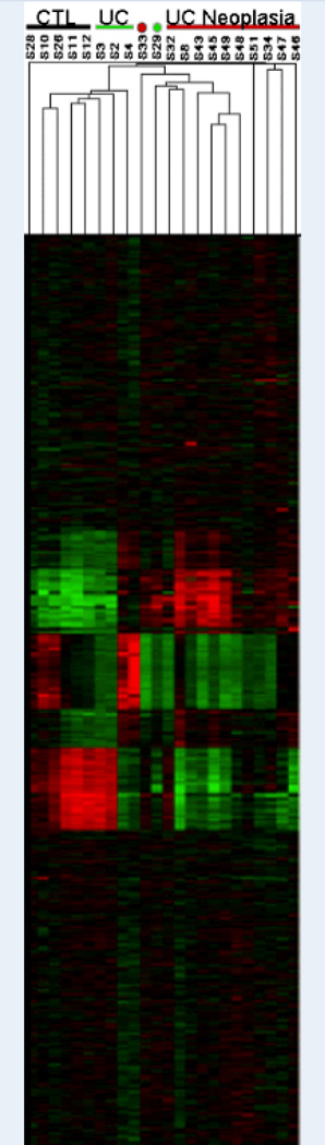
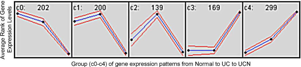
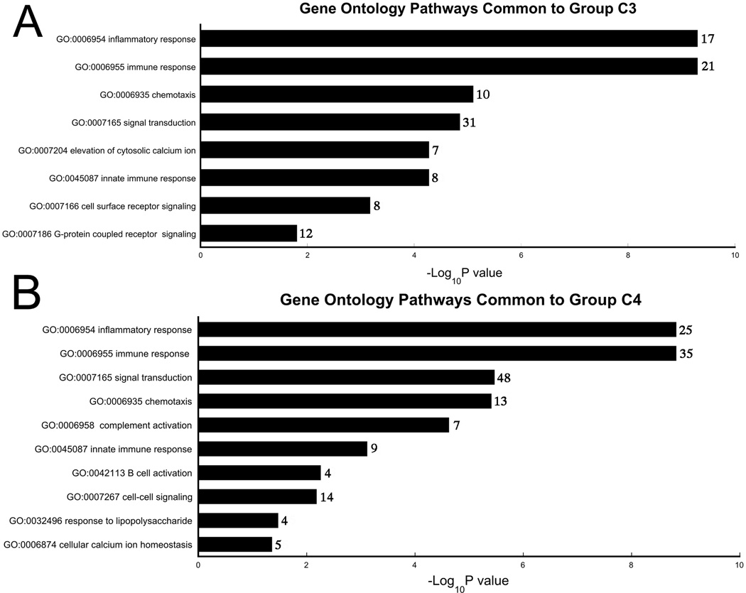
Methods: Gene expression was analyzed by complementary DNA microarray in 5 normal controls, 4 UC patients without dysplasia, and 11 UC patients harboring remote neoplasia. Common gene ontology pathways of significantly differentially expressed genes were identified. Expression of genes which were progressively and significantly upregulated from controls to UC without neoplasia, to UC with remote neoplasia were evaluated by real-time polymerase chain reaction. Several gene products were also examined by immunohistochemistry.
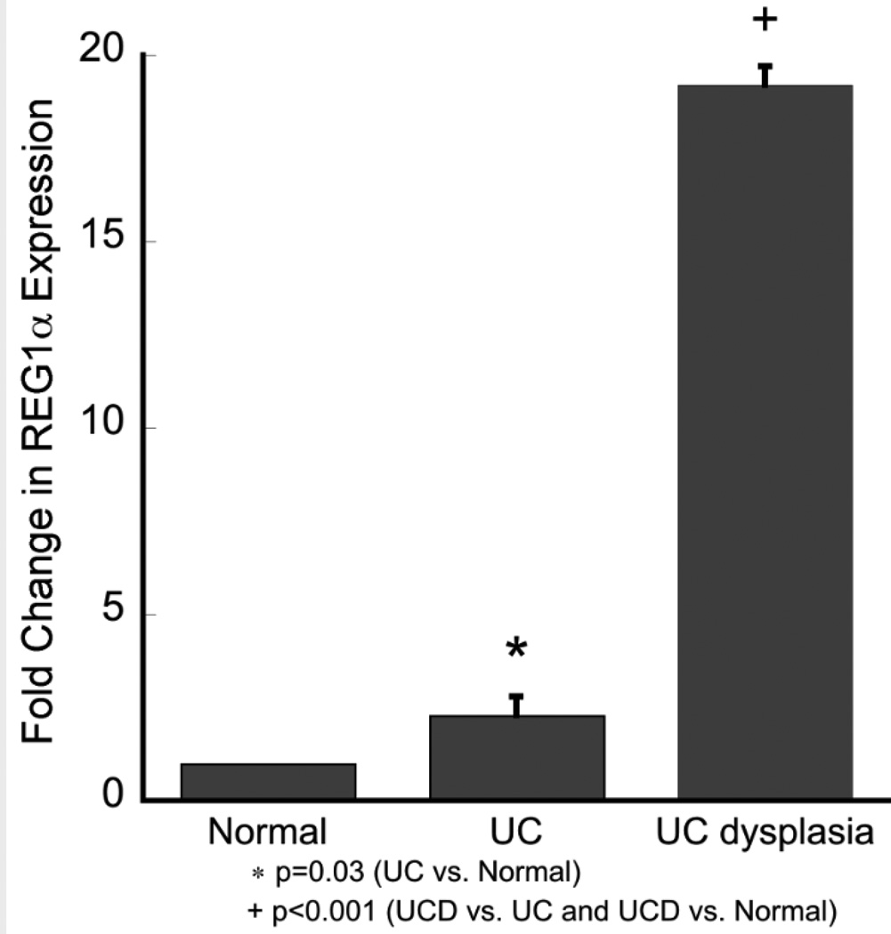
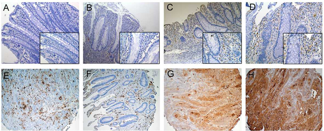
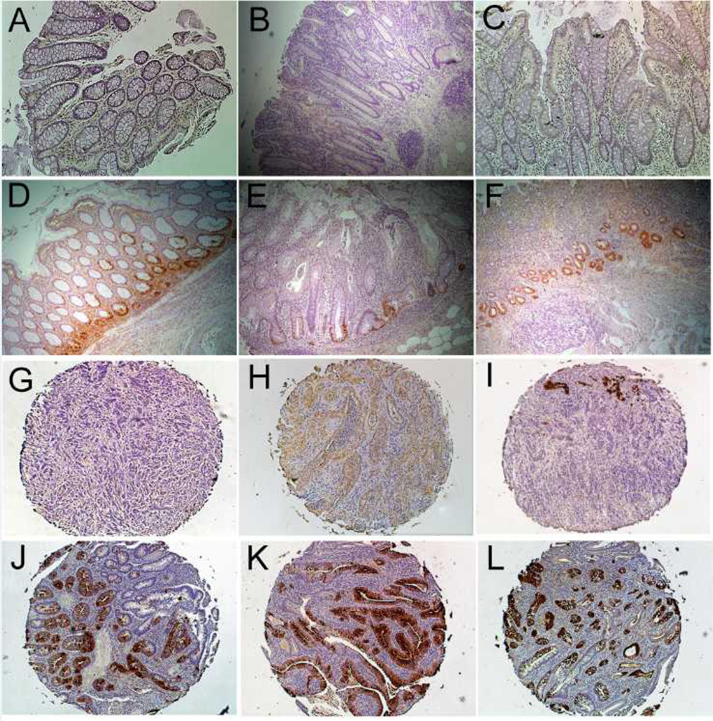
Results: Four hundred and sixty-eight genes were significantly upregulated, and 541 genes were significantly downregulated in UC patients with neoplasia compared with UC patients without neoplasia. Nine genes (ACSL1, BIRC3, CLC, CREM, ELTD1, FGG, S100A9, THBD, and TPD52L1) were progressively and significantly upregulated from controls to nondysplastic UC to UC with neoplasia. Immunostaining of proteins revealed increased expression of S100A9 and REG1α in UC-associated cancer and in nondysplastic tissue from UC patients harboring remote neoplasia compared with UC patients without neoplasia and controls.
Conclusions: Gene expression changes occurring as a field effect in the distal colon of patients with chronic UC identify patients harboring remote neoplastic lesions. These markers may lead to a more accurate and less invasive method of detection of neoplasia in patients with inflammatory bowel disease.
Figures
Similar articles
-
miR-4728-3p Functions as a Tumor Suppressor in Ulcerative Colitis-associated Colorectal Neoplasia Through Regulation of Focal Adhesion Signaling.Inflamm Bowel Dis. 2017 Aug;23(8):1328-1337. doi: 10.1097/MIB.0000000000001104. Inflamm Bowel Dis. 2017. PMID: 28594651 Free PMC article.
-
Up-regulation of mitochondrial chaperone TRAP1 in ulcerative colitis associated colorectal cancer.World J Gastroenterol. 2014 Dec 7;20(45):17037-48. doi: 10.3748/wjg.v20.i45.17037. World J Gastroenterol. 2014. PMID: 25493016 Free PMC article.
-
A Panel of Methylated MicroRNA Biomarkers for Identifying High-Risk Patients With Ulcerative Colitis-Associated Colorectal Cancer.Gastroenterology. 2017 Dec;153(6):1634-1646.e8. doi: 10.1053/j.gastro.2017.08.037. Epub 2017 Aug 25. Gastroenterology. 2017. PMID: 28847750 Free PMC article.
-
Serrated Epithelial Change Is Associated With High Rates of Neoplasia in Ulcerative Colitis Patients: A Case-controlled Study and Systematic Review With Meta-analysis.Inflamm Bowel Dis. 2021 Aug 19;27(9):1475-1481. doi: 10.1093/ibd/izaa312. Inflamm Bowel Dis. 2021. PMID: 33295614
-
Limits of diagnosis and molecular markers for early detection of ulcerative colitis-associated colorectal neoplasia.Digestion. 2008;77 Suppl 1:2-12. doi: 10.1159/000111482. Epub 2008 Jan 18. Digestion. 2008. PMID: 18204256 Review.
Cited by
-
Development and validation of a prognostic model for colon cancer based on mitotic gene signatures and immune microenvironment analysis.Discov Oncol. 2024 Oct 9;15(1):535. doi: 10.1007/s12672-024-01421-2. Discov Oncol. 2024. PMID: 39382813 Free PMC article.
-
Biomarkers for colitis-associated colorectal cancer.World J Gastroenterol. 2016 Sep 21;22(35):7882-91. doi: 10.3748/wjg.v22.i35.7882. World J Gastroenterol. 2016. PMID: 27672285 Free PMC article. Review.
-
Increased mucosal expression of miR-215 precedes the development of neoplasia in patients with long-standing ulcerative colitis.Oncotarget. 2018 Apr 17;9(29):20709-20720. doi: 10.18632/oncotarget.25065. eCollection 2018 Apr 17. Oncotarget. 2018. PMID: 29755683 Free PMC article.
-
miR-193a-3p is a Key Tumor Suppressor in Ulcerative Colitis-Associated Colon Cancer and Promotes Carcinogenesis through Upregulation of IL17RD.Clin Cancer Res. 2017 Sep 1;23(17):5281-5291. doi: 10.1158/1078-0432.CCR-17-0171. Epub 2017 Jun 9. Clin Cancer Res. 2017. PMID: 28600480 Free PMC article.
-
Epidermal growth factor, latrophilin, and seven transmembrane domain-containing protein 1 marker, a novel angiogenesis marker.Onco Targets Ther. 2015 Dec 16;8:3767-74. doi: 10.2147/OTT.S93843. eCollection 2015. Onco Targets Ther. 2015. PMID: 26719704 Free PMC article. Review.
References
-
- Ekbom A, Helmick C, Zack M, et al. Ulcerative colitis and colorectal cancer A population-based study. N Engl J Med. 1990;323:1228–1233. - PubMed
-
- Itzkowitz SH. Molecular biology of dysplasia and cancer in inflammatory bowel disease. Gastroenterol Clin North Am. 2006;35:553–571. - PubMed
-
- Rutter M, Saunders B, Wilkinson K, et al. Severity of inflammation is a risk factor for colorectal neoplasia in ulcerative colitis. Gastroenterology. 2004;126:451–459. - PubMed
Publication types
MeSH terms
Substances
Grants and funding
LinkOut - more resources
Full Text Sources
Other Literature Sources
Medical
Molecular Biology Databases
Miscellaneous