. 2013 Feb 19;110(8):3011-6.
doi: 10.1073/pnas.1222861110. Epub 2013 Feb 6.

Hematopoietic stem cell and progenitor cell mechanisms in myelodysplastic syndromes

Affiliations

Hematopoietic stem cell and progenitor cell mechanisms in myelodysplastic syndromes

Wendy W Pang et al. Proc Natl Acad Sci U S A. .

Abstract

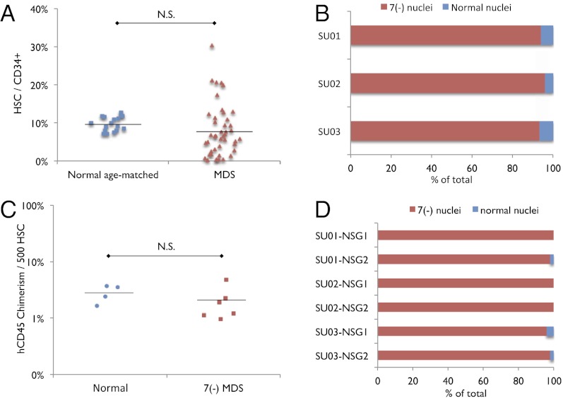
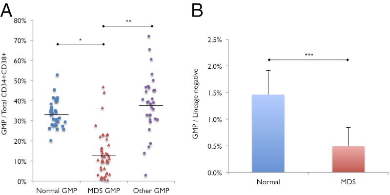
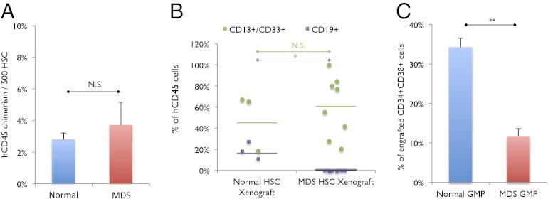
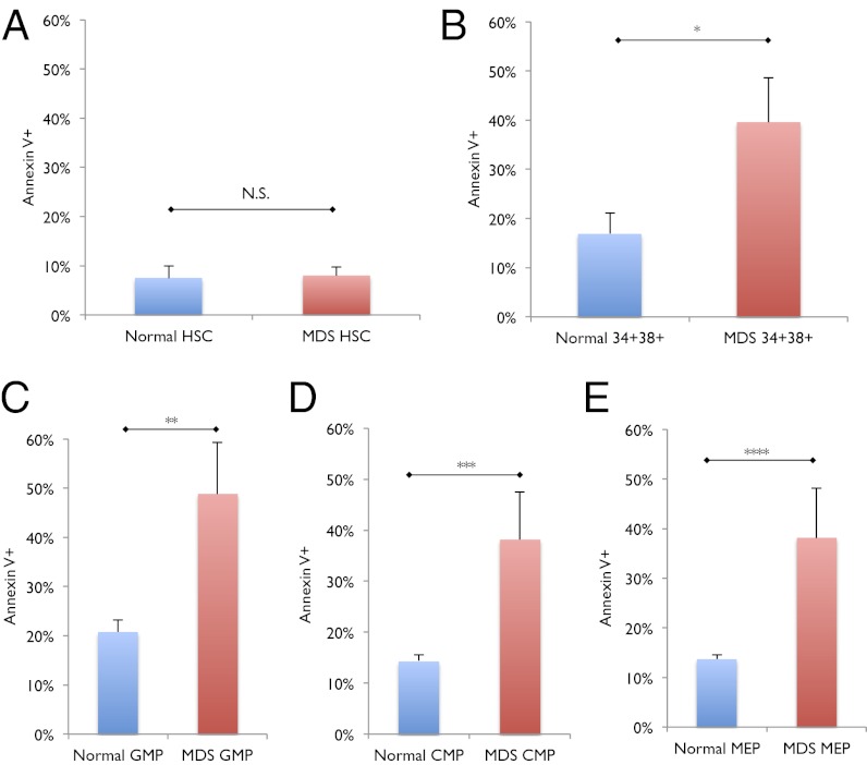
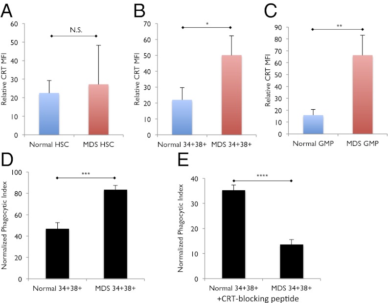
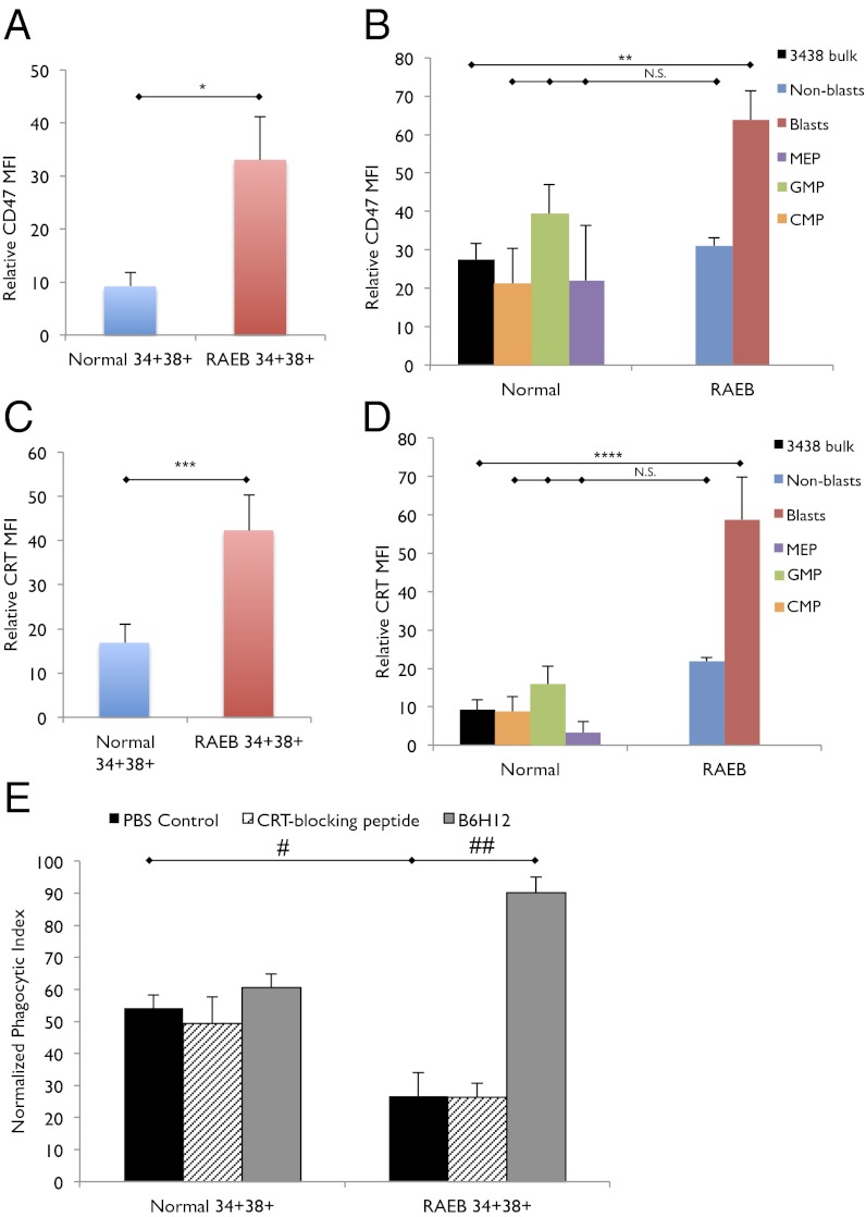
Myelodysplastic syndromes (MDS) are a group of disorders characterized by variable cytopenias and ineffective hematopoiesis. Hematopoietic stem cells (HSCs) and myeloid progenitors in MDS have not been extensively characterized. We transplanted purified human HSCs from MDS samples into immunodeficient mice and show that HSCs are the disease-initiating cells in MDS. We identify a recurrent loss of granulocyte-macrophage progenitors (GMPs) in the bone marrow of low risk MDS patients that can distinguish low risk MDS from clinical mimics, thus providing a simple diagnostic tool. The loss of GMPs is likely due to increased apoptosis and increased phagocytosis, the latter due to the up-regulation of cell surface calreticulin, a prophagocytic marker. Blocking calreticulin on low risk MDS myeloid progenitors rescues them from phagocytosis in vitro. However, in the high-risk refractory anemia with excess blasts (RAEB) stages of MDS, the GMP population is increased in frequency compared with normal, and myeloid progenitors evade phagocytosis due to up-regulation of CD47, an antiphagocytic marker. Blocking CD47 leads to the selective phagocytosis of this population. We propose that MDS HSCs compete with normal HSCs in the patients by increasing their frequency at the expense of normal hematopoiesis, that the loss of MDS myeloid progenitors by programmed cell death and programmed cell removal are, in part, responsible for the cytopenias, and that up-regulation of the "don't eat me" signal CD47 on MDS myeloid progenitors is an important transition step leading from low risk MDS to high risk MDS and, possibly, to acute myeloid leukemia.


Conflict of interest statement

Conflict of interest statement: W.W.P., C.Y.P., and I.L.W. filed US Patent Application Serial No. 13/508,319 entitled “Cell Surface Marker Expression in Hematopoietic Stem Cells and Progenitors for the Diagnosis, Prognosis, and Treatment of Myelodysplastic Syndromes;” I.L.W. filed US Patent Application Serial No. 12/321,215 entitled “Methods for Manipulating Phagocytosis Mediated by CD47;” and I.L.W. filed a patent application regarding therapeutic and diagnostic methods for manipulating phagocytosis through calreticulin and low-density lipoprotein-related receptors.

Figures

Fig. 1.
Fig. 1.
Immunophenotypic HSCs from low risk MDS bone marrow are not increased in frequency and monosomy 7 MDS HSCs reconstitute clonal disease in immunodeficient mice. (A) Frequency of immunophenotypic HSCs within the CD34+ population in normal age-matched (n = 18) and low risk MDS (n = 45) bone marrow samples. N.S., no significance. (B) Percentage of HSCs with nuclei exhibiting deletion of chromosome 7 and normal karyotype in monosomy 7 MDS patients (n = 3; SU001–SU003). One hundred fifty nuclei were analyzed for each sample. (C) Human CD45+ chimerism per 500 transplanted HSCs from normal bone marrow controls (n = 4) and monosomy 7 bone marrow samples (n = 3) into sublethally irradiated NSG newborn mice (one recipient per normal HSC sample; two recipients per monosomy 7 sample; 1,500–3,000 HSCs transplanted per recipient). N.S., no significance. (D) Percentage of successfully engrafted human CD45+ cells at 12–16 wk after transplant, with nuclei exhibiting deletion of chromosome 7 and normal karyotype. HSCs were sorted from three monosomy 7 bone marrow samples (SU001–SU003) and xenotransplanted into two recipients each (NSG1, NSG2). Fifty human CD45+ nuclei were analyzed from each xenotransplant recipient.
Fig. 2.
Fig. 2.
Decreased GMP frequency in low risk MDS. (A) Frequency of GMPs out of total myeloid progenitors (CMPs + GMPs + MEPs) in normal (n = 34), low risk MDS (n = 46; MDS), and non-MDS with at least one cytopenia bone marrow samples (n = 32; Other). *P < 10−13, **P < 10−10. (B) Frequency of GMPs out of total lineage negative bone marrow mononuclear cells in normal and low risk MDS bone marrow samples. ***P < 0.0006. Error bars represent one SD. “MDS” label represents low risk MDS samples.
Fig. 3.
Fig. 3.
Low risk MDS HSCs transplanted into immunodeficient mice yield decreased GMPs. (A) Engraftment of 1,500–3,000 HSCS from normal (n = 4) and low risk MDS (n = 4) bone marrow samples transplanted into NSG newborn mice at 16 wk after transplant (one recipient per normal HSC sample; two recipients per low risk MDS sample), as represented by percentage of human CD45+ chimerism per 500 human HSCs transplanted. N.S., no significance. (B) Lymphoid (CD19+) and myeloid (CD13+/CD33+) engraftment as a percentage of hCD45+ cells in NSG newborn mice at 12–16 wk after transplant. *P < 0.03; N.S. no significance. (C) Frequency of GMPs out of total human CD34+CD38+ cells engrafted in NSG newborn mice at 16 wk after transplant. **P < 0.0007. Error bars represent one SD. “MDS” label represents low risk MDS samples.
Fig. 4.
Fig. 4.
Increased apoptosis in low risk MDS GMPs but not in MDS HSCs. (A) Frequency of Annexin V-positive cells in normal (n = 11) and low risk MDS (n = 14) HSCs. N.S. no significance. (B) Frequency of Annexin V-positive cells in normal (n = 11) and low risk MDS (n = 14) CD34+CD38+ myeloid progenitor cells. *P < 0.03. (C) Frequency of Annexin V-positive cells in normal (n = 11) and low risk MDS (n = 14) GMPs. **P < 0.02. (D) Frequency of Annexin V-positive cells in normal (n = 11) and low risk MDS (n = 14) CMPs. ***P < 0.02. (E) Frequency of Annexin V-positive cells in normal (n = 11) and low risk MDS (n = 14) MEPs. ****P < 0.02. Error bars represent one SD. “MDS” label represents low risk MDS samples.
Fig. 5.
Fig. 5.
Cell-surface CRT is increased on and is necessary for the phagocytosis CD34+CD38+ myeloid progenitors in low risk MDS. (A) Cell-surface CRT expression, as expressed by mean fluorescence intensity (MFI) relative to fluorescence-minus-one (FMO) control, on normal (n = 11) and low risk MDS (n = 14) HSCs. N.S., no significance. (B) Cell-surface CRT expression, as expressed by MFI relative to FMO control, on normal (n = 11) and low risk MDS (n = 14) CD34+CD38+ myeloid progenitor cells. *P < 0.05. (C) Cell-surface CRT expression, as expressed by MFI relative to FMO control, on normal (n = 11) and low risk MDS (n = 14) GMPs. **P < 0.02. (D) Phagocytosis of normal and low risk MDS CD34+CD38+ myeloid progenitors (phagocytic index = no. of engulfed green cells / no. of macrophages × 100). ***P < 0.02. (E) Phagocytosis of normal and low risk MDS CD34+CD38+ myeloid progenitors treated with CRT-blocking peptide. ****P < 0.003. Error bars represent one SD. “MDS” label represents low risk MDS samples.
Fig. 6.
Fig. 6.
CD47 is up-regulated on and necessary for the evasion of phagocytosis by CD34+CD38+ myeloid progenitors in high risk RAEB stage of MDS. (A) CD47 expression, as expressed by MFI relative to FMO control, on normal (n = 9) and RAEB (n = 7) CD34+CD38+ myeloid progenitor cells. *P < 0.03. (B) CD47 expression, as expressed by MFI relative to FMO control, on normal (n = 9) CD34+CD38+ myeloid progenitor cells (3438 bulk), normal MEP, normal GMP, normal CMP, and RAEB (n = 7) CD34+CD38+ blasts and RAEB CD34+CD38+ nonblasts. **P < 0.03; N.S. no significance. (C) Cell surface CRT expression, as expressed by MFI relative to FMO control, on normal (n = 9) and RAEB (n = 7) CD34+CD38+ myeloid progenitor cells. ***P < 0.04. (D) Cell surface CRT expression, as expressed by MFI relative to FMO control, on normal CD34+CD38+ myeloid progenitor cells (n = 9) and RAEB CD34+CD38+ blasts (n = 7) and RAEB CD34+CD38+ nonblasts (n = 7). ****P < 0.04; N.S., no significance. (E) Phagocytosis of normal and RAEB CD34+CD38+ myeloid progenitor cells treated with PBS control, CRT-blocking peptide, or anti-CD47 antibody (B6H12). #P < 0.03, ##P < 0.006. Error bars represent one SD.

References

    1. Brunning R, et al. Myelodysplastic syndromes/neoplasms. In: Swerdlow S, et al., editors. WHO Classification of Tumours and Haematopoietic and Lymphoid Tissues. Vol 2. Lyon, France: Intl Agency Res Cancer; 2008.
    1. Hofmann WK, Koeffler HP. Myelodysplastic syndrome. Annu Rev Med. 2005;56:1–16. - PubMed
    1. Greenberg P, et al. International scoring system for evaluating prognosis in myelodysplastic syndromes. Blood. 1997;89(6):2079–2088. - PubMed
    1. Greenberg PL, et al. Revised international prognostic scoring system for myelodysplastic syndromes. Blood. 2012;120(12):2454–2465. - PMC - PubMed
    1. van de Loosdrecht AA, et al. Standardization of flow cytometry in myelodysplastic syndromes: Report from the first European LeukemiaNet working conference on flow cytometry in myelodysplastic syndromes. Haematologica. 2009;94(8):1124–1134. - PMC - PubMed

Publication types