. 2013 Apr;87(8):4384-94.
doi: 10.1128/JVI.02628-12. Epub 2013 Feb 6.

Severe fever with thrombocytopenia virus glycoproteins are targeted by neutralizing antibodies and can use DC-SIGN as a receptor for pH-dependent entry into human and animal cell lines

Affiliations

Severe fever with thrombocytopenia virus glycoproteins are targeted by neutralizing antibodies and can use DC-SIGN as a receptor for pH-dependent entry into human and animal cell lines

Heike Hofmann et al. J Virol. 2013 Apr.

Abstract

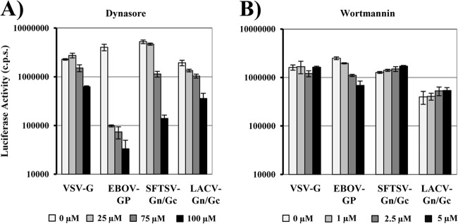
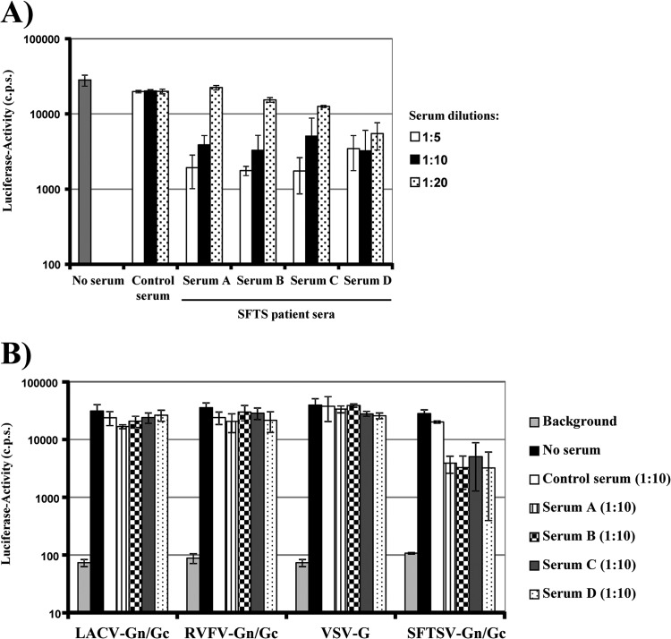
Severe fever with thrombocytopenia syndrome virus (SFTSV) is a novel bunyavirus that recently emerged in China. Infection with SFTSV is associated with case-fatality rates of up to 30%, and neither antivirals nor vaccines are available at present. Development of antiviral strategies requires the elucidation of virus-host cell interactions. Here, we analyzed host cell entry of SFTSV. Employing lentiviral and rhabdoviral vectors, we found that the Gn/Gc glycoproteins (Gn/Gc) of SFTSV mediate entry into a broad range of human and animal cell lines, as well as human macrophages and dendritic cells. The Gn/Gc proteins of La Crosse virus (LACV) and Rift Valley Fever Virus (RVFV), other members of the bunyavirus family, facilitated entry into an overlapping but not identical range of cell lines, suggesting that SFTSV, LACV, and RVFV might differ in their receptor requirements. Entry driven by SFTSV Gn/Gc was dependent on low pH but did not require the activity of the pH-dependent endosomal/lysosomal cysteine proteases cathepsins B and L. Instead, the activity of a cellular serine protease was required for infection driven by SFTSV and LACV Gn/Gc. Sera from convalescent SFTS patients inhibited SFTSV Gn/Gc-driven host cell entry in a dose-dependent fashion, demonstrating that the vector system employed is suitable to detect neutralizing antibodies. Finally, the C-type lectin DC-SIGN was found to serve as a receptor for SFTSV Gn/Gc-driven entry into cell lines and dendritic cells. Our results provide initial insights into cell tropism, receptor usage, and proteolytic activation of SFTSV and will aid in the understanding of viral spread and pathogenesis.


Figures

Fig 1
Fig 1
Expression of the SFTSV Gc glycoprotein and incorporation into virus-like particles. (A) 293T cells were transiently transfected with empty pCAGGS plasmid or pCAGGS encoding SFTSV Gn/Gc with a V5 antigenic tag. At 48 h posttransfection, cell lysates were prepared and analyzed for SFTSV Gn/Gc expression by Western blotting using a V5-specific monoclonal antibody. (B) Schematic overview of the SFTSV Gn/Gc protein. The signal peptide (striped) and the transmembrane domain (black) are shown. The putative proteolytic cleavage site is indicated by an arrow, and consensus signals for N-linked glycosylation are indicated by triangles. (C) Incorporation of SFTSV Gc into virus-like particles was analyzed by expression of pCAGGS SFTSV Gn/Gc V5 or empty plasmid in combination with p96ZM651gag-opt encoding human immunodeficiency virus type 1 Gag (p55), followed by Western blotting of particles released into the culture supernatant by a p55-specific antibody (bottom) or the V5-reactive antibody (top). (D) Rhabdoviral pseudotypes were harvested from the supernatant of 293T cells expressing SFTSV Gn/Gc V5 and a VSVΔG-GFP/luc genome and analyzed by Western blotting using a VSV M-specific antibody (bottom) or the V5-antibody (top). Molecular mass markers (kilodaltons) are indicated.
Fig 2
Fig 2
Cellular entry of rhabdoviral pseudotypes bearing the glycoprotein of SFTSV. Rhabdoviral pseudotypes were generated by expression of VSV G or SFTSV, LACV, or RVFV Gn/Gc in 293T cells, followed by transduction with a VSVΔG-GFP/luc virus. Particles released into the supernatant were normalized for comparable infectivity and used for infection of the indicated human cell lines (A), primary human cells (B), or cell lines derived from nonhuman species (C); particles harboring no glycoprotein (pCAGGS) were used as a control. (B) Spinoculation was used for macrophage infection. After 24 h, the cells were lysed, and infectivity was determined by luciferase assay. The results of representative experiments performed in triplicate are shown and were confirmed in at least three independent experiments performed with different pseudotype preparations. The error bars indicate standard deviations (SD).
Fig 3
Fig 3
Entry driven by the SFTSV glycoproteins depends on low pH. Pseudotypes bearing the glycoproteins of VSV, MLV, and SFTSV were normalized for comparable infectivity and used for infection of U373 cells preincubated with the indicated concentrations of ammonium chloride (NH4Cl) (left), bafilomycin A (right), or solvent (0 mM), followed by transduction with the indicated pseudotypes. Infectivity was determined by luciferase activity in cell extracts. The results shown are representative of three independent experiments; the error bars indicate SD.
Fig 4
Fig 4
SFTSV glycoprotein-mediated entry is not dependent on endosomal cathepsin activity. Vero target cells were preincubated with the indicated concentrations of cathepsin B and L inhibitors (CA074, CA074Me, and CatL III) and a serine protease inhibitor (AEBSF) or solvent as a control. Thereafter, the cells were infected with pseudotypes bearing glycoproteins of VSV, Zaire EBOV, SFTSV, or LACV as indicated, normalized for comparable infectivity. Luciferase activity in cell lysates was measured at 24 h postinfection. The averages of four (three for LACV Gn/Gc) experiments are shown; the error bars indicate standard errors of the mean (SEM).
Fig 5
Fig 5
Dynamin activity is required for efficient cellular entry of pseudotypes bearing the glycoproteins of SFTSV. (A) Vero target cells were preincubated with the indicated concentrations of the dynamin inhibitor dynasore or solvent (0 μM), followed by inoculation with infectivity-normalized pseudotypes bearing the glycoproteins of VSV, Zaire EBOV, SFTSV, or LACV as indicated. (B) Vero target cells were pretreated with wortmannin at the indicated concentrations or with solvent (0 μM) and processed as described for panel A. At 24 h postinfection, luciferase activity was determined in cell extracts. Infections were performed in triplicate; the error bars indicate SD. The experiments shown are representative of three independent experiments.
Fig 6
Fig 6
Sera from convalescent SFTS patients neutralize infectivity of SFTSV glycoprotein-bearing pseudotypes. (A) One hundred TCID50 pseudotypes harboring the SFTSV Gn/Gc protein were preincubated with the indicated dilutions of serum from a healthy donor (control) or convalescent SFTS patients (sera A, B, C, and D) in duplicate. Thereafter, 293T target cells were infected with the pseudotypes in quadruplicate. Infectivity was determined after 48 h by luciferase assay. The results were confirmed in a separate experiment. The error bars indicate SD. (B) The experiment was carried out as in panel A, but pseudotypes bearing VSV G or LACV and RFVF Gn/Gc were included as controls, and a single serum dilution (1:10) was tested. Similar results were obtained in an independent experiment.
Fig 7
Fig 7
DC-SIGN can serve as a host cell receptor for entry mediated by the glycoproteins of SFTSV. (A) Raji B cells engineered to express DC-SIGN or DC-SIGNR were infected with pseudotypes bearing the glycoproteins of VSV, SFTSV, LACV, or RVFV in triplicate. Parental Raji B cells were used as a control. The pseudotypes had been normalized for comparable infectivity on Vero cells. At 24 h postinfection, cell lysates were prepared and analyzed for luciferase activity. The results are representative of three independent experiments; the error bars indicate SD. (B) Parental Raji B cells or DC-SIGN-expressing Raji cells were preincubated with mannan (200 μg/ml), a mouse isotype control antibody (10 μg/ml), or the DC-SIGN/R-specific monoclonal antibody 526 (10 μg/ml) as indicated. The cells were then infected with pseudotypes bearing VSV G or SFTSV Gn/Gc, as described for panel A. Infection was quantified by luciferase activity at 24 h postinfection. Infection of Raji DC-SIGN cells in the absence of inhibitor was set as 100%. The results are representative of three independent experiments; the error bars indicate SD. (C) Primary human dendritic cells were preincubated with mannan (200 μg/ml) or the indicated antibodies (10 μg/ml) as described for panel B, followed by pseudotype infection. Cells were analyzed for luciferase activity at 24 h postinfection. Infection of dendritic cells in the absence of inhibitor (medium) was set as 100%. The error bars indicate SD. The results are representative of three independent experiments using two different donors and three independent pseudotype preparations.

References

    1. Walter CT, Barr JN. 2011. Recent advances in the molecular and cellular biology of bunyaviruses. J. Gen. Virol. 92:2467–2484 - PubMed
    1. Jonsson CB, Figueiredo LT, Vapalahti O. 2010. A global perspective on hantavirus ecology, epidemiology, and disease. Clin. Microbiol. Rev. 23:412–441 - PMC - PubMed
    1. Boshra H, Lorenzo G, Busquets N, Brun A. 2011. Rift Valley fever: recent insights into pathogenesis and prevention. J. Virol. 85:6098–6105 - PMC - PubMed
    1. Hollidge BS, Gonzalez-Scarano F, Soldan SS. 2010. Arboviral encephalitides: transmission, emergence, and pathogenesis. J. Neuroimmune Pharmacol. 5:428–442 - PMC - PubMed
    1. Ikegami T, Makino S. 2009. Rift valley fever vaccines. Vaccine 27(Suppl. 4):D69–D72 - PMC - PubMed

MeSH terms