. 2013 Mar 29;288(13):9363-72.
doi: 10.1074/jbc.M113.453712. Epub 2013 Feb 6.

Vac14 protein multimerization is a prerequisite step for Fab1 protein complex assembly and function

Affiliations

Vac14 protein multimerization is a prerequisite step for Fab1 protein complex assembly and function

Tamadher A Alghamdi et al. J Biol Chem. .

Abstract

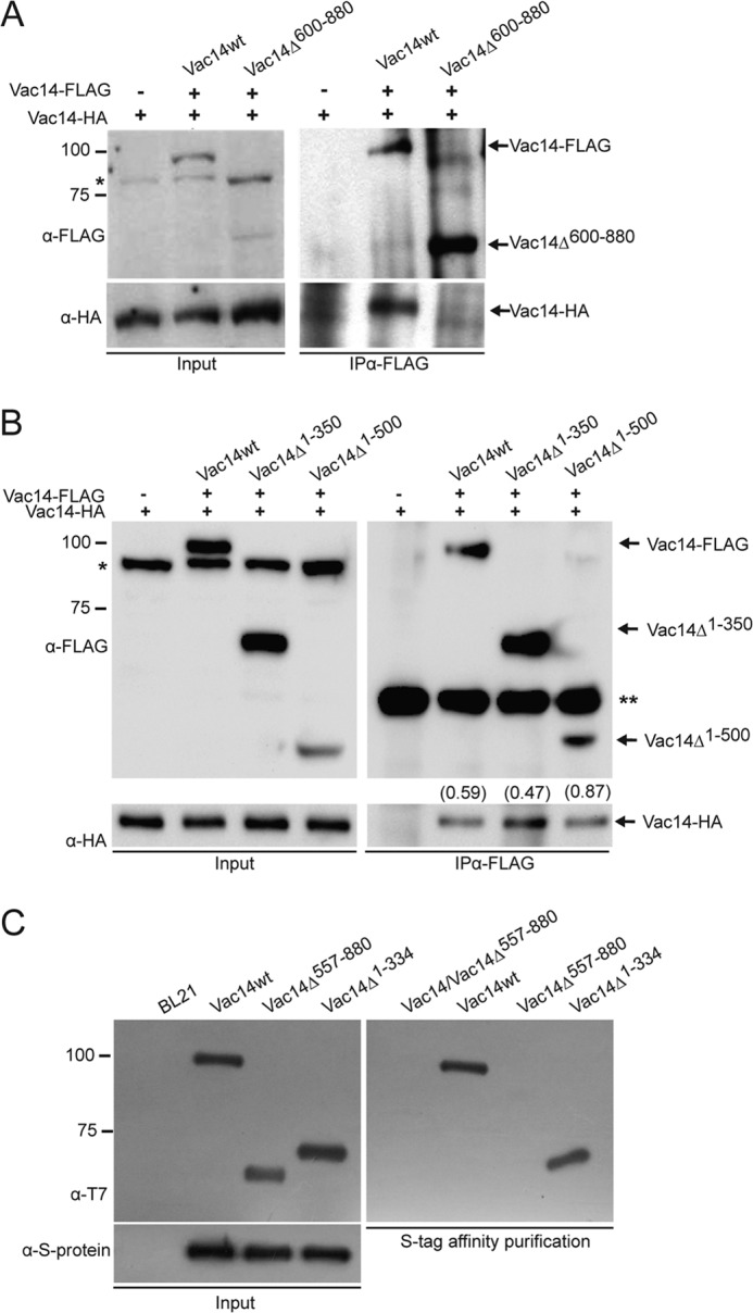
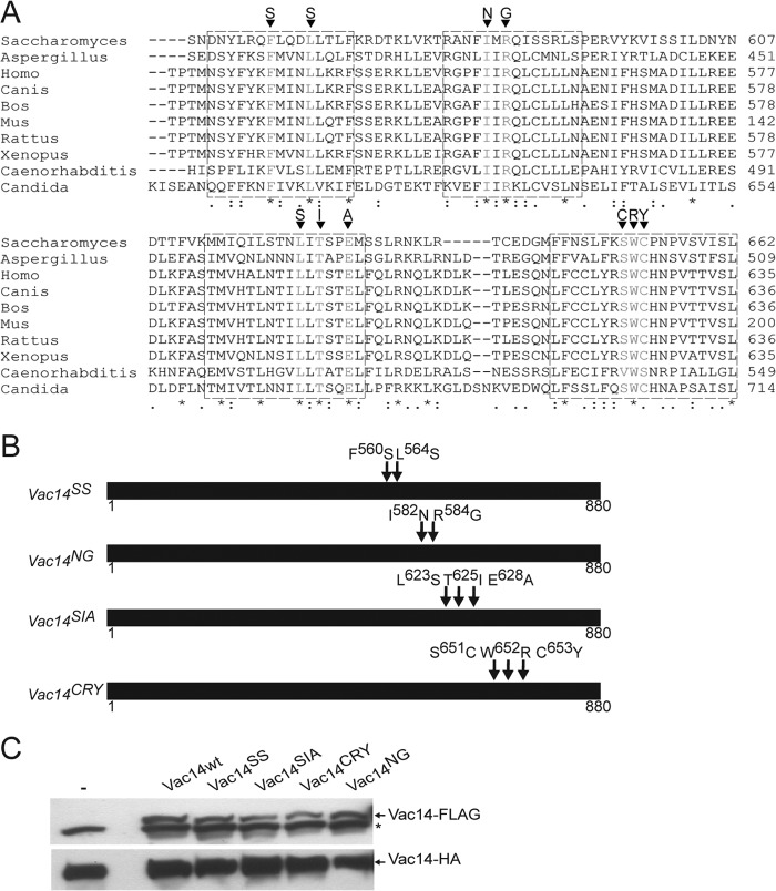
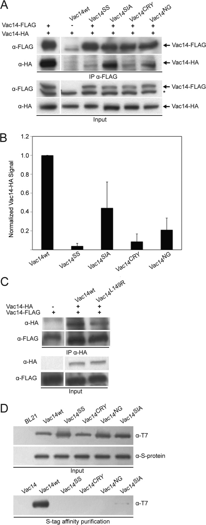
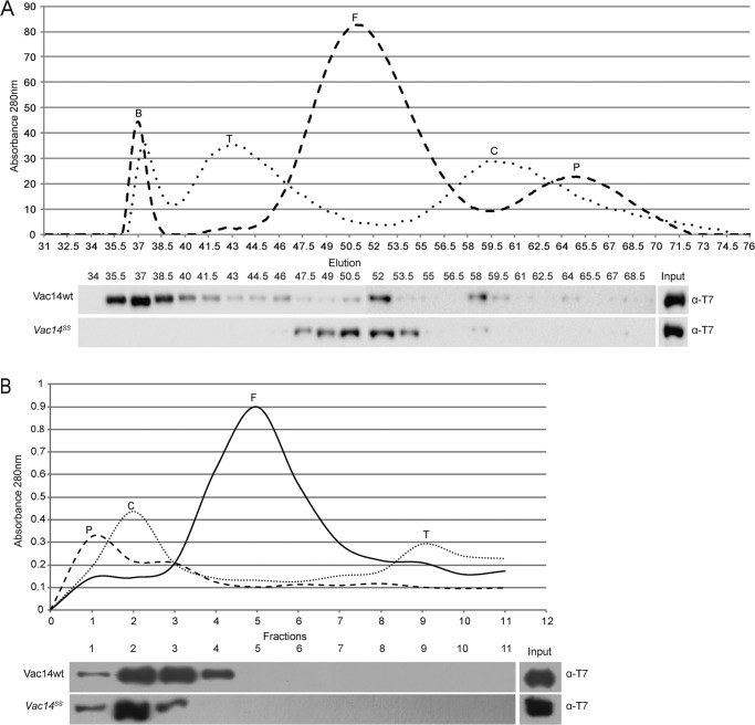
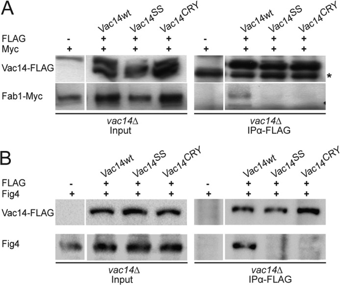
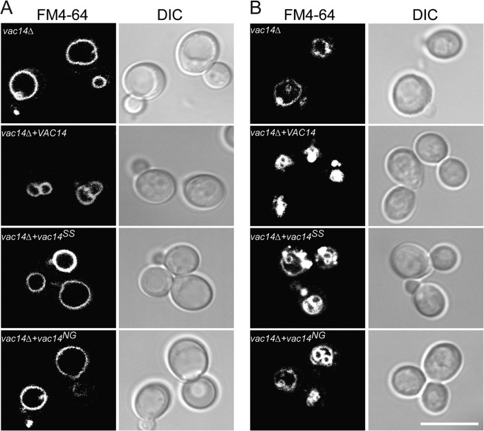
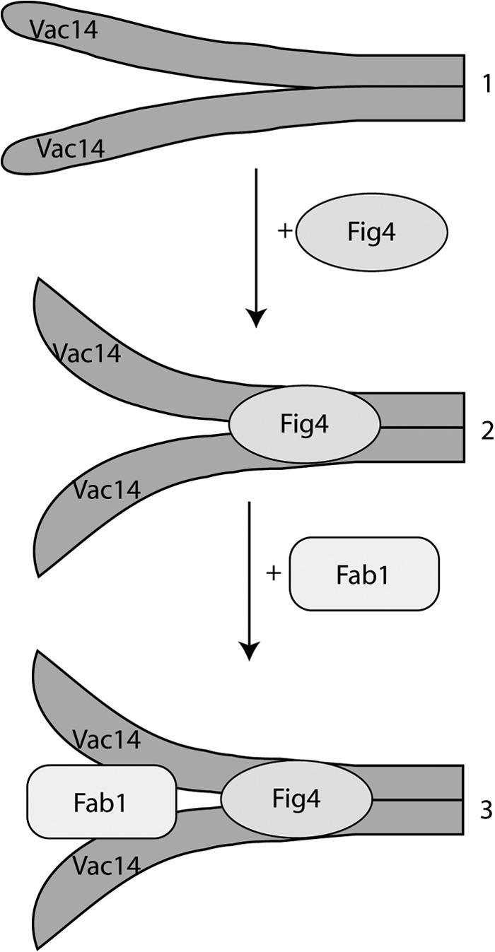
Phosphatidylinositol 3,5-bisphosphate (PtdIns(3,5)P2) helps control various endolysosome functions including organelle morphology, membrane recycling, and ion transport. Further highlighting its importance, PtdIns(3,5)P2 misregulation leads to the development of neurodegenerative diseases like Charcot-Marie-Tooth disease. The Fab1/PIKfyve lipid kinase phosphorylates PtdIns(3)P into PtdIns(3,5)P2 whereas the Fig4/Sac3 lipid phosphatase antagonizes this reaction. Interestingly, Fab1 and Fig4 form a common protein complex that coordinates synthesis and degradation of PtdIns(3,5)P2 by a poorly understood process. Assembly of the Fab1 complex requires Vac14/ArPIKfyve, a multimeric scaffolding adaptor protein that coordinates synthesis and turnover of PtdIns(3,5)P2. However, the properties and function of Vac14 multimerization remain mostly uncharacterized. Here we identify several conserved C-terminal motifs on Vac14 required for self-interaction and provide evidence that Vac14 likely forms a dimer. We also show that monomeric Vac14 mutants do not support interaction with Fab1 or Fig4, suggesting that Vac14 multimerization is likely the first molecular event in the assembly of the Fab1 complex. Finally, we show that cells expressing monomeric Vac14 mutants have enlarged vacuoles that do not fragment after hyperosmotic shock, which indicates that PtdIns(3,5)P2 levels are greatly abated. Therefore, our observations support an essential role for the Vac14 homocomplex in controlling PtdIns(3,5)P2 levels.


Figures

FIGURE 1.
FIGURE 1.
The C terminus of Vac14 is necessary and sufficient for Vac14 self-interaction. A and B, FLAG-Vac14, or its C-terminal (A) and N-terminal truncations (B) were immunoprecipitated with monoclonal anti-FLAG antibodies from whole cell lysates. Input and IP samples were then separated by SDS-PAGE and immunoblotted with anti-FLAG or anti-HA antibodies to, respectively, detect FLAG-tagged Vac14 and recovered Vac14-HA. The values in parentheses in B show the ratio of Vac14-HA signal to the respective FLAG-Vac14 signal obtained by volumetric densitometry. * denotes a nonspecific band whereas ** denotes the antibody heavy chain. C, purified recombinant wild-type Vac14-S·Tag bound to S protein-agarose beads were incubated with purified recombinant T7-Vac14-His6 or its recombinant truncation mutants. As a negative control, recombinant T7-His6-tagged Vac14 proteins were incubated with S protein-agarose beads alone. In all interaction assays, the input lanes represent 20% of protein or cell lysates used.
FIGURE 2.
FIGURE 2.
Identification of conserved Vac14 C-terminal motifs. A, sequence alignment of yeast Vac14 and various eukaryotic orthologs using the ClustalW algorithm. * indicates fully conserved residues, a colon indicates strong conservation between groups of similar properties, and a period indicates partial residue conservation. Boxed regions represent selected conserved motifs for site-directed mutagenesis. The targeted mutations are shown for each motif. B, schematic representation of Vac14 point mutants. Arrows show the relative positions of the point mutations in Vac14. C, whole cell lysates from cells genomically expressing Vac14-HA alone (−) or co-expressing FLAG-tagged wild-type Vac14 (Vac14wt) or Vac14 point mutants, were subjected to SDS-PAGE and Western blotting. Monoclonal anti-FLAG and monoclonal anti-HA antibodies were used to, respectively, detect the FLAG-tagged (top) and HA-tagged (bottom) Vac14 proteins. An * (asterisk) indicates a nonspecific band detected by the anti-FLAG antibody in whole cell lysates.
FIGURE 3.
FIGURE 3.
Identification of four conserved C-terminal motifs necessary for Vac14 self-interaction. A, co-IP from whole cell lysates expressing Vac14-HA and transformed with empty vector (−), or with vectors expressing FLAG-tagged wild-type or Vac14 point mutants. IP was done with monoclonal anti-FLAG antibodies and subjected to SDS-PAGE and Western blotting with anti-FLAG or anti-HA antibodies. The * indicates nonspecific bands. B, semiquantification of Vac14-HA co-immunoprecipitated with FLAG-tagged Vac14. Integrated pixel intensity of Vac14-HA was normalized to integrated pixel intensity of inputted FLAG-tagged Vac14. All pixel intensity values were background-corrected and employed nonsaturated signals. Shown is the normalized mean ± S.D. All mutant Vac14 means were statistically analyzed against wild-type Vac14 using Student's t test (n = 3, p < 0.05). C, IP of HA-tagged wild-type and L149R Vac14 with anti-HA antibodies, followed by SDS-PAGE and immunoblotting against genomically encoded Vac14-FLAG. D, recombinant Vac14-S·Tag bound to S protein-agarose beads were incubated with T7-His6-tagged recombinant wild-type and point mutant Vac14 proteins. In all cases, input lanes represent 20% of total whole cell lysates or protein used in affinity precipitation.
FIGURE 4.
FIGURE 4.
The multimeric state of Vac14 and Vac14SS. A, gel-exclusion chromatography through a Sephacryl S300 column. Three hundred micrograms of each protein standard, purified recombinant wild type T7-Vac14-His6, and purified recombinant T7-Vac14SS-His6 were separately injected and fractionated through the S300 column. Eluted standards were detected by A280 nm (top). The elutions of recombinant T7-Vac14-His6 and T7-Vac14SS-His6 were collected in 2-ml fractions, followed by SDS-PAGE, and immunoblotted with anti-T7 antibodies. Input lane represents 10% of the total recombinant protein injected. B, differential velocity ultracentrifugation through a 10–40% glycerol gradient. Approximately 300 μg of each protein standard and purified recombinant T7-Vac14-His6 or T7-Vac14SS-His6 were layered onto individual glycerol gradients and centrifuged at 30,000 rpm for 5 h. Fractionated protein standards and recombinant Vac14 were collected in 1-ml fractions. Protein standards were detected at A280 nm, and recombinant T7-Vac14-His6 was detected by immunoblotting with anti-T7 antibodies. Input lane represents 10% of the total recombinant protein. The protein standards used were: blue dextran (B), thyroglobulin (T), ferritin (F), catalase (C), and phosphorylase b (P).
FIGURE 5.
FIGURE 5.
Monomeric Vac14 mutants do not interact with Fab1 or Fig4. vac14Δ FAB1-Myc cells were transformed with plasmids encoding FLAG-tagged Vac14, Vac14SS, or Vac14CRY mutants. FLAG-Vac14 proteins were then immunoprecipitated with monoclonal anti-FLAG antibodies and subjected to SDS-PAGE and immunoblotting. A, co-IP of Fab1-Myc with FLAG-tagged wild-type Vac14 but not with corresponding mutants. B, co-IP of Fig4 with FLAG-tagged wild-type Vac14 but not with corresponding Vac14 mutants. vac14Δ FAB1-Myc cells carrying an empty vector (−) were used as a negative control. Input lanes represent 20% of total protein used in IP. * (asterisk) indicates nonspecific.
FIGURE 6.
FIGURE 6.
Monomeric Vac14 mutants display vacuolar defects. A and B, vac14Δ cells transformed with either empty vector or vectors expressing Vac14, Vac14SS, or Vac14NG were grown to log phase. Vacuoles were then labeled with FM4-64 as described under “Experimental Procedures.” A, vacuolar morphology of log-phase cells. B, vacuolar morphology of cells exposed to a 10-min hyperosmotic shock with 0.9 m NaCl. The respective FM4-64 (left) and differential interference contrast (DIC, right) are shown for each condition. The scale bar represents 10 μm.
FIGURE 7.
FIGURE 7.
Model for the assembly of the Fab1 complex. Step 1, Vac14 is dimerized through the C-terminal tails, either in parallel (shown) or anti-parallel (not shown). Step 2, the Vac14 homodimer recruits the Fig4 phosphatase to form the Vac14-Fig4 subcomplex. We postulate that Fig4 association induces a conformational change on the N-terminal regions of the Vac14 dimer that better binds Fab1 because (i) Fig4 is necessary for Fab1-Vac14 association and (ii) there is no evidence to suggest that Fab1 and Fig4 bind directly to each other. Step 3, the Fab1 kinase finally binds to the N termini of the Vac14 dimer. Although not shown, we postulate that Vac7 and possibly Atg18 interact last with the Fab1 complex because neither of these proteins is necessary for the Fab1 complex assembly.

References

    1. Ho C. Y., Alghamdi T. A., Botelho R. J. (2012) Phosphatidylinositol 3,5-bisphosphate: no longer the poor PIP2. Traffic 13, 1–8 - PubMed
    1. Bonangelino C. J., Catlett N. L., Weisman L. S. (1997) Vac7p, a novel vacuolar protein, is required for normal vacuole inheritance and morphology. Mol. Cell. Biol. 17, 6847–6858 - PMC - PubMed
    1. Gary J. D., Wurmser A. E., Bonangelino C. J., Weisman L. S., Emr S. D. (1998) Fab1p is essential for PtdIns(3)P 5-kinase activity and the maintenance of vacuolar size and membrane homeostasis. J. Cell Biol. 143, 65–79 - PMC - PubMed
    1. Ikonomov O. C., Sbrissa D., Mlak K., Shisheva A. (2002) Requirement for PIKfyve enzymatic activity in acute and long-term insulin cellular effects. Endocrinology 143, 4742–4754 - PubMed
    1. Rusten T. E., Rodahl L. M., Pattni K., Englund C., Samakovlis C., Dove S., Brech A., Stenmark H. (2006) Fab1 phosphatidylinositol-3-phosphate 5-kinase controls trafficking but not silencing of endocytosed receptors. Mol. Biol. Cell 17, 3989–4001 - PMC - PubMed

Publication types

MeSH terms

Substances

LinkOut - more resources