. 2013 Mar 29;288(13):9126-34.
doi: 10.1074/jbc.M112.430900. Epub 2013 Feb 6.

X-ray crystal structure of Escherichia coli RNA polymerase σ70 holoenzyme

Affiliations

X-ray crystal structure of Escherichia coli RNA polymerase σ70 holoenzyme

Katsuhiko S Murakami. J Biol Chem. .

Abstract

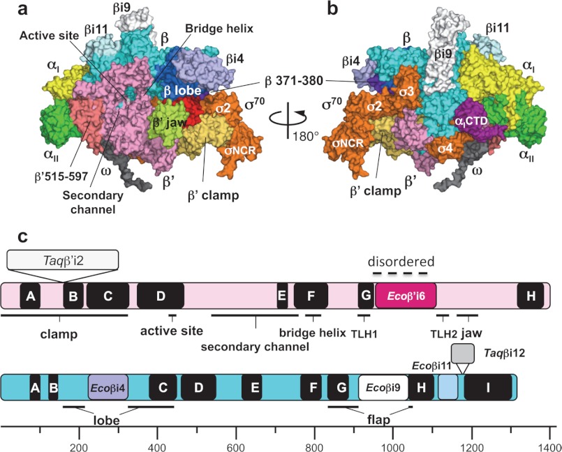
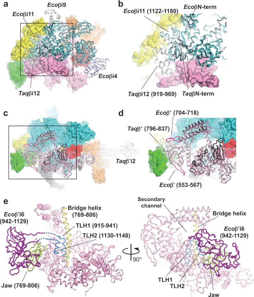
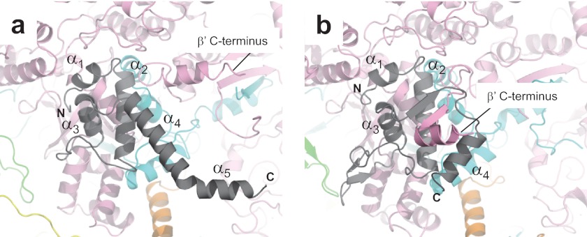
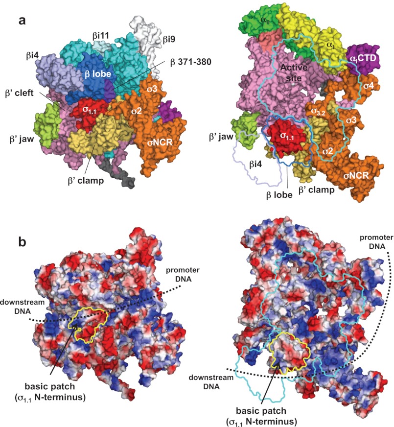
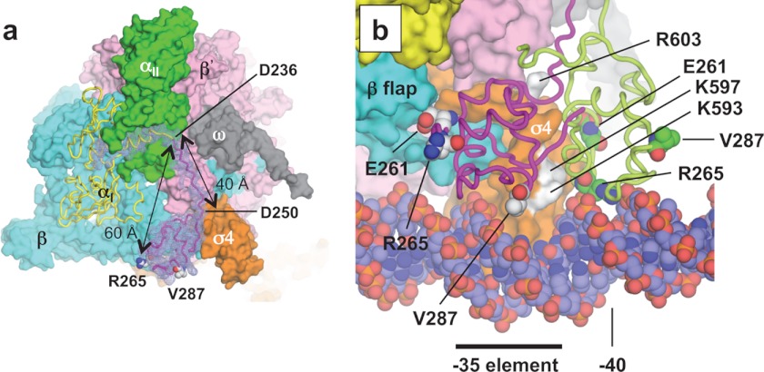
Escherichia coli RNA polymerase (RNAP) is the most studied bacterial RNAP and has been used as the model RNAP for screening and evaluating potential RNAP-targeting antibiotics. However, the x-ray crystal structure of E. coli RNAP has been limited to individual domains. Here, I report the x-ray structure of the E. coli RNAP σ(70) holoenzyme, which shows σ region 1.1 (σ1.1) and the α subunit C-terminal domain for the first time in the context of an intact RNAP. σ1.1 is positioned at the RNAP DNA-binding channel and completely blocks DNA entry to the RNAP active site. The structure reveals that σ1.1 contains a basic patch on its surface, which may play an important role in DNA interaction to facilitate open promoter complex formation. The α subunit C-terminal domain is positioned next to σ domain 4 with a fully stretched linker between the N- and C-terminal domains. E. coli RNAP crystals can be prepared from a convenient overexpression system, allowing further structural studies of bacterial RNAP mutants, including functionally deficient and antibiotic-resistant RNAPs.


Figures

FIGURE 1.
FIGURE 1.
Three-dimensional crystal structure of the E. coli RNAP σ70 holoenzyme. a and b, surface representation of the E. coli RNAP holoenzyme. Panel a shows a view from the RNAP secondary channel leading to the active site, and panel b shows the σ-binding site. Each subunit of RNAP is denoted by a unique color: yellow, αI; green, αII; cyan, β; pink, β′; gray, ω; and orange, σ70. Several domains described under “Results and Discussion” are also denoted by a unique color and are indicated. c, linear maps of the β′ (upper) and β (lower) subunits. Conserved regions of the β′ subunit (A–H) and the β subunit (A–I) are shown as black boxes with the structural domains of RNAP. Specific insertions of the E. coli and Thermus RNAPs are shown by the same colors as in panels a and b and in Fig. 2.
FIGURE 2.
FIGURE 2.
Structure comparisons of the β and β′ subunits of the E. coli and T. aquaticus RNAPs. a, superposition of Ecoβ and Taqβ RNAPs. Ecoβ (cyan) and Taqβ (black) are shown as α-carbon backbones in addition to the molecular surfaces of other E. coli RNAP subunits (αI, yellow; αII, green; β′, pink; σ70, orange; and σ1.1, red). b, magnified view of the boxed region in a. c, superposition of Ecoβ and Taqβ RNAPs. Ecoβ′ (pink and magenta), Taqβ′ (black and white) are shown as α-carbon backbones in addition to the molecular surfaces of other E. coli RNAP subunits (αI, yellow; αII, green; β′, pink; σ70, orange; and σ1.1, red). d, magnified view of the boxed region in c. e, the bridge helix (yellow), TLH (light blue), and jaw (yellow green) are highlighted on the α-carbon backbone of the Ecoβ′ (pink) structure. Ecoβ′i6 (purple) was modeled using the E. coli core enzyme model (24).
FIGURE 3.
FIGURE 3.
Structure comparisons of the ω subunits of the E. coli and Thermus RNAPs. Shown are close-up views of the ω (gray) and β′ (pink) subunit interactions of the E. coli (a) and Thermus (b) RNAPs. The positions of α helices (E. coli, α1–α5; and Thermus, α1–α4) of the ω subunits are indicated, and the C termini of the β′ subunits are also indicated.
FIGURE 4.
FIGURE 4.
Structure and function of σ1.1. a, molecular surface of the holoenzyme with σ1.1. Left, front view; right, side view. In the right panel, β subunit has been removed and outlined for clarity. b, electrostatic distribution of the holoenzyme. Left, front view; right, side view (orientations are the same as in a). Positive electrostatic potential is blue, and negative potential is red. The positions of σ1.1 in these views are indicated by yellow outlines. A basic patch found at the σ1.1 N terminus is shown. The potential DNA pathway during open complex formation is shown by dotted lines.
FIGURE 5.
FIGURE 5.
α-CTD structure. a, difference (FoFc) electron density map between the holoenzyme and holoenzyme without αI residues 215–315 (including the last α-NTD α helix, linker, and α-CTD), shown in mesh, overlaid on the final holoenzyme structure (αII, β, β′, ω, and σ, surface representation; and αI, ribbon model). The positions of two key residues for DNA (Arg-265) and transcription factor (Val-287) interaction on the α-CTD are indicated. Glu-261, another key residue of the α-CTD for the σ4 interaction, positioned between the α-CTD and β subunit, cannot be seen from this view. The distance between the α-NTD and Arg-295 and the length of the linker are shown. b, superimposed holoenzyme crystal structure (this study) and holoenzyme·catabolite activator protein·DNA model (Protein Data Bank code 3IYD). The two structures were aligned by superimposing their σ4 domains. The α-CTD from the crystal structure (magenta) and the model (light green) are shown as ribbons, and key α-CTD residues are shown as spheres. Surface-exposed σ4.2 residues that are predicted for direct interaction with the α-CTD are colored white and are indicated.

References

    1. Werner F., Grohmann D. (2011) Evolution of multisubunit RNA polymerases in the three domains of life. Nat. Rev. Microbiol. 9, 85–98 - PubMed
    1. Gruber T. M., Gross C. A. (2003) Multiple σ subunits and the partitioning of bacterial transcription space. Annu. Rev. Microbiol. 57, 441–466 - PubMed
    1. Murakami K. S., Darst S. A. (2003) Bacterial RNA polymerases: the wholo story. Curr. Opin. Struct. Biol. 13, 31–39 - PubMed
    1. Murakami K. S., Masuda S., Darst S. A. (2002) Structural basis of transcription initiation: RNA polymerase holoenzyme at 4 Å resolution. Science 296, 1280–1284 - PubMed
    1. Vassylyev D. G., Sekine S., Laptenko O., Lee J., Vassylyeva M. N., Borukhov S., Yokoyama S. (2002) Crystal structure of a bacterial RNA polymerase holoenzyme at 2.6 Å resolution. Nature 417, 712–719 - PubMed

Publication types