Review
. 2013 Mar;109(3):407-15.
doi: 10.1160/TH12-09-0678. Epub 2013 Feb 7.

Cellular and molecular regulation of vascular permeability

Affiliations
Review

Cellular and molecular regulation of vascular permeability

Lauren M Goddard et al. Thromb Haemost. 2013 Mar.

Abstract

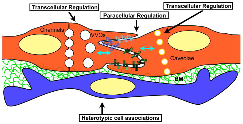
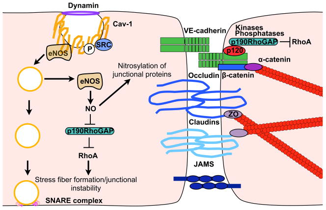
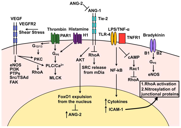
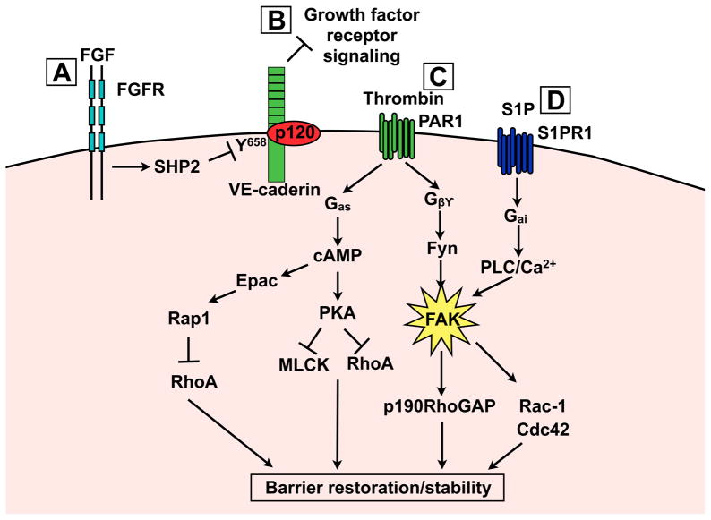
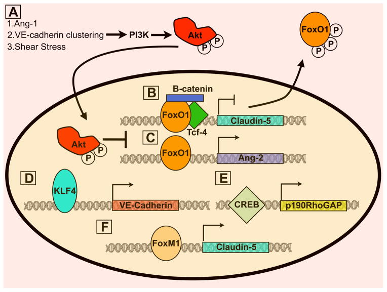
Vascular permeability is a highly coordinated process that integrates vesicular trafficking, complex junctional rearrangements, and refined cytoskeletal dynamics. In response to the extracellular environment, these three cellular activities have been previously assumed to work in parallel to regulate the passage of solutes between the blood and tissues. New developments in the area of vascular permeability, however have highlighted the interdependence between trans- and para-cellular pathways, the cross-communication between adherens and tight junctions, and the instructional role of pericytes on endothelial expression of barrier-related genes. Additionally, significant effort has been placed in understanding the molecular underpinings that contribute to barrier restoration following acute permeability events and in clarifying the importance of context-dependent signaling initiated by permeability mediators. Finally, recent findings have uncovered an unpredicted role for transcription factors in the coordination of vascular permeability and clarified how junctional complexes can transmit signals to the nucleus to control barrier function. The goal of this review is to provide a concise and updated view of vascular permeability, discuss the most recent advances in molecular and cellular regulation, and introduce integrated information on the central mechanisms involved in trans-endothelial transport.


Conflict of interest statement

Conflicts of Interest

None declared.

Figures

Figure 1
Figure 1. Pathways that regulate barrier function in endothelial cells
Scheme shows two endothelial cells and the subendothelial space. Vascular permeability is regulated and maintained through three compartments including: paracellular junctions (adherent and junctional complexes), transcellular pathways (channels, VVOs and caveolae) and heterotypic cell interactions (usually pericytes). The three pathways are interconnected molecularly (blue arrows), however the details of this cross-talk remain largely unclear. BM: Basement membrane.
Figure 2
Figure 2. Cross talk between transcellular trafficking and paracellular junctional complexes
Caveolae fission and loss of Cav-1 enhancee NOS mediated NO production. Nitrosylation of p190RhoGAP impairs inhibition of RhoA, resulting in stress fiber formation, junctional instability, and increased paracellular permeability. Direct nitrosylation of junctional proteins may also regulate junctional disassembly.
Figure 3
Figure 3. Signal transduction pathways that increase paracellular permeability
VEGF activation of VEGFR2 initiates several downstream signalling cascades leading to adherens protein internalisation, calcium release, and stress fiber formation. Thrombin and histamine, via G-protein coupled receptors, results in RhoA activation, calcium release and the development of stress fibers. Ang-2 inhibits the barrier stabilising effect of Ang-1 thus making the barrier vulnerable to permeability enhancing agents. LPS and TNF-α signalling result in NF-κB nuclear translocation where increased ICAM-1 expression leads to RhoA activation and NO-mediated nitrosylation of junctional proteins. Bradykinin promotes eNOS signalling.
Figure 4
Figure 4. Signalling mechanisms leading to enhanced barrier stability and restoration
A) FGF signalling increases basal barrier function through SHP2 phosphatase mediated p120/VEcadherin complex stabilisation. B) VE-cadherin can inhibit growth factor receptors that normally enhance permeability. C) Gβγ and Gas signal transduction results in FAK, Epac/Rap1 and PKA activation. All three of these targets coordinate to increase cortical actin and stabilise junctional complexes. D) S1P signalling leads to FAK phosphorylation via a PLC-dependent mechanism. Differential phosphorylation and cellular recruitment of FAK may explain how thrombin and S1P regulate FAK by distinct mechanisms.
Figure 5
Figure 5. Transcriptional regulation of vascular permeability
A) Akt phosphorylation by several stimuli results in FoxO1 phosphorylation and nuclear translocation. Subsequently, FoxO1 target genes including claudin-5 (B) and Ang-2(C) are expressed and repressed, respectively. Claudin-5 inhibition by FoxO1 requires complex formation between β-catenin and Tcf. Therefore, VE-cadherin/β-catenin complex formation is important for claudin-5 expression as it prevent β-catenin translocation to the nucleus. D) KLF4 stabilises the barrier by enhancing expression of VE-cadherin. E) CREB upregulates p190RhoGAP, which is important for inhibiting RhoA activation at adherens junctions. F) Both estrogen receptor (not depicted) and FoxM1 increase claudin-5 expression, thus promoting barrier stability.

References

    1. Armulik A, Genové G, Mäe M, et al. Pericytes regulate the blood-brain barrier. Nature. 2010;468:557–561. doi: 10.1038/nature09522. - DOI - PubMed
    1. Daneman R, Zhou L, Kebede AA, et al. Pericytes are required for blood-brain barrier integrity during embryogenesis. Nature. 2010;468:562–566. doi: 10.1038/nature09513. - DOI - PMC - PubMed
    1. Armulik A, Genové G, Betsholtz C. Pericytes: developmental, physiological, and pathological perspectives, problems, and promises. Dev Cell. 2011;21:193–215. doi: 10.1016/j.devcel.2011.07.001. - DOI - PubMed
    1. Mehta D. Signalling Mechanisms Regulating Endothelial Permeability. Physiol Rev. 2006;86:279–367. doi: 10.1152/physrev.00012.2005. - DOI - PubMed
    1. Dvorak AM, Feng D. The vesiculo-vacuolar organelle (VVO). A new endothelial cell permeability organelle. J Histochem Cytochem. 2001;49:419–432. doi: 10.1177/002215540104900401. - DOI - PubMed

Publication types

Substances