. 2013 Mar 7;495(7439):98-102.
doi: 10.1038/nature11847. Epub 2013 Feb 6.

NFIB is a governor of epithelial-melanocyte stem cell behaviour in a shared niche

Affiliations

NFIB is a governor of epithelial-melanocyte stem cell behaviour in a shared niche

Chiung-Ying Chang et al. Nature. .

Abstract

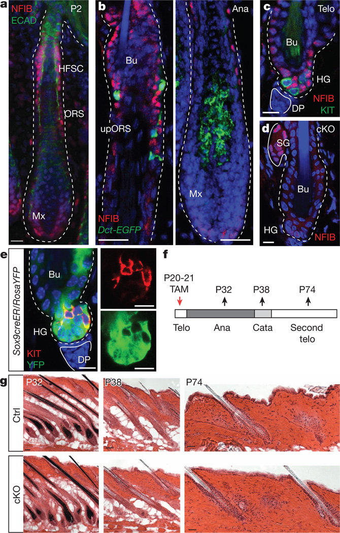
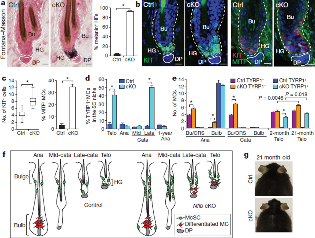
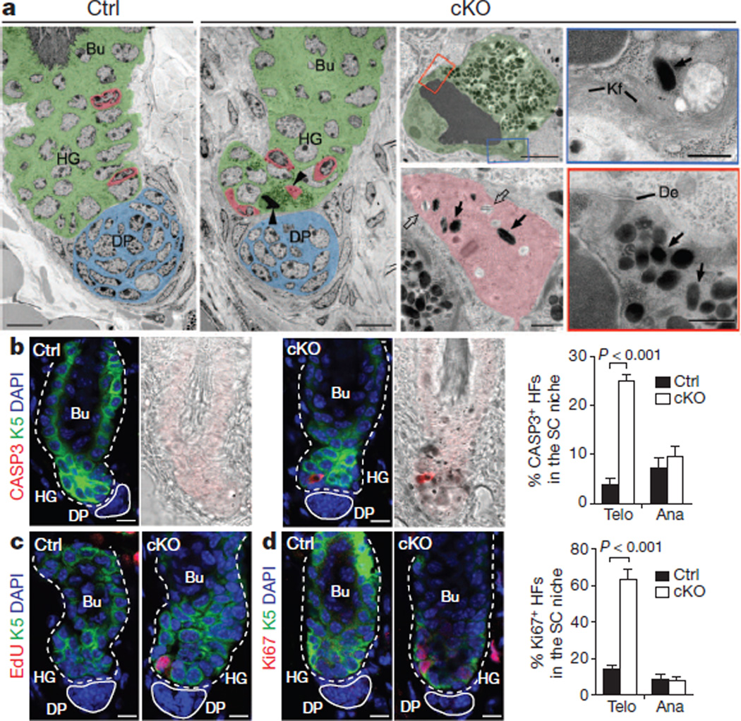
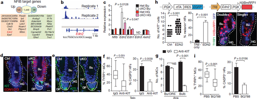
Adult stem cells reside in specialized niches where they receive environmental cues to maintain tissue homeostasis. In mammals, the stem cell niche within hair follicles is home to epithelial hair follicle stem cells and melanocyte stem cells, which sustain cyclical bouts of hair regeneration and pigmentation. To generate pigmented hairs, synchrony is achieved such that upon initiation of a new hair cycle, stem cells of each type activate lineage commitment. Dissecting the inter-stem-cell crosstalk governing this intricate coordination has been difficult, because mutations affecting one lineage often affect the other. Here we identify transcription factor NFIB as an unanticipated coordinator of stem cell behaviour. Hair follicle stem-cell-specific conditional targeting of Nfib in mice uncouples stem cell synchrony. Remarkably, this happens not by perturbing hair cycle and follicle architecture, but rather by promoting melanocyte stem cell proliferation and differentiation. The early production of melanin is restricted to melanocyte stem cells at the niche base. Melanocyte stem cells more distant from the dermal papilla are unscathed, thereby preventing hair greying typical of melanocyte stem cell differentiation mutants. Furthermore, we pinpoint KIT-ligand as a dermal papilla signal promoting melanocyte stem cell differentiation. Additionally, through chromatin-immunoprecipitation with high-throughput-sequencing and transcriptional profiling, we identify endothelin 2 (Edn2) as an NFIB target aberrantly activated in NFIB-deficient hair follicle stem cells. Ectopically induced Edn2 recapitulates NFIB-deficient phenotypes in wild-type mice. Conversely, endothelin receptor antagonists and/or KIT blocking antibodies prevent precocious melanocyte stem cell differentiation in the NFIB-deficient niche. Our findings reveal how melanocyte and hair follicle stem cell behaviours maintain reliance upon cooperative factors within the niche, and how this can be uncoupled in injury, stress and disease states.


Figures

Figure 1
Figure 1. Conditional Nfib targeting in hair follicle stem cells does not perturb hair cycle or follicle architecture
a–c, Immunofluorescence. a, Enrichment of nuclear NFIB in hair follicle stem cells and ORS of developing hair follicles. ECAD, E-cadherin; HFSC, hair follicle stem cells; Mx, matrix. b, NFIB in anagen hair follicles from adult Dct-EGFP BAC transgenic mice. NFIB is not seen in EGFP+ melanocytes. Ana, anagen; Bu, bulge; upORS, upper ORS. c, Absence of NFIB in KIT+ melanocyte stem cells of telogen hair follicles.DP, dermal papilla;HG, hair germ; Telo, telogen. d–f, Tamoxifen (TAM) was administered to Sox9-CreER/Nfibfl/fl/RosaYFP-cKO mice and analysed at various times thereafter. d, Note hair follicle stem-cell-specific Nfib targeting in bulge and hair germ. SG, sebaceous gland. e, Note YFP reporter activity in hair follicle stem cells but not in KIT+ melanocyte stem cells. f, Schematic. Cata, catagen; P20, postnatal day 20. g, Haematoxylin- and eosin-stained back skins reveal normal hair cycle and follicle architecture upon NFIB loss. Ctrl, control. Scale bars: 50 µm(P32,P38ing); 25 µm(a,b,P74ing); 10 µm(c–e).
Figure 2
Figure 2. NFIB loss enhances melanocyte stem-cell self-renewal and perturbs melanocyte stem cells activity in the hair follicle stem cell niche
a, Ectopic pigmentation in telogen-phase cKO hair germ as detected by Fontana–Masson melanin staining and quantifications (n=3 mice per experiment, >40 hair follicles per mouse). b–e, Immunofluorescence and quantifications. b, c, Increased melanocytes and ectopic differentiation in telogen cKO hair follicles (30–50 hair follicles; 2 mice per experiment). KIT marks melanocyte stem cells and differentiated melanocytes; MITF andTYRP1 are differentiation-specific melanocyte markers. MCs, melanocytes. d, Precocious melanocyte differentiation begins in the niche at late catagen. Quantifications of TYRP1+ among KIT+ or Dct-EGFP+ melanocytes (>40 hair follicles; 2 mice per experiment). Analyses shown start at second telogen. SC, stem cell. e, Quantifications of melanocyte stem cells (TYRP1) and differentiated melanocytes (TYRP1+) at different hair follicle stages (>30 hair follicles; 2 mice per experiment). f, Summary of data. McSC, melanocyte stemcell;MC, melanocyte. g, Lack of hair greying in ageing Nfib-cKO mice. Scale bars, 10 µm (a, b). *P<0.001. Error bars indicate s.e.m.
Figure 3
Figure 3. Premature transfer of pigment promotes apoptotic cell death in hair follicle stem cells in the NFIB-deficient niche
a, Hair follicle ultrastructure. Pseudo-colouring highlights distinct stem cell niche cell types: green, hair follicle stem cell; red, melanocyte; blue, dermal papilla. Arrowheads mark cells shown at higher magnification. Open and solid arrows denote immature and mature melanosomes, respectively. Note pigment-laden apoptotic hair follicle stem cells whose boxed regions at higher magnification reveal condensed chromatin, degenerating mitochondrion, keratin filaments (Kf), desmosomes (De) and melanosomes. b, Activated caspase 3 antibody marks K5+ apoptotic cKO hair follicle stem cells, seen in telogen but not anagen (n=3 mice; >30 hair follicles per mouse). HFs, hair follicles; SC, stem cell. c, d, Increased hair follicle stem cell proliferation in telogen but not anagen NfibcKO hair follicles. Assessment was by EdU (5-ethynyl-2′-deoxyuridine) incorporation (administered twice, 24 h before analysis) and Ki67 immunostaining (n=3 mice; >30 hair follicles per mouse). Scale bars: 10 µm (low magnifications in a and b–d); 2 µm (apoptotic hair follicle stem cell in a); 0.5 µm(cytoplasmic segments and melanocyte in a). Error bars indicate s.e.m.
Figure 4
Figure 4. RNA-seq and ChIP-seq analyses identify Edn2 as a direct NFIB regulated gene mediating inter-stem cell crosstalk
a, Identification of direct NFIB-regulated genes as those that bind NFIB (from ChIP-seq) and are up/down regulated in Nfib-cKO hair follicle stem cells (from RNA-seq). b, NFIB ChIP-seq profiles of Edn2 promoter. NFIB palindromic-binding-sequence exists within the peak. c, qPCR on FACS-isolated populations confirm RNA-seq results (Het, normalizations to heterozygote values). d, Immunofluorescence shows ectopic endothelins in Nfib-cKO hair follicles. e, Immunofluorescence reveals KITL in dermal papilla. f, Reductions in TYRP1+ melanocytes (>60 hair follicles; 3mice) and apoptosis (n = 4 mice;>30 hair follicles permouse) in cKO hair follicles after injecting anti-KIT blocking antibody (anti-KIT) during catagen/telogen. IgG, control. g, Anti-KIT-mediated inhibition of KIT signalling during anagen (>45 hair follicles in two mice). h, Overhead schematic: lentiviral constructs. Quantifications show increased KIT+ melanocytes (32 hair follicles; two mice per experiment) and melanin+ hair follicles (n=3 mice;>60 hair follicles per mouse) upon EDN2 induction. Immunofluorescence showsTYRP1+ melanocyte differentiation in doubly- (Edn2) and not singly-transduced/uninfected (Ctrl) hair follicle stem cells. i, Reduced TYRP1+ melanocytes (>46 hair follicles in two mice) and apoptosis (n=3 mice; >25 hair follicles per mouse) in Nfib-cKO hair follicles after injection of EDNRB inhibitor BQ788. Scale bars: 10 µm (d, e, h). ND, not determined; NS, not significant. Error bars indicate s.e.m.

References

    1. Tumbar T, et al. Defining the epithelial stem cell niche in skin. Science. 2004;303:359–363. - PMC - PubMed
    1. Nishimura EK, et al. Dominant role of the niche in melanocyte stem cell fate determination. Nature. 2002;416:854–860. - PubMed
    1. Blanpain C, Fuchs E. Epidermal homeostasis: a balancing act of stem cells in the skin. Nature Rev. Mol. Cell Biol. 2009;10:207–217. - PMC - PubMed
    1. Cotsarelis G. Gene expression profiling gets to the root of human hair follicle stem cells. J. Clin. Invest. 2006;116:19–22. - PMC - PubMed
    1. Hirobe T. How are proliferation and differentiation of melanocytes regulated? Pigment Cell Melanoma Res. 2011;24:462–478. - PubMed

Publication types

MeSH terms

Associated data