. 2013 Apr 15;304(8):F1066-75.
doi: 10.1152/ajprenal.00543.2012. Epub 2013 Feb 6.

Tamm-Horsfall protein translocates to the basolateral domain of thick ascending limbs, interstitium, and circulation during recovery from acute kidney injury

Affiliations

Tamm-Horsfall protein translocates to the basolateral domain of thick ascending limbs, interstitium, and circulation during recovery from acute kidney injury

Tarek M El-Achkar et al. Am J Physiol Renal Physiol. .

Abstract

Tamm-Horsfall protein (THP) is a glycoprotein normally targeted to the apical membrane domain of the kidney's thick ascending limbs (TAL). We previously showed that THP of TAL confers protection to proximal tubules against acute kidney injury (AKI) via a possible cross talk between the two functionally distinct tubular segments. However, the extent, timing, specificity, and functional effects of basolateral translocation of THP during AKI remain unclear. Using an ischemia-reperfusion (IRI) model of murine AKI, we show here that, while THP expression in TAL is downregulated at the peak of injury, it is significantly upregulated 48 h after IRI. Confocal immunofluorescence and immunoelectron microscopy reveal a major redirection of THP during recovery from the apical membrane domain of TAL towards the basolateral domain, interstitium, and basal compartment of S3 segments. This corresponds with increased THP in the serum but not in the urine. The overall epithelial polarity of TAL cells does not change, as evidenced by correct apical targeting of Na(+)-K(+)-2Cl cotransporter (NKCC2) and basolateral targeting of Na(+)-K(+)-ATPase. Compared with the wild-type, THP(-/-) mice show a significantly delayed renal recovery after IRI, due possibly to reduced suppression by THP of proinflammatory cytokines and chemokines such as monocyte chemoattractant protein-1 during recovery. Taken together, our data suggest that THP redistribution in the TAL after AKI is a protein-specific event and its increased interstitial presence negatively regulates the evolving inflammatory signaling in neighboring proximal tubules, thereby enhancing kidney recovery. The increase of serum THP may be used as a prognostic biomarker for recovery from AKI.


Figures

Fig. 1.
Fig. 1.
Changes in Tamm-Horsfall protein (THP) and neutrophil gelatinase-associated lipocalin (NGAL) mRNA expression after ischemia-reperfusion injury (IRI) in mice. Real-time PCR was performed to quantify THP (A) or NGAL (B) mRNAs using as templates total RNAs extracted from wild-type mouse kidneys of sham or those of post-IRI at 6, 24, or 48 h. Measurements were normalized to simultaneously amplified, endogenous control, GAPDH. Readings from sham for each time point were set at 1, and those from post-IRI were expressed as fold changes in reference to the corresponding sham (only 24-h sham is shown in the figure for clarity). Bar graphs represent averages ± SE (n = 6 mice/group). *P < 0.05, groups with statistical significance compared with the corresponding sham or control group.
Fig. 2.
Fig. 2.
Localization and semiquantitation of THP protein after acute kidney injury (AKI). AD: representative confocal immunofluorescent microscopic images (×20 objective) of the outer medulla of THP+/+ mouse kidneys stained for THP (red) after 24 h sham (A) and 6, 24, or 48 h after IRI (BD, respectively). Brush borders were stained green with Oregon-phalloidin, allowing the detection of S3 proximal tubular segments. E and F: high-power fields (×60 objective, nuclei stained blue with DAPI) from sham and 48 h post-IRI showing the shift in subcellular distribution of THP. Arrows mark the basolateral regions of thick ascending limb (TAL) where no THP staining was seen in sham (E), whereas a significant signal was detected at 48 h post-IRI (F). G: Western blot probing for THP on whole kidney extracts from sham (Sh), 24, and 48 h post-IRI (a different animal per lane). All ischemic kidneys at 48 h showed a marked increase in THP, as was also illustrated by densitometry quantification (fold increase compared with sham as reference) after adjustment for β-actin (H). *P < 0.05, statistical significance.
Fig. 3.
Fig. 3.
Serum THP level after AKI. Serum THP in mice was measured using ELISA at baseline, 24, and 48 h after sham and IRI surgeries (n = 5 for sham; n = 6 for IRI). Serum levels of THP in sham mice were not statistically different at any time point. In IRI mice, THP started to increase at 24 h (P = 0.1 compared with its baseline but P < 0.05 compared with sham 24 h) and continued its increase significantly at 48 h. *Statistical significance (P < 0.05) vs. corresponding sham. #Significance (P < 0.05) vs. IRI 24h.
Fig. 4.
Fig. 4.
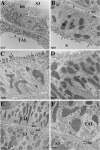
Immunogold electron microscopy of THP in sham kidneys. AC, E, and F: representative fields from sham kidney sections (outer stripe) from THP+/+ mice stained for THP with colloidal gold. D (×25,000): THP−/− serving as a negative control. A (×7,500): TAL separated by vascular endothelium from an S3 segment with a brush border (bb). THP can be seen at the apex of TAL. *Area shown at higher magnification in B (×20,000). Vascular endothelium (V) separating the basement membranes (bm) of TAL incidentally harbors particles that contains THP (arrowhead). C (×25,000): another TAL expressing THP predominantly at the apical membrane (arrows) but with occasional THP particles seen in the cytoplasm or basolateral domains (double arrows). E (×25,000): physical proximity between TAL and S3. In most sham fields, there was very little or no THP labeling in these areas. However, we do see rarely THP gold particles at the basal domain of TAL (F; ×30,000, double arrows) and even more rarely at the basal aspect of S3 segments (arrowheads).
Fig. 5.
Fig. 5.
Immunogold electron microscopy of THP 48 h after IRI. A: low-power field (×3000) showing TAL next to an S3 segment. Note the disarrayed mitochondria in S3 after ischemia. A collecting duct is also noted (CD). *Area is shown at a higher power (×25,000) in B. Note the marked expression of THP along the apical membrane as seen in sham. However, there is significant increase in THP expression now seen in the cytoplasm (arrowheads). #Area in A is magnified in C (×25,000) denoting the basal domain of TAL and the interstitial area between TAL and S3. Note the significant THP presence at the basal domain of TAL (arrowheads) and around the TAL basement membrane and the interstitium (arrows). THP could also be markedly observed around interstitial cells (IC). Furthermore, THP gold particles can be clearly observed at the basolateral surface of S3 (double arrows).
Fig. 6.
Fig. 6.
Quantitation of colloidal gold density. Bar graphs represent averages (±SE) of colloidal gold densities (number of particles per unit surface area expressed in μm2) in random 24 high-power fields (×25,000 to ×30,000) from sham and IRI animals. Area was computed using ImageJ software (National Institutes of Health). Basolateral area was defined as the lower one third (computed from the distance between basal to apical membranes) of each TAL cell. *P < 0.05, statistical significance compared with sham.
Fig. 7.
Fig. 7.
Effect of Injury on Na+-K+-ATPase localization in the outer medulla. AF: representative high power (× 40 objective) confocal microscopy images of outer medulla sections from kidneys 48 h after sham (AC) or IRI surgery (DF). Na+-K+-ATPase immunofluorescent staining is shown in the red channel; Phalloidin (brush border stain, to identify tubules) is in the green channel. A: merge channel that includes DAPI (blue staining for nuclei) is shown in C and F. In sham, Na+-K+-ATPase is localized to the basolateral membrane of S3 proximal tubules (arrows in A) and TAL (double arrows). After IRI, proximal tubules lose the basolateral staining with occasional staining appearing on the apical aspect (arrowheads in D) of injured/recovering S3 segments (marked as S3* in E). TAL retain the same pattern of basal membrane distribution of Na+-K+-ATPase (double arrows in D).
Fig. 8.
Fig. 8.
Effect of injury on Na+-K+-2Cl cotransporter (NKCC2) localization in TAL tubules. AD: representative low power (×20 objective, A and B) and high power (×40 objective, C and D) light microscopy images from the outer medulla of sections from kidneys (48 h after surgery) stained using immunohistochemistry for NKCC2. Arrows in sham (A) and IRI (B) show that NKCC2 is localized to the apical domain of TAL tubules. Higher power images confirm that ischemia does not alter the apical localization of NKCC2 (arrowheads in C and D).
Fig. 9.
Fig. 9.
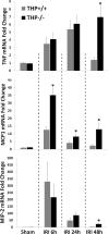
THP and suppression of inflammation in vivo. Wild-type (THP+/+) and THP knockout (THP−/−) mice were subject to sham or renal ischemia with reperfusion for 6, 24, or 48 h after surgery, and real-time PCR quantification of TNF-α mRNA expression was then carried out using whole kidney RNA extracts (top). Parallel experiments were carried out to quantify monocyte chemoattractant protein-1 (MCP-1) mRNA (middle) and macrophage inflammatory protein-2 (MIP-2) mRNA expression (bottom). Bar graphs represented averages ± SE. Each group within each time point consisted of 5–6 animals per genotype. Values were expressed as fold changes compared with wild-type sham set as reference for each time point, and only the sham at 24 h is shown for clarity (there was no difference between the sham groups at any of the other time points). *P < 0.05, statistical significance between strains.
Fig. 10.
Fig. 10.
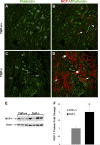
MCP-1 expression 48 h after IRI in THP+/+ and THP−/−kidneys. AD: representative confocal microscopy images (×20 objective) of outer medulla sections from THP+/+ and THP−/− kidneys 48 h after IRI. MCP-1 (red) immunofluorescent staining is shown in the merge channel. Phallodin staining (green) is also shown in a separate channel. In THP−/−, MCP-1 staining was not only noted in S3 segments that were recovering (S3*with partial brush border in C, arrows in D) but also in normal appearing S3 segments (S3 in C with full brush border, double arrows in D). In THP+/+, tubular MCP-1 expression was less prominent (arrow in B). MCP-1 staining was also observed in few interstitial cells (arrowhead in B). E: Western blot for MCP-1 in total kidney extracts from THP+/+ and THP−/− mice 48 h after IRI (each lane represents a separate animal). Because of some variability between animals, quantitation using band densitometry after normalization to actin was performed in two independent experiments. Average is shown in F, expressed as fold change compared with THP+/+ (set as reference). *P < 0.05, significant difference vs. THP+/+.
Fig. 11.
Fig. 11.
Effect of THP deficiency on long-term recovery after AKI. A and B: serial measurements of serum creatinine (Cr) from THP−/− (A; n = 5 for each sham and IRI groups) and THP+/+ (B; n = 5 for sham, and n = 4 for IRI) mice subjected to sham surgeries or to an equivalent degree of renal ischemia-reperfusion injury (IRI 22 min for THP−/− and 30 min in THP+/+; peak Cr at 24 h not statistically different between the groups). *P < 0.05, statistical significance compared with sham. #P < 0.05, statistical significance compared with THP+/+.

References

    1. Abbate M, Bonventre JV, Brown D. The microtubule network of renal epithelial cells is disrupted by ischemia and reperfusion. Am J Physiol Renal Fluid Electrolyte Physiol 267: F971–F978, 1994 - PubMed
    1. Bachmann S, Koeppen-Hagemann I, Kriz W. Ultrastructural localization of Tamm-Horsfall glycoprotein (THP) in rat kidney as revealed by protein A-gold immunocytochemistry. Histochemistry 83: 531–538, 1985 - PubMed
    1. Bates JM, Raffi HM, Prasadan K, Mascarenhas R, Laszik Z, Maeda N, Hultgren SJ, Kumar S. Tamm-Horsfall protein knockout mice are more prone to urinary tract infection: rapid communication. Kidney Int 65: 791–797, 2004 - PubMed
    1. Bleyer AJ, Hart TC, Shihabi Z, Robins V, Hoyer JR. Mutations in the uromodulin gene decrease urinary excretion of Tamm-Horsfall protein. Kidney Int 66: 974–977, 2004 - PubMed
    1. Chertow GM, Burdick E, Honour M, Bonventre JV, Bates DW. Acute kidney injury, mortality, length of stay, and costs in hospitalized patients. J Am Soc Nephrol 16: 3365–3370, 2005 - PubMed

Publication types

MeSH terms

LinkOut - more resources