. 2013 Apr;24(7):933-44.
doi: 10.1091/mbc.E12-06-0481. Epub 2013 Feb 6.

Tetraspanin CD9 links junctional adhesion molecule-A to αvβ3 integrin to mediate basic fibroblast growth factor-specific angiogenic signaling

Affiliations

Tetraspanin CD9 links junctional adhesion molecule-A to αvβ3 integrin to mediate basic fibroblast growth factor-specific angiogenic signaling

Swetha S D Peddibhotla et al. Mol Biol Cell. 2013 Apr.

Abstract

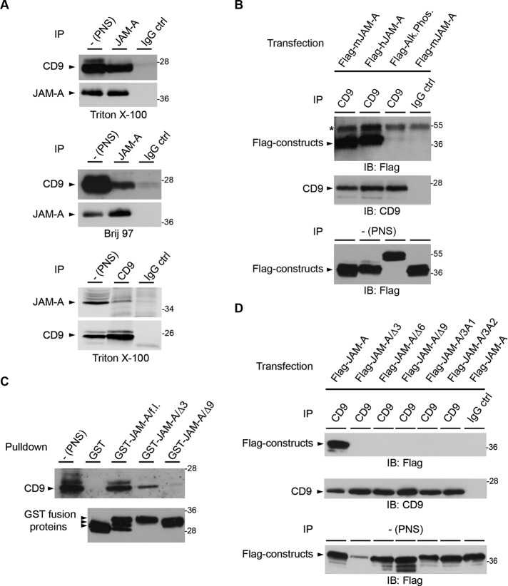
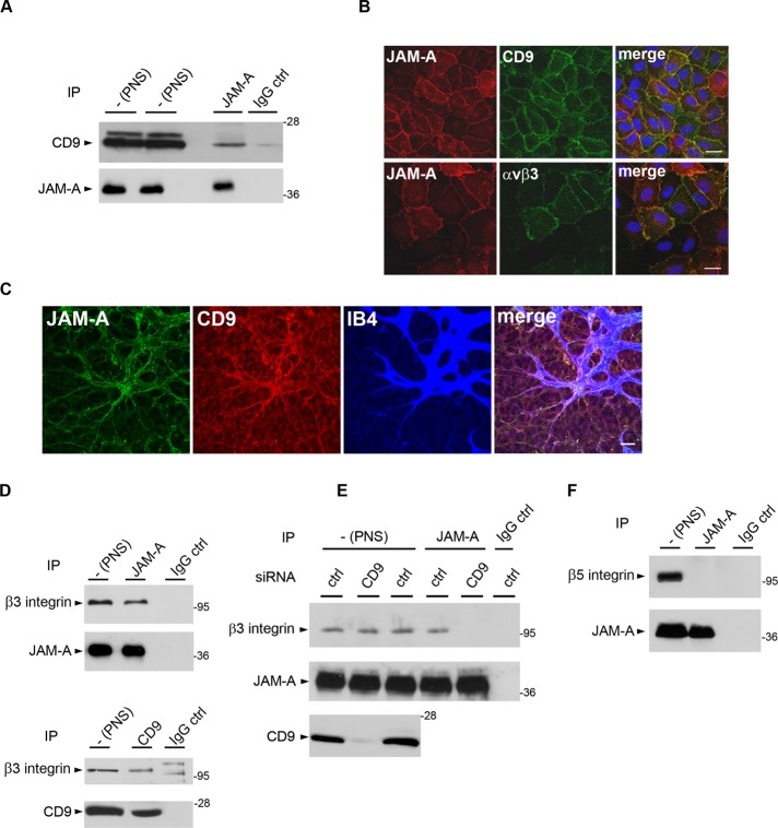
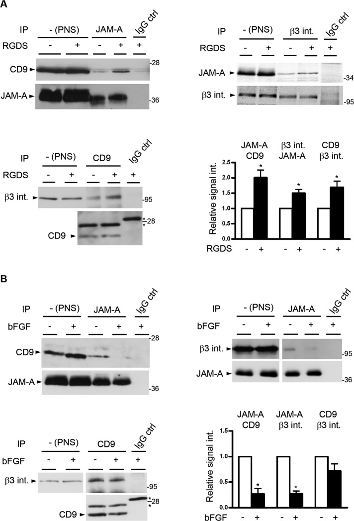
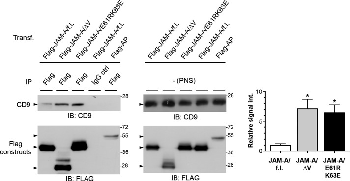
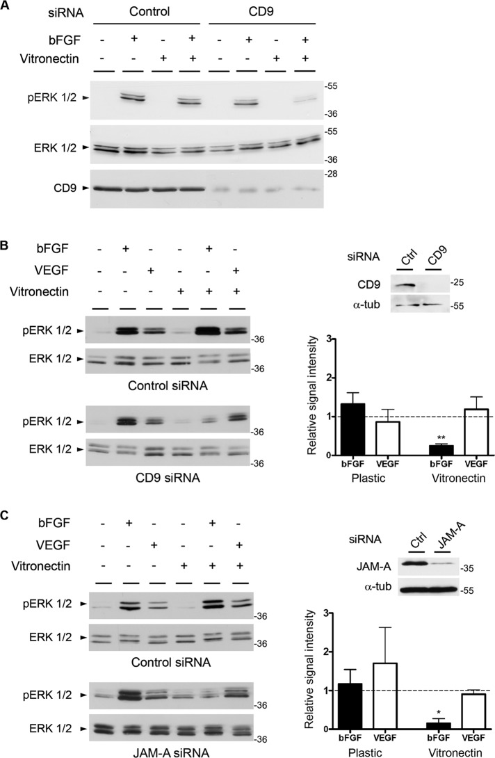
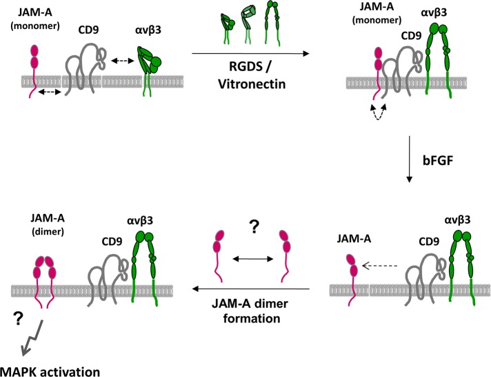
Junctional adhesion molecule-A (JAM-A) is a member of the immunoglobulin family with diverse functions in epithelial cells, including cell migration, cell contact maturation, and tight junction formation. In endothelial cells, JAM-A has been implicated in basic fibroblast growth factor (bFGF)-regulated angiogenesis through incompletely understood mechanisms. In this paper, we identify tetraspanin CD9 as novel binding partner for JAM-A in endothelial cells. CD9 acts as scaffold and assembles a ternary JAM-A-CD9-αvβ3 integrin complex from which JAM-A is released upon bFGF stimulation. CD9 interacts predominantly with monomeric JAM-A, which suggests that bFGF induces signaling by triggering JAM-A dimerization. Among the two vitronectin receptors, αvβ3 and αvβ5 integrin, which have been shown to cooperate during angiogenic signaling with bFGF and vascular endothelial growth factor (VEGF), respectively, CD9 links JAM-A specifically to αvβ3 integrin. In line with this, knockdown of CD9 blocks bFGF- but not VEGF-induced ERK1/2 activation. JAM-A or CD9 knockdown impairs endothelial cell migration and tube formation. Our findings indicate that CD9 incorporates monomeric JAM-A into a complex with αvβ3 integrin, which responds to bFGF stimulation by JAM-A release to regulate mitogen-activated protein kinase (MAPK) activation, endothelial cell migration, and angiogenesis. The data also provide new mechanistic insights into the cooperativity between bFGF and αvβ3 integrin during angiogenic signaling.


Figures

FIGURE 1:
FIGURE 1:
JAM-A interacts with CD9. (A) JAM-A interacts with CD9 by way of a primary tetraspanin interaction. Top and middle panels, HeLa cells were lysed in either Triton X-100– or Brij97-containing lysis buffer as indicated. JAM-A immunoprecipitates were immunoblotted for CD9 (90% of input) and for JAM-A (10% of input). Bottom panel, Immunoprecipitation was performed in the reverse order: CD9 was immunoprecipitated, and immunoprecipitates were immunoblotted for JAM-A (90% of input) or CD9 (10% of input). Note that JAM-A efficiently interacts with CD9 under both lysis conditions. (B) JAM-A strongly interacts with CD9 in HEK293T cells. CD9 immunoprecipitates obtained from Flag-JAM-A–transfected HEK293T cells were immunoblotted with anti-Flag antibodies (top, 90% of input) or anti-CD9 antibodies (middle, 10% of input). Postnuclear supernatants (PNS) were immunoblotted with anti-Flag antibodies (bottom, 2.5% of total lysate). The asterisk denotes signals resulting from IgG heavy chains. (C) CD9 interacts with the cytoplasmic tail of JAM-A. GST precipitates obtained from HEK293T cells with GST fusion proteins containing the full-length cytoplasmic tail of JAM-A (GST-JAM-A/f.l.) or deletion mutants lacking 3 or 9 C-terminal amino acid residues (GST-JAM-A/Δ3, GST-JAM-A/Δ9) were immunoblotted for CD9 (top panel). Equal loading of GST fusion proteins was verified by immunoblotting aliquots with anti-GST antibodies (bottom panel). Arrowheads indicate GST-JAM-A/f.l. constructs resulting from proteolytic cleavage. (D) The interaction between JAM-A and CD9 requires the PDZ domain-binding motif of JAM-A. CD9 immunoprecipitates obtained from HEK293T cells transfected with full-length JAM-A (Flag-JAM-A), C-terminal truncation mutants (Flag-JAM-A/Δ3, -/Δ6, -/Δ9), or triple alanine substitutions (Flag-JAM-A/3A1, Flag-JAM-A/3A2) were immunoblotted with anti-Flag antibodies (top, 90% of input), or with anti-CD9 antibodies (middle, 10% of input). Expression levels of Flag constructs were analyzed by immunoblotting the PNS with anti-Flag antibodies (bottom, 2.5% of total lysate). In addition, all Flag constructs localize to the cell surface as analyzed by flow cytometry (Supplemental Figure S5). Experiments shown in this figure are representative of at least three independent experiments. IP, immunoprecipitation; IB, immunoblotting.
FIGURE 2:
FIGURE 2:
CD9 assembles a ternary complex by linking JAM-A to αvβ3 integrin in endothelial cells. (A) JAM-A interacts with CD9 in endothelial cells. JAM-A immunoprecipitates obtained from HUVECs were immunoblotted with mAbs against CD9 (top, 90% of input) or against JAM-A (bottom, 10% of input). (B) Both CD9 and αvβ3 integrin colocalize with JAM-A at endothelial cell–cell junctions. HUVECs were costained for JAM-A and CD9 (top panels) and for JAM-A and for αvβ3 integrin (bottom panels). Scale bars: 10 μm. (C) JAM-A and CD9 are coexpressed in the vasculature of the P6 mouse retina at the angiogenic front. Whole-mount preparations of a mouse retina were stained with antibodies against JAM-A (green) and CD9 (red), and with isolectin B4 (IB4, blue) to visualize endothelial cells. Scale bar: 24 μm. (D) JAM-A and CD9 interact with β3 integrin. Immunoprecipitates obtained from HUVECs with antibodies against JAM-A (top panel) or CD9 (bottom panel) were immunoblotted for β3 integrin. Specific IP was verified by immunoblotting 10% of the precipitated material with antibodies against the precipitated protein. The two bands present in the IP performed with control IgG in the bottom panel represent unspecific bands as they do not match the molecular weight of β3 integrin and as they did not appear in other IPs performed with the same IgG (see also Figure 3, A and B). (E) CD9 links JAM-A to αvβ3 integrin. JAM-A immunoprecipitates obtained from CD9 knockdown HUVECs were immunoblotted with anti-β3 integrin antibodies (top panel, 90% of input) or JAM-A antibodies (middle panel, 10% of input). Postnuclear supernatants were also blotted with CD9 antibodies to control for knockdown efficiency of CD9 (bottom panel, 2.5% of total PNS). Note that β3 integrin does not interact with JAM-A in the absence of CD9. (F) JAM-A does not interact with β5 integrins in HUVECs. JAM-A immunoprecipitates obtained from HUVECs were analyzed for the presence of β5 integrin (top, 90% of input) or JAM-A (bottom, 10% of input). All biochemical experiments are representative of three independent experiments.
FIGURE 3:
FIGURE 3:
Ternary complex formation is dependent on integrin activation and negatively regulated by bFGF. (A) Integrin activation promotes ternary complex formation. HUVECs were stimulated with RGDS peptide (100 μg/ml, 20 min). After lysis, JAM-A IPs were analyzed for the presence of CD9 (top, left panel), CD9 IPs were analyzed for the presence of β3 integrin (top, right panel), and β3 integrin IPs were analyzed for JAM-A (bottom, left panel). In all cases, equal and specific IP was verified by immunoblotting 10% of the precipitated material with antibodies against the precipitated protein. The asterisks denote unspecific bands derived from Ig light chains. Bottom, right panel, densitometric analysis of the binary interactions; y-axis: relative signal intensity. Densitometric values obtained from unstimulated cells (no RGDS) were arbitrarily set as 1. Error bars denote the mean ± SE from four separate experiments. Statistical significance was evaluated using one-sample t tests; *, p < 0.05. (B) bFGF dissociates JAM-A from the ternary complex. HUVECs were stimulated with bFGF (10 ng/ml, 10 min). After lysis, JAM-A IPs were analyzed for CD9 (top, left panel) or for β3 integrin (top, right panel), and CD9 IPs were analyzed for β3 integrin (bottom, left panel). In all cases, equal and specific IP was verified by immunoblotting 10% of the precipitated material with antibodies against the precipitated protein. The asterisks denote unspecific bands derived from Ig light chains. Bottom, right panel, densitometric analysis of JAM-A–CD9, JAM-A–β3 integrin and CD9–β3 integrin CoIPs; y-axis: relative signal intensity. Densitometric values obtained from unstimulated cells (no bFGF) were arbitrarily set as 1. Error bars denote the mean ± SE from three separate experiments. Statistical significance was evaluated using one-sample t tests; *, p < 0.05.
FIGURE 4:
FIGURE 4:
CD9 interacts preferentially with monomeric JAM-A. HEK293T cells were transfected with either full-length Flag-JAM-A (Flag-JAM-A/f.l.) or dimerization-defective JAM-A mutants (Flag-JAM-A/ΔV, Flag-JAM-A/E61RK63E); Flag-alkaline phosphatase (Flag-AP) served as negative control. Left panels, Flag constructs were immunoprecipitated with anti-Flag antibodies or IgG control antibodies as indicated, and the immunoprecipitates were immunoblotted with anti-CD9 antibodies (top, 90% of input) or anti-Flag antibodies (bottom, 10% of input). Middle panels, PNS from the samples used for IP were analyzed for the levels of CD9 (top) and of Flag constructs (bottom). Right panel, densitometric analysis of the amount of CD9 associated with JAM-A and JAM-A mutants; y-axis: relative signal intensity. Densitometric values obtained from the interaction of CD9 with wild-type JAM-A (JAM-A/f.l., left bar) was arbitrarily set as 1. Error bars denote the mean ± SE from four separate experiments. Statistical significance was evaluated using one-sample t tests; *, p < 0.05.
FIGURE 5:
FIGURE 5:
Both CD9 and JAM-A specifically cooperate with bFGF in angiogenic signaling. (A) CD9 is required for ERK1/2 phosphorylation in cells grown on vitronectin. CD9 siRNA-treated HUVECs grown either on plastic or on vitronectin were stimulated with bFGF (10 ng/ml, 20 min) as indicated. Cell lysates were analyzed for total ERK1/2 and phosphorylated ERK1/2. Note that the absence of CD9 blocks bFGF-induced Erk1/2 phosphorylation only when cells are grown on vitronectin. (B) CD9 mediates bFGF- but not VEGF-induced ERK1/2 phosphorylation. CD9 siRNA-treated HUVECs were grown on plastic or on vitronectin and stimulated with bFGF (10 ng/ml, 10 min) or VEGF (20 ng/ml, 10 min) as indicated. Cell lysates were analyzed for total ERK1/2 and phosphorylated ERK1/2. Top, right panel, CD9 and α-tubulin (α-tub) levels in ctrl siRNA- and CD9 siRNA-transfected cells. Bottom, right panel, quantification of ERK1/2 phosphorylation; y-axis: relative signal intensity. Bars represent ERK1/2 phosphorylation in CD9 siRNA-treated cells relative to ERK1/2 phosphorylation in control siRNA-treated cells. Phosphorylation levels were quantified as detailed in the Materials and Methods section. Error bars denote the mean ± SE from three independent experiments. Statistical significance was evaluated using one-sample t tests; **, p < 0.01. (C) JAM-A mediates bFGF- but not VEGF-induced ERK1/2 phosphorylation. JAM-A siRNA-transfected HUVECs were grown on plastic or on vitronectin and stimulated with bFGF (10 ng/ml, 10 min) or VEGF (20 ng/ml, 10 min) as indicated. Cell lysates were analyzed for total ERK1/2 and phosphorylated ERK1/2. Top, right panel, JAM-A and α-tubulin (α-tub) levels in ctrl siRNA- and JAM-A siRNA-transfected cells. Bottom, right panel, quantification of ERK1/2 phosphorylation performed as described in (B). Error bars denote the mean ± SE from three independent experiments. Statistical significance was evaluated using one-sample t tests; *, p < 0.05.
FIGURE 6:
FIGURE 6:
CD9 and JAM-A are required for invasive growth and in vitro tube formation. (A) HUVECs were transfected with siRNAs against JAM-A or CD9. Left panel, knockdown efficiencies were analyzed by immunoblotting. Right panel, siRNA-transfected HUVECs were allowed to invade a Matrigel matrix for 16 h in the presence of bFGF. Invasion was analyzed by counting the number of cells at the bottom surface of the filter. Statistical significance was evaluated using one-way ANOVA with Dunnett's post hoc test. **, p < 0.01. (B) siRNA-transfected HUVECs were seeded on basement membrane extracts and incubated for 24 h in the presence of bFGF. Top panel, knockdown efficiency was analyzed by indirect immunofluorescence. Scale bars: 20 μm. Bottom, left panel, representative phase-contrast micrographs of tube-like structures 24 h after seeding in Matrigel. Original magnification: 10×. Bottom, right panel, total tube length after 24 h. Statistical significance was evaluated using repeated-measures ANOVA with Dunnett's post hoc test. *, p < 0.05; **, p < 0.01.
FIGURE 7:
FIGURE 7:
Model of angiogenic signaling regulated by JAM-A and CD9. JAM-A, CD9, and αvβ3 integrin form a ternary complex in the membrane of endothelial cells that contains predominantly monomeric JAM-A. JAM-A is linked to CD9 via its cytoplasmic domain, and this interaction is probably indirect. Complex formation is promoted by αvβ3 integrin activation, most likely by lateral association of the extended conformation of the integrin with CD9. This complex is signaling-competent, yet not active. Stimulation with bFGF releases monomeric JAM-A from the ternary complex through an unknown mechanism. We speculate that once monomeric JAM-A is released from the complex it forms homodimers that mediate MAPK activation.

Similar articles

Cited by

References

    1. Arnaoutova I, Kleinman HK. In vitro angiogenesis: endothelial cell tube formation on gelled basement membrane extract. Nature Protoc. 2010;5:628–635. - PubMed
    1. Barreiro O, Yanez-Mo M, Sala-Valdes M, Gutierrez-Lopez MD, Ovalle S, Higginbottom A, Monk PN, Cabanas C, Sanchez-Madrid F. Endothelial tetraspanin microdomains regulate leukocyte firm adhesion during extravasation. Blood. 2005;105:2852–2861. - PubMed
    1. Barreiro O, Zamai M, Yanez-Mo M, Tejera E, Lopez-Romero P, Monk PN, Gratton E, Caiolfa VR, Sanchez-Madrid F. Endothelial adhesion receptors are recruited to adherent leukocytes by inclusion in preformed tetraspanin nanoplatforms. J Cell Biol. 2008;183:527–542. - PMC - PubMed
    1. Bazzoni G. The JAM family of junctional adhesion molecules. Curr Opin Cell Biol. 2003;15:525–530. - PubMed
    1. Bazzoni G, Martinez-Estrada OM, Orsenigo F, Cordenonsi M, Citi S, Dejana E. Interaction of junctional adhesion molecule with the tight junction components ZO-1, cingulin, and occludin. J Biol Chem. 2000;275:20520–20526. - PubMed

Publication types

MeSH terms

LinkOut - more resources