. 2013 Apr;24(7):995-1006.
doi: 10.1091/mbc.E12-10-0723. Epub 2013 Feb 6.

Investigating lasp-2 in cell adhesion: new binding partners and roles in motility

Affiliations

Investigating lasp-2 in cell adhesion: new binding partners and roles in motility

Katherine T Bliss et al. Mol Biol Cell. 2013 Apr.

Abstract

Focal adhesions are intricate protein complexes that facilitate cell attachment, migration, and cellular communication. Lasp-2 (LIM-nebulette), a member of the nebulin family of actin-binding proteins, is a newly identified component of these complexes. To gain further insights into the functional role of lasp-2, we identified two additional binding partners of lasp-2: the integral focal adhesion proteins vinculin and paxillin. Of interest, the interaction of lasp-2 with its binding partners vinculin and paxillin is significantly reduced in the presence of lasp-1, another nebulin family member. The presence of lasp-2 appears to enhance the interaction of vinculin and paxillin with each other; however, as with the interaction of lasp-2 with vinculin or paxillin, this effect is greatly diminished in the presence of excess lasp-1. This suggests that the interplay between lasp-2 and lasp-1 could be an adhesion regulatory mechanism. Lasp-2's potential role in metastasis is revealed, as overexpression of lasp-2 in either SW620 or PC-3B1 cells-metastatic cancer cell lines-increases cell migration but impedes cell invasion, suggesting that the enhanced interaction of vinculin and paxillin may functionally destabilize focal adhesion composition. Taken together, these data suggest that lasp-2 has an important role in coordinating and regulating the composition and dynamics of focal adhesions.


Figures

FIGURE 1:
FIGURE 1:
Lasp-2 is expressed in HEK 293 cells. RT-PCR analysis detects lasp-2 transcript in HEK 293 cells. (A) Lasp-2–specific primers amplify a single band of the expected size. (B) HEK 293 cell protein lysates were analyzed by Western blot analysis. Our anti–Lasp-2-specific antibody recognizes a single protein band migrating slightly below 35 kDa, whereas an anti–lasp-1 antibody detects a protein band above 35 kDa.
FIGURE 2:
FIGURE 2:
Lasp-2 interacts with vinculin. (A) Lasp-2 (green) colocalizes with vinculin (red) at focal adhesions. HEK 293 cells were transfected with GFP–lasp-2 and stained for vinculin. Areas of colocalization are highlighted with arrows. Scale bar, 10 μm. (B) For analysis with the yeast two-hybrid (Y2H) system, yeast strain AH109 was cotransfected with a bait coding for lasp-2 and a prey coding for vinculin. Various vinculin prey fragments were then cotransformed with lasp-2, and the binding site for lasp-2 on vinculin was determined to be within the vinculin-tail. (C) Also using the Y2H system, the binding site for vinculin on lasp-2 was found to be the SH3 domain of lasp-2 using different lasp-2 bait constructs. (D) Lasp-2 binds to vinculin-tail in a saturable manner in ELISA. A 10-pmol amount of vinculin-tail or His-peptide (negative control) was immobilized on microtiter plates and incubated with increasing amounts of lasp-2. Bound lasp-2 was detected with an anti–lasp-2 antibody. Lasp-2 bound to vinculin-tail but much less to His-peptide. (E) Lasp-2 coimmunoprecipitates with vinculin. Endogenous vinculin was immunoprecipitated from HEK 293 cell lysates expressing either GFP or GFP–lasp-2. GFP–lasp-2 coimmunoprecipitated with vinculin.
FIGURE 3:
FIGURE 3:
Lasp-2 interacts with paxillin. (A) Lasp-2 (green) colocalizes with paxillin (red) at focal adhesions. HEK 293 cells were transfected with GFP–lasp-2 and stained for paxillin. Areas of colocalization are highlighted with arrows. Scale bar, 10 μm. (B) For analysis with the Y2H, yeast strain AH109 was cotransfected with a bait vector coding for lasp-2 and a prey vector coding for paxillin. The binding site for paxillin on lasp-2 was found to be the SH3 domain of lasp-2 using different lasp-2 bait constructs. (C) Paxillin binds to lasp-2 in a saturable manner in ELISA. A 10-pmol amount of GST–lasp-2 or GST alone was immobilized on microtiter plates and incubated with increasing amounts of paxillin. Bound paxillin was detected with an anti-paxillin antibody. (D) Lasp-2 coimmunoprecipitates with paxillin. Endogenous paxillin was immunoprecipitated from HEK 293 cell lysates expressing either GFP or GFP–lasp-2. GFP–lasp–2 coimmunoprecipitated with paxillin.
FIGURE 4:
FIGURE 4:
The lasp-2 interaction with paxillin and vinculin is greatly reduced in the presence of lasp-1. (A) GFP–lasp-2 coimmunoprecipitates with endogenous vinculin from HEK 293 cell lysates. Very little, if any, Cherry–lasp-1 is found to coimmunoprecipitate. However, when GFP–lasp-2 and Cherry–lasp-1 are coexpressed in HEK 293 cells, the interaction of GFP–lasp-2 with vinculin is diminished. (B) Similarly, GFP–lasp-2 coimmunoprecipitates with endogenous paxillin from HEK 293 cell lysates, whereas Cherry–lasp-1 is hardly detected. When GFP–lasp-2 and Cherry–lasp-1 are coexpressed, the interaction of lasp-2 with paxillin is nearly undetectable. (C) The potential interaction of lasp-2 with lasp-1 was evaluated via yeast two-hybrid analysis. Lasp-2 as a bait construct interacted with lasp-1 as a prey construct, as indicated by yeast growth on growth selection plates. (D) The interaction of lasp-2 with lasp-1 was confirmed using ELISA. GST–lasp-1 bound to His–lasp-2 in a saturable manner.
FIGURE 5:
FIGURE 5:
The interaction of vinculin and paxillin is enhanced in the presence of lasp-2 and diminished in the presence of lasp-1. Vinculin and paxillin are known binding partners, and the presence of GFP–lasp-2 enhances the amount of paxillin protein that is coimmunoprecipitated with vinculin in HEK 293 lysates when compared with controls. Endogenous vinculin was immunoprecipitated from cell lysates expressing GFP-paxillin and GFP, GFP–lasp-2, Cherry-lasp-1, or GFP–lasp-2 and Cherry–lasp-1 together. GFP-paxillin is more robustly coimmunoprecipitated in lysates with GFP–lasp-2. This enhancement is not detectable when lasp-2 is lost from the complex as a result of Cherry–lasp-1 expression.
FIGURE 6:
FIGURE 6:
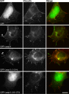
The LIM domain and nebulin repeats are necessary for localizing lasp-2 in HEK 293 cells. GFP–Lasp-2 and GFP–Lasp-2 1–214 assemble in similar patterns at focal adhesions (arrows) in HEK 293 cells, whereas GFP–Lasp-2 161–273 and GFP are predominantly nuclear, with some diffuse cytoplasmic distribution. Staining for endogenous vinculin marks sites of adhesion. These results indicate that the linker–SH3 domain (161–273) of lasp-2 is not necessary for its proper localization. Scale bar, 10 μm.
FIGURE 7:
FIGURE 7:
The knockdown of lasp-2 increases cell spreading rates. (A) siRNA to human lasp-2 was used to reduce lasp-2 RNA and protein levels in HEK 293 cells. RT-PCR shows that lasp-2 transcript is greatly reduced with lasp-2 siRNA. Lasp-2 protein levels are also significantly reduced with lasp-2 siRNA treatment. (B) In cell-spreading assays, the spread area of lasp-2 siRNA–treated cells was ∼15% increased in comparison to controls.
FIGURE 8:
FIGURE 8:
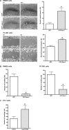
The overexpression of lasp-2 in SW620 and PC-3B1 cells increases cell migration rates but reduces cell invasion. (A) SW620 cells, derived from metastatic colorectal cancer, and PC-3B1 cells, derived from metastatic prostate cancer, migrated at a faster rate when GFP–lasp-2 is expressed. There was a threefold increase in the wound-healing rate in cells expressing GFP–lasp-2 in the SW620 cells. There was a 1.4-fold increase in the wound-healing rate in cells expressing GFP–lasp-2 in the PC-3B1 cells. *p < 0.05. (B) Cell invasion is reduced in cells expressing GFP–lasp-2. GFP–lasp-2–expressing cells invaded the chamber an average of 11-fold less than control cells in SW620 cells and invaded the chamber an average of fourfold less than control cells in PC-3B1 cells. *p < 0.005. (C) Loss of lasp-2 protein leads to an increase in cell invasion. Two different siRNA sequences to human lasp-2 were used to reduce lasp-2 protein levels in PC-3 cells. Cells with lasp-2 protein knocked down invaded the chamber approximately twofold more than controls. Data from one of the siRNA sequences are shown. *p < 0.05.

Similar articles

Cited by

References

    1. Beckerle MC. Zyxin: zinc fingers at sites of cell adhesion. Bioessays. 1997;19:949–957. - PubMed
    1. Carpenter AE, et al. CellProfiler: image analysis software for identifying and quantifying cell phenotypes. Genome Biol. 2006;7:R100. - PMC - PubMed
    1. Chew CS, Chen X, Parente JA, Jr, Tarrer S, Okamoto C, Qin HY. Lasp-1 binds to non-muscle F-actin in vitro and is localized within multiple sites of dynamic actin assembly in vivo. J Cell Sci. 2002;115:4787–4799. - PubMed
    1. Crawford AW, Beckerle MC. Purification and characterization of zyxin, an 82,000-dalton component of adherens junctions. J Biol Chem. 1991;266:5847–5853. - PubMed
    1. Deng XA, Norris A, Panaviene Z, Moncman CL. Ectopic expression of LIM-nebulette (LASP2) reveals roles in cell migration and spreading. Cell Motil Cytoskeleton. 2008;65:827–840. - PMC - PubMed

Publication types

MeSH terms

LinkOut - more resources