. 2013 Apr;24(7):901-13.
doi: 10.1091/mbc.E12-06-0458. Epub 2013 Feb 6.

Evidence for dynein and astral microtubule-mediated cortical release and transport of Gαi/LGN/NuMA complex in mitotic cells

Affiliations

Evidence for dynein and astral microtubule-mediated cortical release and transport of Gαi/LGN/NuMA complex in mitotic cells

Zhen Zheng et al. Mol Biol Cell. 2013 Apr.

Abstract

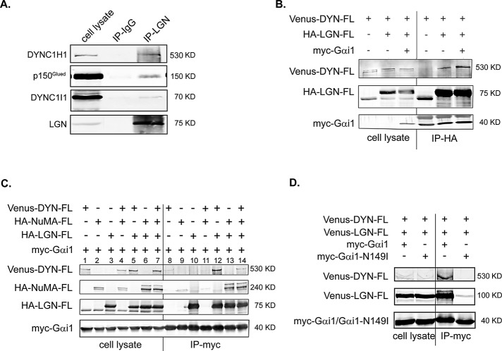
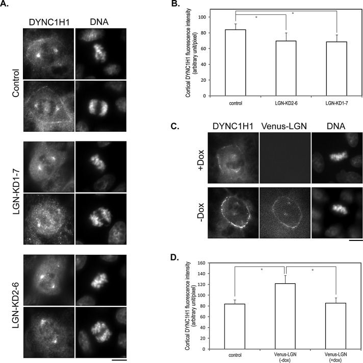
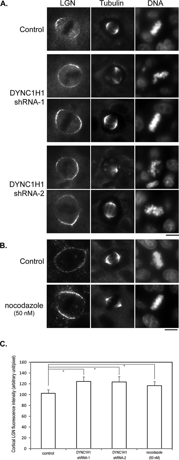
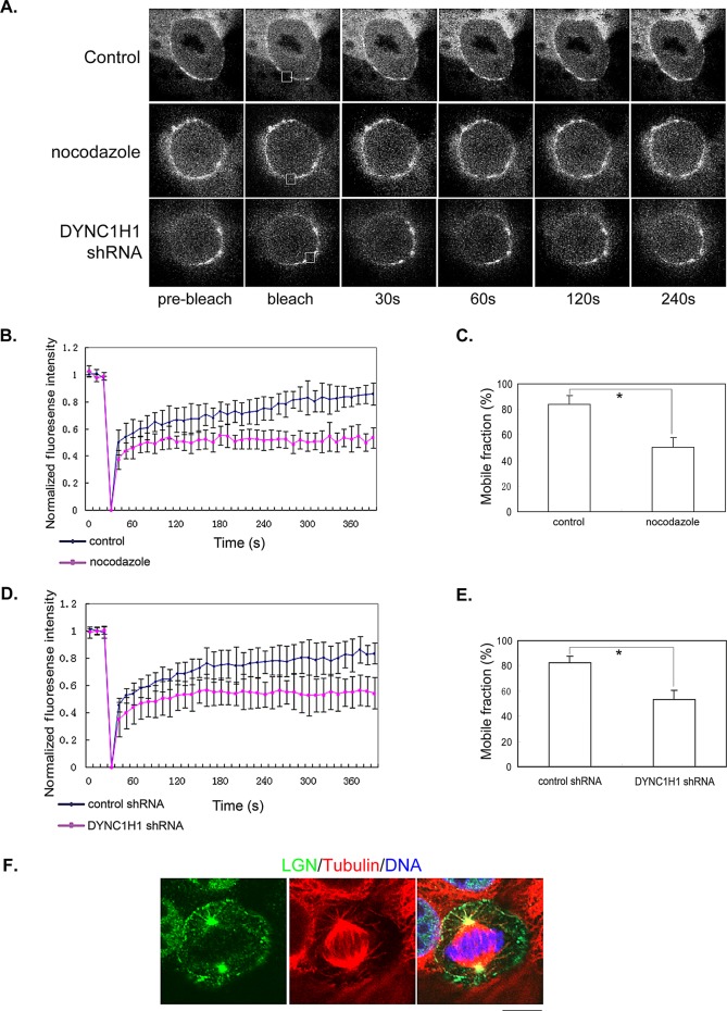
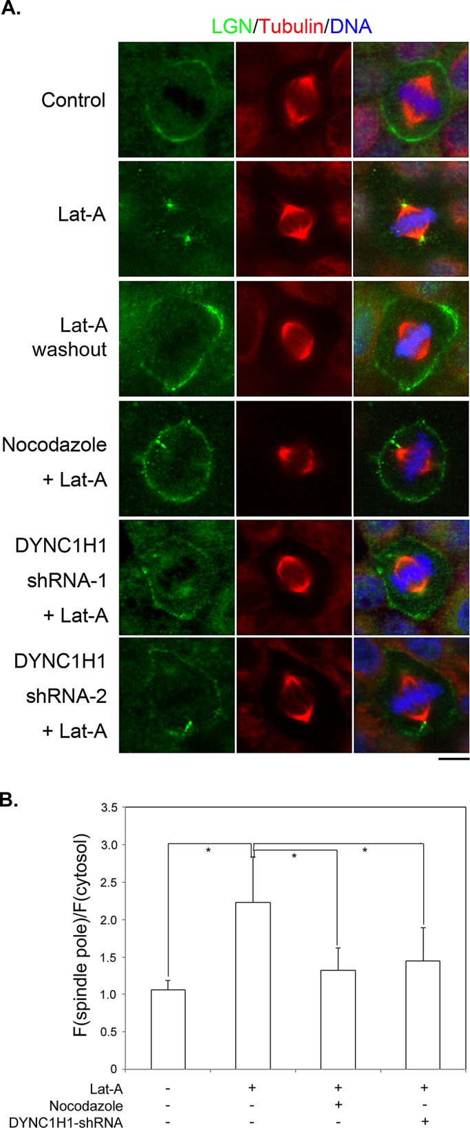
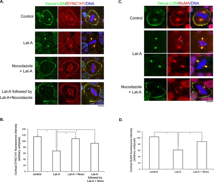
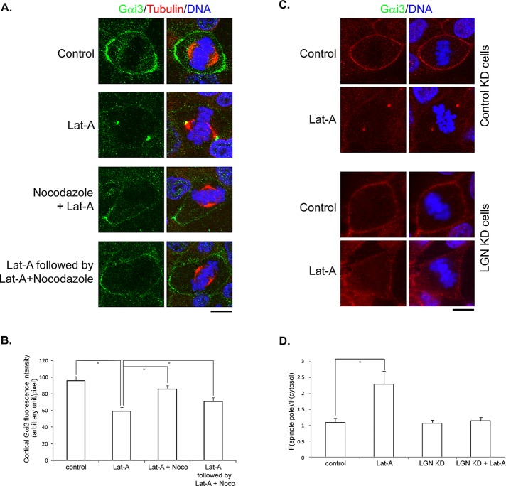
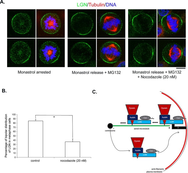
Spindle positioning is believed to be governed by the interaction between astral microtubules and the cell cortex and involve cortically anchored motor protein dynein. How dynein is recruited to and regulated at the cell cortex to generate forces on astral microtubules is not clear. Here we show that mammalian homologue of Drosophila Pins (Partner of Inscuteable) (LGN), a Gαi-binding protein that is critical for spindle positioning in different systems, associates with cytoplasmic dynein heavy chain (DYNC1H1) in a Gαi-regulated manner. LGN is required for the mitotic cortical localization of DYNC1H1, which, in turn, also modulates the cortical accumulation of LGN. Using fluorescence recovery after photobleaching analysis, we show that cortical LGN is dynamic and the turnover of LGN relies, at least partially, on astral microtubules and DYNC1H1. We provide evidence for dynein- and astral microtubule-mediated transport of Gαi/LGN/nuclear mitotic apparatus (NuMA) complex from cell cortex to spindle poles and show that actin filaments counteract such transport by maintaining Gαi/LGN/NuMA and dynein at the cell cortex. Our results indicate that astral microtubules are required for establishing bipolar, symmetrical cortical LGN distribution during metaphase. We propose that regulated cortical release and transport of LGN complex along astral microtubules may contribute to spindle positioning in mammalian cells.


Figures

FIGURE 1:
FIGURE 1:
i-regulated interaction between LGN and DYNC1H1. (A) Endogenous LGN and cytoplasmic dynein form a complex. MDCK II cells were partially synchronized by treating with nocodazole (200 nM) for 12 h. After being released from the treatment for 40 min, cells were harvested and subjected to immunoprecipitation using anti-LGN antibodies or rabbit IgG. The immunoprecipitates were separated by SDS–PAGE and blotted using specific antibodies. (B) Gαi enhances the association between LGN and DYNC1H1. Cos 7 cells were transfected as indicated. Cell lysates (1/30 of total) and immunoprecipitates were separated in a 6% SDS–PAGE gel and blotted using specific antibodies. Note that in 6% gel, myc-Gαi1 comigrated with and was masked by the light chain of anti-HA antibody in the HA-LGN immunoprecipitates. (C) Gαi, LGN, and DYNC1H1 form a complex in vivo, independent of NuMA. Cos 7 cells were transfected as indicated. Cell lysates (1/30 of total) and immunoprecipitates were separated in a 7% SDS–PAGE gel and blotted. (D) The GoLoco-insensitive Gαi1 cannot associate with LGN and DYNC1H1. Cos 7 cells were transfected as indicated. Cell lysates were subjected to analysis as in C.
FIGURE 2:
FIGURE 2:
LGN is required for the cortical localization of DYNC1H1 during mitosis. (A) LGN depletion results in reduced cortical localization of DYNC1H1. MDCK cells transduced by control lentivirus (control) or lentivirus expressing shRNAs targeting different regions of LGN (LGN-KD1-7 and LGN-KD2-6) were stained with anti-DYNC1H1 antibody and DNA dye. Bar, 10 μm. (B) Quantitation of the fluorescence intensity of cortical DYNC1H1 from images acquired in A. n = 50 for each set; *p < 0.01. (C) Slight overexpression of LGN leads to enhanced cortical localization of DYNC1H1. Stable Tet-Off MDCK cells expressing Venus-LGN were cultured in the presence (+Dox) or absence (–Dox) of doxycycline. At 24 h later, cells were stained as in A. Bar, 10 μm. (D) Quantitation of the fluorescence intensity of cortical DYNC1H1 from images acquired in C. n = 50 for each set; *p < 0.01.
FIGURE 3:
FIGURE 3:
Knocking down DYNC1H1 or disrupting astral MTs leads to enhanced cortical localization of LGN. (A) MDCK cells were transfected with plasmids expressing control shRNA (control) or shRNAs targeting DYNC1H1 (shRNA1 and shRNA2). At 48 h later, cells were fixed and stained with anti-LGN, anti–α-tubulin antibodies, and DNA dye. Bar, 10 μm. (B) MDCK cells were cultured in media containing 50 nM nocodazole for 40 min. Cells were then fixed and stained as in A. Bar, 10 μm. (C) Quantitation of the fluorescence intensity of cortical LGN from images acquired in A and B. n = 50 for each set; *p < 0.01.
FIGURE 4:
FIGURE 4:
Dynamic turnover of cortical LGN relies on astral MTs and DYNC1H1. (A) FRAP analysis of cortical LGN. Representative images from live-cell time-lapse series were shown. The photobleaching areas were marked by squares. Stable MDCK cells expressing Venus-LGN were either untreated (top), treated with 50 nM nocodazole for 1 h (middle), or transfected with DYNC1H1 shRNA for 48 h (bottom) before being subjected to FRAP analysis. (B–E) Quantitative analysis of FRAP experiments. (B, D) Plots of normalized fluorescence intensity of cortical Venus-LGN in cells treated with DMSO (B, blue diamonds), 50 nM nocodazole (B, pink squares), or transfected with control shRNA (D, blue diamonds) or DYNC1H1 shRNA (D, pink squares) vs. time (in seconds) after photobleaching. Data are expressed as mean ±SEM (n = 8 for each set). (C, E) The mobile fractions of cortical Venus-LGN in control, nocodazole-treated, or DYNC1H1-knockdown cells were calculated from the fluorescence recovery curves shown in B and D. Data are expressed as mean ±SEM. *p < 0.01. (F) Association of Venus-LGN with astral MTs. MDCK cells expressing Venus-LGN were preextracted with microtubule stabilization buffer containing 0.2% Triton X-100 and then fixed with 4% PFA. Fixed cells were stained with anti-LGN (green), anti–α-tubulin (red) antibodies, and DNA dye (blue). Bar, 10 μm.
FIGURE 5:
FIGURE 5:
Disruption of actin filaments leads to astral microtubule– and dynein-dependent cortical dissociation and spindle pole accumulation of LGN. (A) MDCK II cells were either untreated (Control) or treated as labeled. LatA: 1 μM of LatA for 45 min; Nocodazole + LatA: 50 nM of nocodazole plus 1 μM of LatA for 45 min; DYNC1H1 shRNA-1, -2 + LatA: transfected with DYNC1H1 shRNA-1 or -2 for 48 h and then treated with 1 μM LatA for 45 min. MG132, 5 μM, was added 1 h before treatments and maintained during treatments. Cells were fixed after treatments and stained with anti-LGN (green), anti–α-tubulin (red) antibodies, and DNA dye (blue). (B) Quantitation of LGN signals at spindle poles as described in Materials and Methods. n = 50 for each set; *p < 0.01.
FIGURE 6:
FIGURE 6:
Disruption of actin filaments leads to astral microtubule–dependent cortical dissociation and spindle pole accumulation of DYNC1H1 and NuMA. (A) Venus-LGN–expressing cells were treated as in Figure 5A except for the results at the bottom, for which cells were first treated with 1 μM LatA for 45 min and then treated with 1 μM LatA plus 50 nM nocodazole for another 45 min. Cells were stained with anti-DYNC1H1 antibody (red) and DNA dye (blue). (B) Quantitation of cortical DYNC1H1 fluorescence intensity as described in Materials and Methods. n = 50 for each set; *p < 0.01. (C) Venus-LGN–expressing cells were treated as in A. Cells were stained with anti-NuMA antibody (red) and DNA dye (blue). Second from top, arrow points to the original crescent-shaped NuMA, and arrowhead points to NuMA accumulated at the spindle pole. (D) Quantitation of cortical NuMA fluorescence intensity. n = 50 for each set; *p < 0.01. Bars, 10 μm.
FIGURE 7:
FIGURE 7:
Disruption of actin filaments leads to astral microtubule– and LGN-dependent cortical release and spindle pole accumulation of Gαi3. (A) MDCK II cells were treated as in Figure 5A. Cells were fixed after treatments and stained with anti-Gαi3 (green), anti–α-tubulin (red) antibodies, and DNA dye (blue). (B) Quantitation of cortical Gαi3 fluorescence intensity. n = 50 for each set; *p < 0.01. (C) Stable MDCK cells expressing control shRNA (top two) or shRNA against LGN (bottom two) were either untreated (control) or treated with 1 μM of LatA for 45 min (LatA). Cells were fixed and stained with anti-Gαi3 (red) and DNA dye (blue). (D) Quantitation of Gαi3 signals at spindle poles as described in Materials and Methods. n = 50 for each set; *p < 0.01.
FIGURE 8:
FIGURE 8:
Astral MTs are required for establishing bipolar, symmetrical cortical LGN distribution during metaphase. (A) HeLa cells were partially synchronized by treating with 2 mM thymidine for 16 h. After release in normal medium for 8 h, cells were treated with 50 mM monastrol for 3 h. Monastrol-arrested cells were either directly fixed (left) or washed and released in normal medium containing 5 μM MG132 (middle) or 5 μM MG132 plus 20 nM nocodazole (right) for 1 h and fixed. Fixed cells were stained with anti-LGN (green), anti–α-tubulin (red) antibodies, and DNA dye (blue). Bar, 10 μm. (B) Quantitation of bipolar, symmetrical cortical LGN localization. Only cells with normal chromosome condensation and alignment were included in the quantification. n = 100 from three independent experiments; *p < 0.001. (C) Schematic working model illustrating astral microtubule–mediated transport and recycling of the Gα/LGN/dynein/NuMA complex during mitosis.

Similar articles

Cited by

References

    1. Adames NR, Cooper JA. Microtubule interactions with the cell cortex causing nuclear movements in Saccharomyces cerevisiae. J Cell Biol. 2000;149:863–874. - PMC - PubMed
    1. Ahringer J. Control of cell polarity and mitotic spindle positioning in animal cells. Curr Opin Cell Biol. 2003;15:73–81. - PubMed
    1. Ben-Yair R, Kahane N, Kalcheim C. LGN-dependent orientation of cell divisions in the dermomyotome controls lineage segregation into muscle and dermis. Development. 2011;138:4155–4166. - PubMed
    1. Bowman SK, Neumuller RA, Novatchkova M, Du Q, Knoblich JA. The Drosophila NuMA homolog Mud regulates spindle orientation in asymmetric cell division. Dev Cell. 2006;10:731–742. - PubMed
    1. Busson S, Dujardin D, Moreau A, Dompierre J, De Mey JR. Dynein and dynactin are localized to astral microtubules and at cortical sites in mitotic epithelial cells. Curr Biol. 1998;8:541–544. - PubMed

Publication types

MeSH terms

LinkOut - more resources