. 2013 Jun;18(6):61227.
doi: 10.1117/1.JBO.18.6.061227.

Confocal microscopy with strip mosaicing for rapid imaging over large areas of excised tissue

Affiliations

Confocal microscopy with strip mosaicing for rapid imaging over large areas of excised tissue

Sanjee Abeytunge et al. J Biomed Opt. 2013 Jun.

Abstract

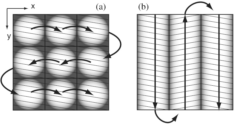
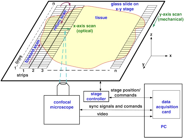
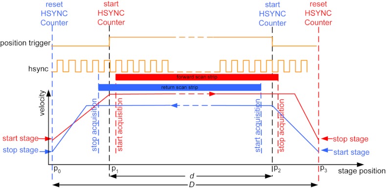
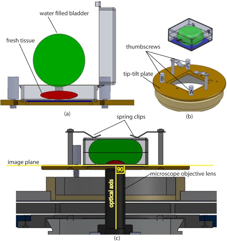
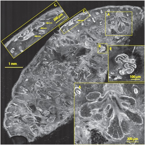
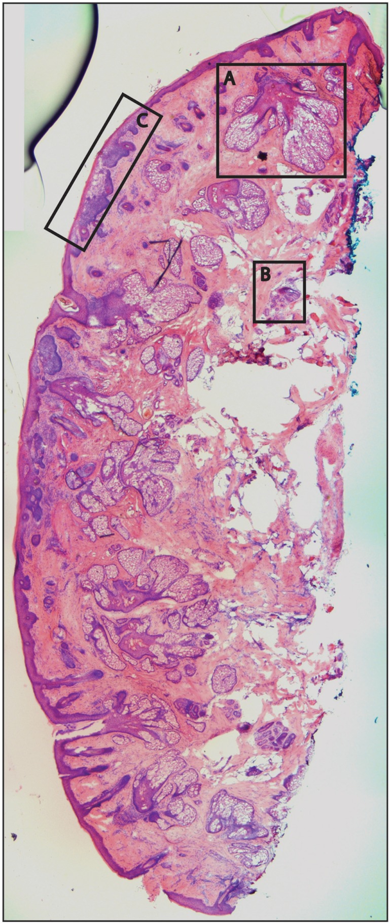
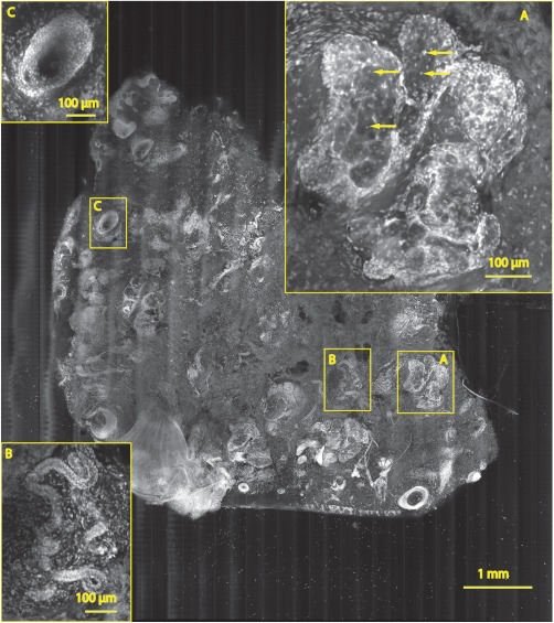
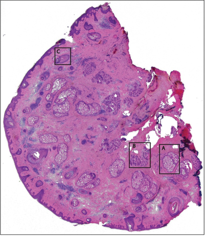
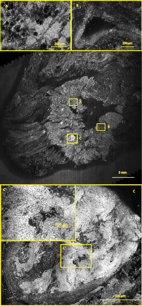
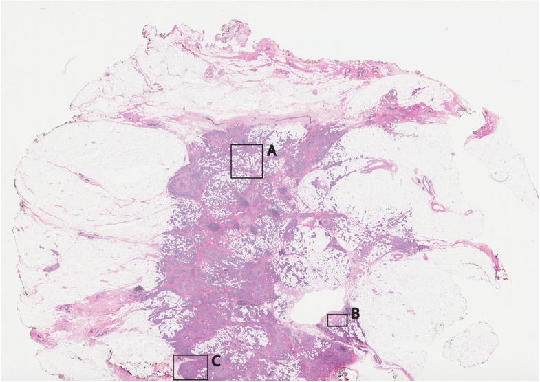
Confocal mosaicing microscopy is a developing technology platform for imaging tumor margins directly in freshly excised tissue, without the processing required for conventional pathology. Previously, mosaicing on 12-×-12 mm² of excised skin tissue from Mohs surgery and detection of basal cell carcinoma margins was demonstrated in 9 min. Last year, we reported the feasibility of a faster approach called "strip mosaicing," which was demonstrated on a 10-×-10 mm² of tissue in 3 min. Here we describe further advances in instrumentation, software, and speed. A mechanism was also developed to flatten tissue in order to enable consistent and repeatable acquisition of images over large areas. We demonstrate mosaicing on 10-×-10 mm² of skin tissue with 1-μm lateral resolution in 90 s. A 2.5-×-3.5 cm² piece of breast tissue was scanned with 0.8-μm lateral resolution in 13 min. Rapid mosaicing of confocal images on large areas of fresh tissue potentially offers a means to perform pathology at the bedside. Imaging of tumor margins with strip mosaicing confocal microscopy may serve as an adjunct to conventional (frozen or fixed) pathology for guiding surgery.


Figures

Fig. 1
Fig. 1
Comparison of mosaicing concepts: (a) mosaicing of square-shaped images in two dimensions; (b) mosaicing of rectangular-shaped long strips in one dimension (this figure is reproduced from Ref. 20).
Fig. 2
Fig. 2
Schematic of the strip-scanning mechanism. The fast optical scan (x axis scan) defines the width of a strip. The slow mechanical scan along the length of the strip (y axis scan) is performed by translating the stage in the direction orthogonal to the x axis. When the acquisition of a strip image is completed (“forward scan”), the stage moves in the x direction a distance equal to or less than the width of the strip. Then the stage moves along the strip length, but in the opposite direction (“return scan”), and another strip image is acquired. This process of acquiring strip images is repeated until the entire tissue is imaged.
Fig. 3
Fig. 3
The synchronizing scheme for the strips. A y translation stage cycle consists of two mechanical movements that produce two image strips: the “forward scan” P0 to P3, and the “return scan” P3 to P0. The velocity profiles are depicted in red for the forward movement, and blue for the return. The image is acquired within the constant velocity portion (d), between P1 and P2, of the scans. This avoids distortion of the mosaic due to compression or elongation of pixels. Therefore, we choose a scan distance (D) for the y translation stage such that the region of constant velocity (d) is larger than the size of the tissue sample. After the forward scan is initiated and the stage reaches P1, the “position trigger” signal is asserted to arm the counter that monitors horizontal synchronization pulses (HSYNC) from the asynchronous optical scanner. When the HSYNC counter receives the next HSYNC pulse, the data acquisition begins. When the stage reaches P2, the HSYNC counter resets and the acquisition continues until the last line is complete in the strip image. Then the stage decelerates and stops at P3. Once the x translation stage moves in the x direction to a predetermined position that sets the width of the strip and the overlap between adjacent strips, the return scan is initiated from P3 to P0. It follows a similar mechanism to the forward scan. This cyclical process, forward scan and return scan, is repeated until the entire tissue is imaged.
Fig. 4
Fig. 4
Schematic representation of the tissue-flattening device: (a) 2-D view of the tissue (in red) inside the cassette in its natural shape, as often seen in Mohs surgical excisions, with the edges curved up from the image plane and the lower (to be imaged) surface not flattened. The cassette is shown to be open with no force is applied to the water-filled bladder; (b) view of the closed tissue-cassette before mounting on the tip-tilt plate; the thumbscrews A, B, and C can be used to adjust the tip and tilt of the image plane relative to the optical axis, so as to orient the two at exactly 90 deg to each other; (c) schematic representation shows how the tissue is flattened, relative to the image plane and optical axis, by the water filled bladder when force is applied to it.
Fig. 5
Fig. 5
A 7.7-×-7.7  mm2 mosaic consisting of 24 fluorescence image strips of excised tissue from Mohs surgery. Typical features such as sebaceous glands (a) and eccrine glands (b) can be seen. Nests of superficial basal cell carcinomas (c) shown with arrows are observed, showing increased density of nuclei. The mosaic dimensions are 8100-×-8100  pixels (width x height) with 8  bits/pixel. Note that the magnified areas are display zooms obtained from the original image showing the detail and resolution of the mosaic. The features in the mosaic compare well, in general, to the pathology (Fig. 6) in terms of location, shape, size, nuclei of cells (shown in bright white) and overall morphology of both basal cell carcinomas and normal features.
Fig. 6
Fig. 6
Frozen H&E-stained pathology of excised tissue from Mohs surgery. This frozen section corresponds to a tissue slice that is adjacent to (but not exactly of) the imaged surface shown in Fig. 5. The features in inset areas A, B, and C are clearly identified across the two figures.
Fig. 7
Fig. 7
A 6.8-×-8  mm2 mosaic consisting of 17 fluorescence image strips of excised tissue from Mohs surgery. Typical features such as sebaceous glands (a), eccrine glands (b), and hair follicle (c) are seen. The mosaic dimensions are 7300-×-8200  pixels (width×height) with 8bits/pixel. The features in the mosaic compare well to the pathology (Fig. 8), in terms of location, shape, size, nuclei of cells (shown with arrows), and overall morphology.
Fig. 8
Fig. 8
Frozen H&E-stained pathology of excised tissue from Mohs surgery. The frozen section corresponds to a tissue slice adjacent to (but not exactly of the) imaged surface shown in Fig. 7. The features in inset areas A, B, and C are clearly identified across the two figures.
Fig. 9
Fig. 9
A 2.5×3.5  cm2 mosaic, consisting of 85 fluorescence image strips, of excised tissue from breast cancer surgery. The mosaic shows a central bright-appearing region of tumor and outlying somewhat darker-appearing regions of fat and fibrous tissue. The nucleus of cells appear to be bright. Closer inspection (insets) shows: (a) cancer cells invading a region of fat cells (b) a breast duct with hollow lumen (c) cancer cells proliferating in a duct. (Continued on next page.)
Fig. 10
Fig. 10
Fixed H&E-stained pathology of excised tissue from breast surgery. Labels indicate matching regions to the confocal mosaic in Fig. 9: (a) a duct; (b) ductal carcinoma in situ; and (c) invasive cancer cells.

References

    1. Rajadhyaksha M., et al. , “Confocal examination of nonmelanoma cancers in thick skin excisions to potentially guide mohs micrographic surgery without frozen histopathology,” J. Investig. Dermatol. 117(5), 1137–1143 (2001). 10.1046/j.0022-202x.2001.01524.x - DOI - PubMed
    1. Haque R., et al. , “Surgical margins and survival after head and neck cancer surgery,” BMC Ear Nose Throat Disord. 6, 2 (2006). 10.1186/1472-6815-6-2 - DOI - PMC - PubMed
    1. Jacobs L., “Positive margins: the challenge continues for breast surgeons,” Ann. Surg. Oncol. 15(5), 1271–1272 (2008). 10.1245/s10434-007-9766-0 - DOI - PMC - PubMed
    1. Al-Arashi M. Y., Salomatina E., Yaroslavsky A. N., “Multimodal confocal microscopy for diagnosing nonmelanoma skin cancers,” Laser. Surg. Med. 39(9), 696–705 (2007). 10.1002/(ISSN)1096-9101 - DOI - PubMed
    1. Jain M., et al. , “Multiphoton microscopy in the evaluation of human bladder biopsies,” Arch. Pathol. Lab. Med. 136(5), 517–526 (2012). 10.5858/arpa.2011-0147-OA - DOI - PMC - PubMed

Publication types

MeSH terms