

Case Reports
doi: 10.1136/bcr-2012-010573.
Basilar artery pseudoaneurysm presenting at 5-month follow-up after traumatic atlanto-occipital dislocation in a 7-year-old girl treated with intracranial stent placement and coiling
Affiliations
- PMID: 23389747
- PMCID: PMC3604333
- DOI: 10.1136/bcr-2012-010573
Item in Clipboard
Case Reports
Basilar artery pseudoaneurysm presenting at 5-month follow-up after traumatic atlanto-occipital dislocation in a 7-year-old girl treated with intracranial stent placement and coiling
BMJ Case Rep.
.
Abstract
Atlanto-occipital dislocation (AOD) is a grave injury that is rarely survivable. Patients who do survive often have long-term sequelae resulting from the intracranial damage sustained during the traumatic event. The high impact needed to cause AOD is translated to the intracranial vessels, which can lead to vascular injury. Pseudoaneurysm is one of the possible outcomes of damage to the vessel wall. We present a case of basilar artery pseudoaneurysm diagnosed 5 months after a traumatic AOD who was treated with intracranial stent placement and coiling.
Figures
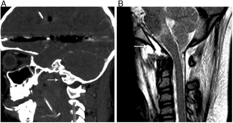
(A) Sagittal CT of the head and cervical spine in bone windows at the time of injury demonstrating atlanto-occipital dislocation (arrow). (B) Sagittal T2-weighted MRI showing prevertebral edema (arrow).
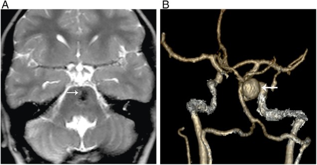
(A) Axial T2-weighted MRI 5 months after the initial trauma showing a dark signal lesion distorting the pons (arrow). (B) Three-dimensional reconstruction CT angiogram 5 months after the initial trauma showing development of a large saccular aneurysm arising from the mid-distal basilar artery (arrow).
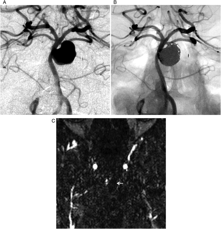
(A) Anterior posterior right vertebral artery angiogram confirms the findings of the CT angiogram. (B) Post-intervention right vertebral angiogram showing normal filling of the parent vessel with only minimal filling of the aneurysm neck. (C) Axial contrast enhanced magnetic resonance angiogram 3 months after coil placement showing no filling of the pseudoaneurysm with metal artifact from the coils.
References
-
- Parkinson D, West M. Traumatic intracranial aneurysms. J Neurosurg 1980;52:11–20 - PubMed
-
- Chaput CD, Torres E, Davis M, et al. Survival of atlanto-occipital dissociation correlates with atlanto-occipital distraction, injury severity score, and neurologic status. J Trauma 2011;71:393–5 - PubMed
-
- Labbe JL, Leclair O, Duparc B. Traumatic atlanto-occipital dislocation with survival in children. J Pediatr Orthop B 2001;10:319–27 - PubMed
-
- Bank WO, Nelson PB, Drayer BP, et al. Traumatic aneurysm of the basilar artery. AJR Am J Roentgenol 1978;130:975–7 - PubMed
Publication types
MeSH terms
LinkOut - more resources
Full Text Sources
Other Literature Sources
Medical