

Early diffusion-weighted imaging and perfusion-weighted imaging lesion volumes forecast final infarct size in DEFUSE 2
- PMID: 23390119
- PMCID: PMC3625664
- DOI: 10.1161/STROKEAHA.111.000135
Early diffusion-weighted imaging and perfusion-weighted imaging lesion volumes forecast final infarct size in DEFUSE 2
Abstract
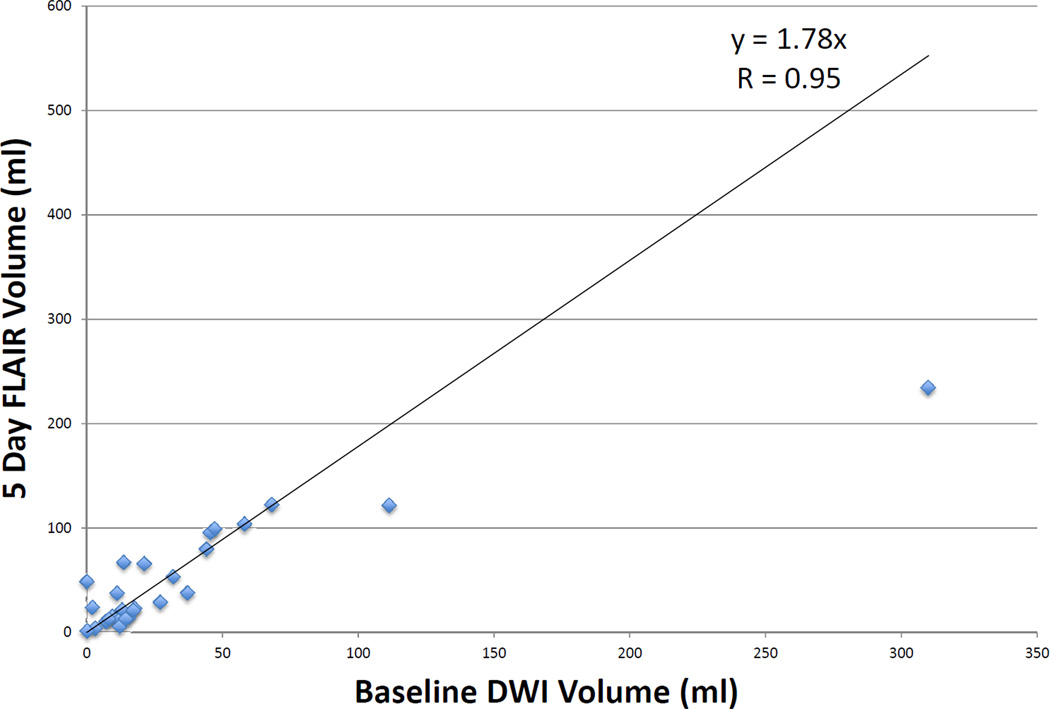
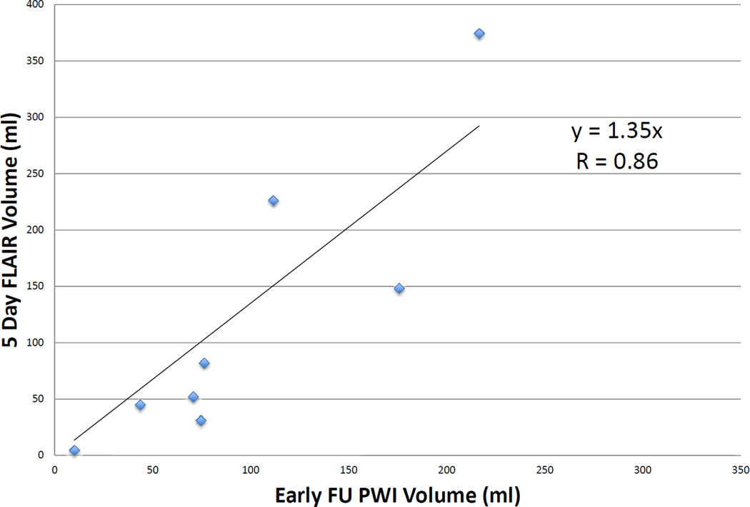
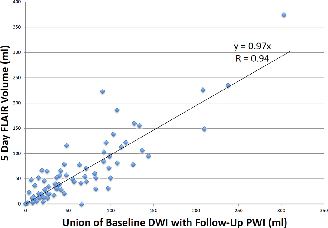
Background and purpose: It is hypothesized that early diffusion-weighted imaging (DWI) lesions accurately estimate the size of the irreversibly injured core and thresholded perfusion-weighted imaging (PWI) lesions (time to maximum of tissue residue function [Tmax] >6 seconds) approximate the volume of critically hypoperfused tissue. With incomplete reperfusion, the union of baseline DWI and posttreatment PWI is hypothesized to predict infarct volume.
Methods: This is a substudy of Diffusion and Perfusion Imaging Evaluation for Understanding Stroke Evolution Study 2 (DEFUSE 2); all patients with technically adequate MRI scans at 3 time points were included. Baseline DWI and early follow-up PWI lesion volumes were determined by the RAPID software program. Final infarct volumes were assessed with 5-day fluid-attenuated inversion recovery and were corrected for edema. Reperfusion was defined on the basis of the reduction in PWI lesion volume between baseline and early follow-up MRI. DWI and PWI volumes were correlated with final infarct volumes.
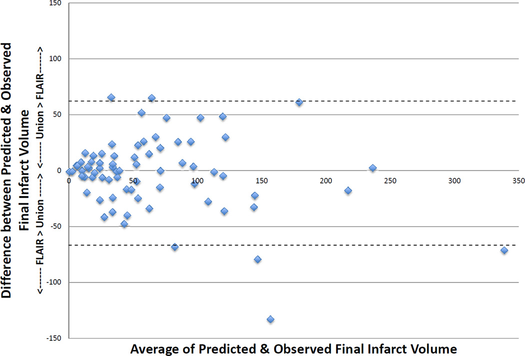
Results: Seventy-three patients were eligible. Twenty-six patients with >90% reperfusion show a high correlation between early DWI volume and final infarct volume (r=0.95; P<0.001). Nine patients with <10% reperfusion have a high correlation between baseline PWI (Tmax >6 seconds) volume and final infarct volume (r=0.86; P=0.002). Using all 73 patients, the union of baseline DWI and early follow-up PWI is highly correlated with final infarct volume (r=0.94; P<0.001). The median absolute difference between observed and predicted final volumes is 15 mL (interquartile range, 5.5-30.2).
Conclusions: Baseline DWI and early follow-up PWI (Tmax >6 seconds) volumes provide a reasonable approximation of final infarct volume after endovascular therapy.
Conflict of interest statement
G. Albers has equity interest in iSchemaView and has worked as a consultant for Covidien and Stryker. R Bammer has equity interest in iSchemaView. G. Zaharchuk receives modest research funding support from GE Healthcare. All other authors report no conflicts of interest.
Figures
References
-
- Albers GW, Thijs VN, Wechsler L, Kemp S, Schlaug G, Skalabrin E, et al. Magnetic resonance imaging profiles predict clinical response to early reperfusion: the diffusion and perfusion imaging evaluation for understanding stroke evolution (DEFUSE) study. Annals of Neurology. 2006;60:508–517. - PubMed
-
- Lansberg MG, Lee J, Christensen S, Straka M, De Silva DA, Mlynash M, et al. RAPID automated patient selection for reperfusion therapy: a pooled analysis of the Echoplanar Imaging Thrombolytic Evaluation Trial (EPITHET) and the Diffusion and Perfusion Imaging Evaluation for Understanding Stroke Evolution (DEFUSE) Study. Stroke. 2011;42:1608–1614. - PMC - PubMed
Publication types
MeSH terms
Grants and funding
LinkOut - more resources
Full Text Sources
Other Literature Sources
Medical
