

Structural and functional brain connectivity in presymptomatic familial frontotemporal dementia
- PMID: 23390180
- PMCID: PMC3598452
- DOI: 10.1212/WNL.0b013e31828407bc
Structural and functional brain connectivity in presymptomatic familial frontotemporal dementia
Erratum in
- Neurology. 2014 Apr 29;82(17):1570
Abstract
Objective: We aimed to investigate whether cognitive deficits and structural and functional connectivity changes can be detected before symptom onset in a large cohort of carriers of microtubule-associated protein tau and progranulin mutations.
Methods: In this case-control study, 75 healthy individuals (aged 20-70 years) with 50% risk for frontotemporal dementia (FTD) underwent DNA screening, neuropsychological assessment, and structural and functional MRI. We used voxel-based morphometry and tract-based spatial statistics for voxelwise analyses of gray matter volume and diffusion tensor imaging measures. Using resting-state fMRI scans, we assessed whole-brain functional connectivity to frontoinsula, anterior midcingulate cortex (aMCC), and posterior cingulate cortex.
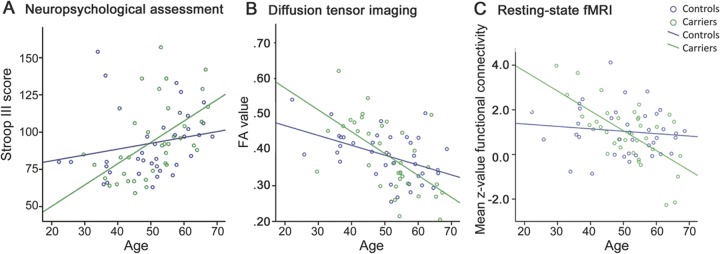
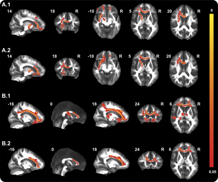
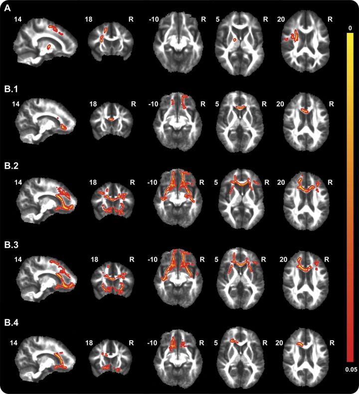
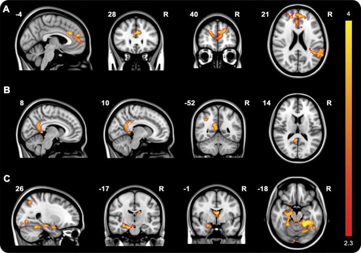
Results: Although carriers (n = 37) and noncarriers (n = 38) had similar neuropsychological performance, worse performance on Stroop III, Ekman faces, and Happé cartoons correlated with higher age in carriers, but not controls. Reduced fractional anisotropy and increased radial diffusivity throughout frontotemporal white matter tracts were found in carriers and correlated with higher age. Reductions in functional aMCC connectivity were found in carriers compared with controls, and connectivity between frontoinsula and aMCC seeds and several brain regions significantly decreased with higher age in carriers but not controls. We found no significant differences or age correlations in posterior cingulate cortex connectivity. No differences in regional gray matter volume were found.
Conclusions: This study convincingly demonstrates that alterations in structural and functional connectivity develop before the first symptoms of FTD arise. These findings suggest that diffusion tensor imaging and resting-state fMRI may have the potential to become sensitive biomarkers for early FTD in future clinical trials.
Figures
References
-
- Neary D, Snowden JS, Gustafson L, et al. Frontotemporal lobar degeneration: a consensus on clinical diagnostic criteria. Neurology 1998;51:1546–1554 - PubMed
-
- Snowden JS, Gibbons ZC, Blackshaw A, et al. Social cognition in frontotemporal dementia and Huntington's disease. Neuropsychologia 2003;41:688–701 - PubMed
-
- Boccardi M, Sabattoli F, Laakso MP, et al. Frontotemporal dementia as a neural system disease. Neurobiol Aging 2005;26:37–44 - PubMed
Publication types
MeSH terms
LinkOut - more resources
Full Text Sources
Other Literature Sources