. 2013;8(2):e55116.
doi: 10.1371/journal.pone.0055116. Epub 2013 Feb 4.

Occludin is involved in adhesion, apoptosis, differentiation and Ca2+-homeostasis of human keratinocytes: implications for tumorigenesis

Affiliations

Occludin is involved in adhesion, apoptosis, differentiation and Ca2+-homeostasis of human keratinocytes: implications for tumorigenesis

Susanne Rachow et al. PLoS One. 2013.

Abstract

Tight junction (TJ) proteins are involved in a number of cellular functions, including paracellular barrier formation, cell polarization, differentiation, and proliferation. Altered expression of TJ proteins was reported in various epithelial tumors. Here, we used tissue samples of human cutaneous squamous cell carcinoma (SCC), its precursor tumors, as well as sun-exposed and non-sun-exposed skin as a model system to investigate TJ protein alteration at various stages of tumorigenesis. We identified that a broader localization of zonula occludens protein (ZO)-1 and claudin-4 (Cldn-4) as well as downregulation of Cldn-1 in deeper epidermal layers is a frequent event in all the tumor entities as well as in sun-exposed skin, suggesting that these changes result from chronic UV irradiation. In contrast, SCC could be distinguished from the precursor tumors and sun-exposed skin by a frequent complete loss of occludin (Ocln). To elucidate the impact of down-regulation of Ocln, we performed Ocln siRNA experiments in human keratinocytes and uncovered that Ocln downregulation results in decreased epithelial cell-cell adhesion and reduced susceptibility to apoptosis induction by UVB or TNF-related apoptosis-inducing ligand (TRAIL), cellular characteristics for tumorigenesis. Furthermore, an influence on epidermal differentiation was observed, while there was no change of E-cadherin and vimentin, markers for epithelial-mesenchymal transition. Ocln knock-down altered Ca(2+)-homeostasis which may contribute to alterations of cell-cell adhesion and differentiation. As downregulation of Ocln is also seen in SCC derived from other tissues, as well as in other carcinomas, we suggest this as a common principle in tumor pathogenesis, which may be used as a target for therapeutic intervention.


Conflict of interest statement

Competing Interests: The authors have declared that no competing interests exist.

Figures

Figure 1
Figure 1. Schematic drawing of a squamous cell carcinoma denoting the different areas of the tumor.
Figure 2
Figure 2. Immunolocalization of Cldn-1, JAM-A, and Cldn-4 in healthy, non-sun- exposed skin and SCC.
Immunolocalization of Cldn-1 (A1, A2, B1, B2), JAM-A (C1, C2, D1, D2), and Cldn-4 (E1, E2, F1, F2) in healthy, non-sun-exposed skin (A1, A2, C1, C2, E1, E2) and SCC (B1, B2, D1, D2, F1, F2). (A1, B1, C1, D1, E1, F1: epifluorescence pictures; A2, B2, C2, D2, E2, F2: overlay of epifluorescence and phase contrast pictures). Note that Cldn-1 is downregulated in the uppermost and lowermost layers whereas JAM-A is primarily downregulated in the uppermost layers of SCC. Furthermore Cldn-4 demonstrates broader localization in SCC compared to healthy skin. Bars: 50 µm.
Figure 3
Figure 3. Immunolocalization of ZO-1 and Ocln in healthy, non-sun-exposed skin and SCC.
Immunolocalization of ZO-1 (A1, A2, B1, B2, C1, C2), and Ocln (D1, D2, E1, E2, F1, F2), in healthy, non-sun-exposed skin (A1, A2, D1, D2) and SCC (B1, B2, C1, C2, E1, E2, F1, F2). (A1, B1, C1, D1, E1, F1: epifluorescence pictures; A2, B2, C2, D2, E2, F2: overlay of epifluorescence and phase contrast pictures). For both molecules an example of broader localization (B1, B2, E1, E2) and loss of the protein (C1, C2, F1, F2) are shown. Note that exposure time in C1, C2, F1, F2 was substantially higher than in the other figures in order to show that also with very high exposure time no Ocln or ZO-1 staining was seen in these samples. Bars: 50 µm.
Figure 4
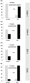
Figure 4. Distribution patterns of TJ proteins in normal skin from sun-exposed and non-sun-exposed areas.
Percentages denote distribution patterns of the various TJ proteins in sun-exposed (sun; e. g. face, lower arms) and non-sun-exposed areas (no-sun; e. g. abdomen, bottom). ***: p<0.001 between sun-exposed and non-sun-exposed skin.
Figure 5
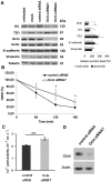
Figure 5. Influence of Ocln on epidermal differentiation and EMT markers, cell-cell adhesion and paracellular Ca2+ permeability.
(A) (left side) Western Blot analysis of 3D skin models after silencing of Ocln with 2 different siRNAs shows downregulation of Ocln and of the epidermal differentiation marker involucrin and an upregulation of TG1. There is no alteration for the EMT markers E-cadherin and vimentin. Same amounts of protein were loaded and actin or tubulin were used as gel loading controls. A representative experiment is shown (n = 3). (right side) Semiquantitative analysis of Ocln, involucrin, TG1, E-cadherin and vimentin. Band intensities were normalized to actin (Inv, TG1) or tubulin (E-cad, vim). Subsequently, the values were normalized to control siRNA treated cells (n = 3; mean±SEM; *p<0.05, ***p<0.001 compared to control siRNA). (B) Calcium induced cell-cell adhesion was investigated with a hanging drop assay at the indicated time points. Significantly greater numbers of particles (indicating less cell-cell adhesion) were found in the suspensions of Ocln knock-down cells compared to the controls (*: p<0.05 n = 3), (C) Electrophysiological studies of paracellular Ca2+-permeability in Ocln siRNA-treated cultured keratinocytes revealed an increase in paracellular permeability for Ca2+ in Ocln knock-down cells compared to cells treated with control siRNA (n = 6, mean ± SEM). (D) Example for the knock-down of Ocln in siRNA treated submerged cells. Mean knock-down of Ocln in the cells used for experiments in Figure 5 B, C and 6 was 76% +/−9%.
Figure 6
Figure 6. Influence of Ocln on DNA fragmentation after induction of apoptosis by TRAIL and UVB.
Apoptotic response at 20 h of TRAIL treatment (25 ng/ml) (A+B) or 16 h after UVB-irradiation (C) in human keratinocyte cultures with or without Ocln knock-down due to Ocln siRNA7 (A+C+D) or Ocln siRNA9 treatment (B). A significant decrease of apoptotic DNA fragmentation induced by TRAIL and UV light was observed after downregulation of Ocln. (D) Cytotoxicity measured by LDH assay at 3 h and 20 h of TRAIL treatment (100% corresponds to complete lysis of the cells). Note that 3 h after TRAIL treatment (when apoptotic fragmentation was already detectable) cytotoxicity was not primarily influenced indicating an apoptotic rather than necrotic mechanism. Mean +/− SEM. *: p<0.05; ** p<0.01. Example of one out of at least 10 independent cell lines. All experiments were performed in triplicates.

References

    1. Aijaz S, Balda MS, Matter K (2006) Tight junctions: molecular architecture and function. Int Rev Cytol 248: 261–298. - PubMed
    1. Balda MS, Matter K (2009) Tight junctions and the regulation of gene expression. Biochim Biophys Acta 1788: 761–767. - PubMed
    1. Ebnet K, Suzuki A, Ohno S, Vestweber D (2004) Junctional adhesion molecules (JAMs): more molecules with dual functions? J Cell Sci 117: 19–29. - PubMed
    1. Furuse M, Tsukita S (2006) Claudins in occluding junctions of humans and flies. Trends Cell Biol 16: 181–188. - PubMed
    1. Shen L, Weber CR, Raleigh DR, Yu D, Turner JR (2011) Tight junction pore and leak pathways: a dynamic duo. Annu Rev Physiol 73: 283–309. - PMC - PubMed

Publication types

MeSH terms