Randomized Controlled Trial
. 2013 Feb 7:10:24.
doi: 10.1186/1742-2094-10-24.

Cerebral ischemia initiates an immediate innate immune response in neonates during cardiac surgery

Affiliations
Randomized Controlled Trial

Cerebral ischemia initiates an immediate innate immune response in neonates during cardiac surgery

Selma O Algra et al. J Neuroinflammation. .

Abstract

Background: A robust inflammatory response occurs in the hours and days following cerebral ischemia. However, little is known about the immediate innate immune response in the first minutes after an ischemic insult in humans. We utilized the use of circulatory arrest during cardiac surgery to assess this.

Methods: Twelve neonates diagnosed with an aortic arch obstruction underwent cardiac surgery with cardiopulmonary bypass and approximately 30 minutes of deep hypothermic circulatory arrest (DHCA, representing cerebral ischemia). Blood samples were drawn from the vena cava superior immediately after DHCA and at various other time points from preoperatively to 24 hours after surgery. The innate immune response was assessed by neutrophil and monocyte count and phenotype using FACS, and concentrations of cytokines IL-1β, IL-6, IL-8, IL-10, TNFα, sVCAM-1 and MCP-1 were assessed using multiplex immunoassay. Results were compared to a simultaneously drawn sample from the arterial cannula. Twelve other neonates were randomly allocated to undergo the same procedure but with continuous antegrade cerebral perfusion (ACP).

Results: Immediately after cerebral ischemia (DHCA), neutrophil and monocyte counts were higher in venous blood than arterial (P = 0.03 and P = 0.02 respectively). The phenotypes of these cells showed an activated state (both P <0.01). Most striking was the increase in the 'non-classical' monocyte subpopulations (CD16(intermediate); arterial 6.6% vs. venous 14%; CD16+ 13% vs. 22%, both P <0.01). Also, higher IL-6 and lower sVCAM-1 concentrations were found in venous blood (both P = 0.03). In contrast, in the ACP group, all inflammatory parameters remained stable.

Conclusions: In neonates, approximately 30 minutes of cerebral ischemia during deep hypothermia elicits an immediate innate immune response, especially of the monocyte compartment. This phenomenon may hold important clues for the understanding of the inflammatory response to stroke and its potentially detrimental consequences.

Trial registration: ClinicalTrial.gov: NCT01032876.


Figures

Figure 1
Figure 1
Sample collection during CPB. Upper picture shows all time points at which blood samples were collected. The first was before CPB (‘Pre-op’). Once on CPB, patients were cooled to deep hypothermia and DHCA or ACP was initiated (‘Start’). At the end of DHCA or ACP (‘End’), full-flow CPB was recommenced and the patient was rewarmed to normothermia, during which the ‘30 min’ sample was drawn. Four and 24 hours after surgery, the final samples were collected (‘4 h post-operative’ and ‘24 h post-operative’). The lower picture depicts the arterial and venous samples drawn at the ‘End’ time point. Samples were simultaneously drawn from the arterial and venous cannulae, transporting blood to and from the brain, respectively. ACP, antegrade cerebral perfusion; CPB, cardiopulmonary bypass; DHCA, deep hypothermic circulatory arrest.
Figure 2
Figure 2
Monocyte subpopulations. (a) Gating of monocytes into CD16+, CD16int (intermediates) and CD16+ subpopulations. (b) CD16- monocytes show a low expression of HLA-DR, whereas CD16int and CD16+ show a high expression (MFIs of 663 (IQR 337 to 867), 2174 (1189 to 4884) and 3903 (1372 to 8393), respectively). IQR, interquartile range; HLA-DR, human leukocyte antigen D-related; MFI, median fluorescence intensity.
Figure 3
Figure 3
Deep hypothermic CPB decreases neutrophil and monocyte counts. Perioperative time course of neutrophil and monocyte and counts, in DHCA and ACP groups separately (DHCA; (a) and (c); ACP; (b) and (d)). During CPB, counts were corrected for the dilution of the CPB circuit (shown in grey). ACP, antegrade cerebral perfusion; CPB, cardiopulmonary bypass; DHCA, deep hypothermic circulatory arrest.
Figure 4
Figure 4
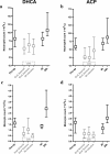
Cerebral ischemia increases neutrophils and monocyte counts. (a) Immediately after cerebral ischemia (i.e., DHCA), neutrophil and monocyte counts are increased in the venous sample compared to arterial (P = 0.03 and P = 0.02, respectively). (b) In continuous cerebral perfusion (i.e., ACP) there is no difference in neutrophil and monocyte counts. (c) To rule out the effect of hyperviscosity in the venous sample after cerebral ischemia, hematocrit and thrombocyte numbers were assessed revealing no difference after DHCA. (d) Also after ACP there is no difference in hematocrit and thrombocyte number. ACP, antegrade cerebral perfusion; DHCA, deep hypothermic circulatory arrest.
Figure 5
Figure 5
Cerebral ischemia leads to an immediate activation of neutrophils and monocytes. (a) After cerebral ischemia (i.e., DHCA), CD62L expression is the same in arterial and venous blood, but CD11b expression is higher in the venous sample compared to arterial (P <0.01). (b) After continuous cerebral perfusion (i.e., ACP), neutrophil CD11b and CD62L expressions remain stable. (c) After cerebral ischemia (DHCA), in the venous sample, the proportion of monocytes that is CD16- is decreased compared to arterial, due to an increase in CD16intermediate and CD16+ monocyte subpopulations (all P <0.01). (d) After continuous cerebral perfusion (ACP), the monocyte subpopulations remain unchanged.
Figure 6
Figure 6
Cerebral ischemia increases IL-6, and decreases sVCAM-1 concentrations. (a) After cerebral ischemia (i.e., DHCA), serum IL-6 is mildly increased, and sVCAM-1 decreased, in the venous sample compared to arterial (both P = 0.03). (b) After continuous cerebral perfusion (ACP), all cytokine levels remain stable.

References

    1. Denker SP, Ji S, Dingman A, Lee SY, Derugin N, Wendland MF, Vexler ZS. Macrophages are comprised of resident brain microglia not infiltrating peripheral monocytes acutely after neonatal stroke. J Neurochem. 2007;100:893–904. doi: 10.1111/j.1471-4159.2006.04162.x. - DOI - PMC - PubMed
    1. Garcia JH, Liu KF, Yoshida Y, Chen S, Lian J. Brain microvessels: factors altering their patency after the occlusion of a middle cerebral artery (Wistar rat) Am J Pathol. 1994;145:728–740. - PMC - PubMed
    1. Offner H, Subramanian S, Parker SM, Afentoulis ME, Vandenbark AA, Hurn PD. Experimental stroke induces massive, rapid activation of the peripheral immune system. J Cereb Blood Flow Metab. 2006;26:654–665. doi: 10.1038/sj.jcbfm.9600217. - DOI - PubMed
    1. Perego C, Fumagalli S, De Simoni MG. Temporal pattern of expression and colocalization of microglia/macrophage phenotype markers following brain ischemic injury in mice. J Neuroinflammation. 2011;8:174. doi: 10.1186/1742-2094-8-174. - DOI - PMC - PubMed
    1. Wang LW, Chang YC, Lin CY, Hong JS, Huang CC. Low-dose lipopolysaccharide selectively sensitizes hypoxic ischemia-induced white matter injury in the immature brain. Pediatr Res. 2010;68:41–47. doi: 10.1203/PDR.0b013e3181df5f6b. - DOI - PMC - PubMed

Publication types

Associated data