. 2013 May 1;377(1):188-201.
doi: 10.1016/j.ydbio.2013.01.031. Epub 2013 Feb 4.

Constitutive activation of NOTCH1 signaling in Sertoli cells causes gonocyte exit from quiescence

Affiliations

Constitutive activation of NOTCH1 signaling in Sertoli cells causes gonocyte exit from quiescence

Thomas Xavier Garcia et al. Dev Biol. .

Abstract

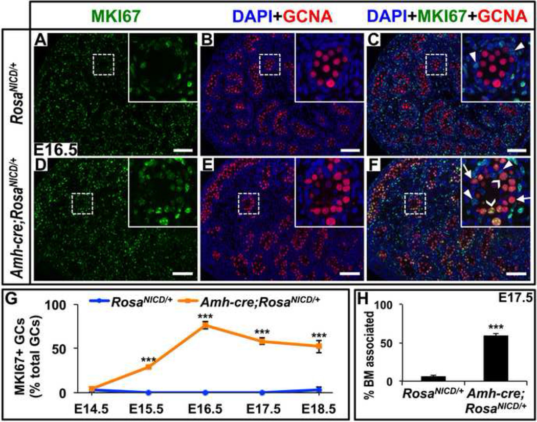
Notch signaling components have long been detected in Sertoli and germ cells in the developing and mature testis. However, the role of this pathway in testis development and spermatogenesis remains unknown. Using reporter mice expressing green fluorescent protein following Notch receptor activation, we found that Notch signaling was active in Sertoli cells at various fetal, neonatal, and adult stages. Since Notch signaling specifies stem cell fate in many developing and mature organ systems, we hypothesized that maintenance and differentiation of gonocytes and/or spermatogonial stem cells would be modulated through this pathway in Sertoli cells. To this end, we generated mutant mice constitutively expressing the active, intracellular domain of NOTCH1 (NICD1) in Sertoli cells. We found that mutant Sertoli cells were morphologically normal before and after birth, but presented a number of functional changes that drastically affected gonocyte numbers and physiology. We observed aberrant exit of gonocytes from mitotic arrest, migration toward cord periphery, and premature differentiation before birth. These events, presumably unsupported by the cellular microenvironment, were followed by gonocyte apoptosis and near complete disappearance of the gonocytes by day 2 after birth. Molecular analysis demonstrated that these effects are correlated with a dysregulation of Sertoli-expressed genes that are required for germ cell maintenance, such as Cyp26b1 and Gdnf. Taken together, our results demonstrate that Notch signaling is active in Sertoli cells throughout development and that proper regulation of Notch signaling in Sertoli cells is required for the maintenance of gonocytes in an undifferentiated state during fetal development.


Figures

Figure 1
Figure 1. Relative expression of Notch1 and Hes1 in Sertoli cells and undifferentiated and early differentiating spermatogonia
(A–D) Real-time PCR analysis of gene expression in Sertoli cells, undifferentiated spermatogonia (GFP positive cells), and early differentiating spermatogonia (GFP negative cells) isolated from P10 Pou5f1-GFP testes. Vim is a marker for Sertoli cells, Pou5f1 is a marker for undifferentiated spermatogonia, and Hes1 is a canonical Notch target gene. Results are given as means ± standard error means (SEMs). Statistical analysis was performed by single factor ANOVA.
Figure 2
Figure 2. Sertoli cells undergo active Notch signaling in fetal, juvenile, and adult stages
Immunofluorescence images of testes from Transgenic Notch Reporter GFP (TNR-GFP) mice in fetal (A-C), postnatal/juvenile (D-F), and adult (G) stages. Exact stages are noted in the first and second panels of each row. In the first column, germ cells are labeled in blue with PECAM1 in A-D and with Mouse Vasa Homolog (MVH) in E-G. The three right-most columns are high-magnification images focusing on individual testis cords, highlighting strong expression of TNR-GFP (green) in Sertoli cells (red) and absence of TNR-GFP expression in germ cells (blue). In the high-magnification images, germ cells are labeled with PECAM1 (membrane stain) in A-D and Germ cell nuclear antigen (GCNA) in E-G. Sertoli cells are labeled with SOX9 (nuclear stain) in A-B and E-G and with AMH (cytoplasmic stain) in C-D. Scale bars represent 50 µm.
Figure 3
Figure 3. Notch1, RosaNICD, and canonical Notch target gene expression following Amh-cre mediated recombination
(A) Schematic representation of the mouse Notch1 transcript (Nm_008714.3; nucleotides 1-9497), highlighting the CDS (265-7860), NICD (5509-7144), and TaqMan probe [Mm00627192_m1 (1819 ± 44), Mm00435249_m1 (6165 ± 38)] positions, which sit inside (in) and outside (out) of the NICD. (B) Real-time PCR analysis of gene expression in RosaNICD/+ (black bars) and Amh-cre;RosaNICD/+ (grey bars) whole testes at E14.5 and E17.5, and primary cultures of Sertoli cells (SC) isolated from P10 testes. Notch1 (in) (Mm00435249_m1) represents relative transcript levels of Notch1 and RosaNICD, whereas Notch1 (out) (Mm00627192_m1) represents relative transcript levels of Notch1 only. Hey1 and Heyl are canonical Notch target genes. Results are given as means ± standard error means (SEMs). Statistical analysis was performed by Student’s t test. n≥3, **P<0.05 and ***P<0.001.
Figure 4
Figure 4. Constitutive expression of NICD1 in fetal Sertoli cells results in progressive germ cell loss
(A–B) Stereomicroscopy images of testes from RosaNICD/+ (A) and Amh-cre;RosaNICD/+ (B) males at P2, P10, and P180. (C–D) Brightfield images of hematoxylin and eosin-stained RosaNICD/+ (C) and Amh-cre;RosaNICD/+ (D) testis sections at P60. (E–J) Immunofluorescence images of RosaNICD/+ (E,G,I) and Amh-cre;RosaNICD/+ (F,H,J) testis sections at E15.5 (E-F), E18.5 (G-H), and P2 (I-J). Germ cells are marked by Germ cell nuclear antigen (GCNA) in red and nuclei are marked by DAPI in blue. Scale bars represent 1 mm (A-B) and 100 µm (C-J). Higher magnification insets (E-J) are 100 µm × 100 µm. (K) Quantitation of GCNA positive cells (mean ± SEM) of circular tubule cross-sections at the indicated time points. The number of GCNA positive cells in oval and oblong tubule cross-sections was normalized to circular tubule cross-sections based on tubule cross-sectional area. **P<0.05; ***P<0.001.
Figure 5
Figure 5. Sertoli cell differentiation is normal following constitutive expression of NICD1
(A–D) Immunofluorescence images of RosaNICD/+ (A,C) and Amh-cre;RosaNICD/+ (B,D) testis sections at E17.5 (A-B) and P2 (C-D), showing SOX9 (green), GCNA (red), and DAPI (blue) staining. Scale bars represent 100 µm. Higher magnification insets are 100 µm × 100 µm. Representative SOX9 positive Sertoli cells marked with arrowheads. (E) Real-time PCR analysis of Dhh, Sox9, and Vim gene expression in RosaNICD/+ and Amh-cre;RosaNICD/+ whole testes at the indicated time points, E14.5 and E17.5. Results are given as means ± standard error means (SEMs). Means are not significantly different. Statistical analysis was performed by Student’s t test. n≥3.
Figure 6
Figure 6. Sertoli cell proliferation is normal, however germ cell mitosis and tubule position is abnormal
(A–F) Immunofluorescence images of RosaNICD/+ (A-C) and Amh-cre;RosaNICD/+ (B-F) testis sections at E16.5, showing MKI67 (green), GCNA (red), and DAPI (blue) staining. Scale bars represent 100 µm. Higher magnification insets are 100 µm × 100 µm. Examples of basement membrane-associated germ cells (arrows, F); MKI67 positive Sertoli cells (arrowheads, C,F); and MKI67 positive germ cells (chevrons, F) as marked. (G) Quantitation of percent total MKI67 positive germ cells at the indicated time points, E14.5-E18.5. (H) Quantitation of percent total GCNA positive cells associated with the basement membrane at E17.5. Results are given as means ± standard error means (SEMs). Statistical analysis was performed by Student’s t test. n≥3, ***P<0.001.
Figure 7
Figure 7. Gonocytes express the meiosis specific proteins, STRA8 and SYCP3, in Amh-cre; RosaNICD testes
(A–L) Immunofluorescence images of RosaNICD/+ (A-C,G-I) and Amh-cre; RosaNICD/+ (D-F,J-L) testis sections at E15.5. STRA8 and SYCP3 (green), GCNA (red), and DAPI (blue). Scale bars represent 100 µm. Higher magnification insets are 100 µm × 100 µm.
Figure 8
Figure 8. Germ cells undergo apoptosis in Amh-cre;RosaNICD/+ testes
(A–F) Immunofluorescence images of RosaNICD/+ (A,C,E) and Amh-cre;RosaNICD/+ (B,D,F) testis sections at E15.5 (A-B) and E17.5 (C-F) showing SOX9 (green), TUNEL (red), and DAPI (blue). Hashed lines in A-D denotes boundary between testis cords and interstitial compartment. Scale bars represent 100 µm. Higher magnification insets are 100 µm × 100 µm.
Figure 9
Figure 9. CYP26B1, Cyp26b1, and Gdnf expression is decreased in Amh-cre;RosaNICD testes
(A–B) Immunofluorescence images of RosaNICD/+ (A) and Amh-cre;RosaNICD/+ (B) testis sections at E15.5. CYP26B1 (green), GCNA (red), and DAPI (blue). Hashed line in insets denotes boundary between testis cord (T) and interstitial compartment (I). Examples of CYP26B1+ Sertoli cells (arrowheads) and CYP26B1+ Leydig cells (chevrons) as marked. Scale bars represent 100 µm. Higher magnification insets are 50 µm × 50 µm. (C) Real-time PCR analysis of Cyp26b1 gene expression in RosaNICD/+ and Amh-cre;RosaNICD/+ whole testes at E14.5 (D) Real-time PCR analysis of Gdnf gene expression in RosaNICD/+ (black bars) and Amh-cre;RosaNICD/+ (grey bars) whole testes at E14.5 and E17.5, and primary cultures of Sertoli cells isolated from P10 testes. (E) Cultures of primarily isolated Sertoli cells from P2 testes were treated with 0, 1, 5, and 25 µM concentrations of the γ-secretase inhibitor, DAPT, incubated for 18 hours, then assayed for the mRNA expression levels of Cyp26b1, Gdnf, Hes1, Hey1, and Heyl. The Notch target genes Hes1, Hey1, and Heyl were significantly decreased as expected, and Cyp26b1 and Gdnf were significantly increased. Results are given as means ± standard error means (SEMs). Statistical analysis was performed by Student’s t test. n≥3, **P<0.05; ***P<0.001.

References

    1. Artavanis-Tsakonas S, Muskavitch MA. Notch: the past, the present, and the future. Curr Top Dev Biol. 2010;92:1–29. - PubMed
    1. Barrionuevo F, Georg I, Scherthan H, Lecureuil C, Guillou F, Wegner M, Scherer G. Testis cord differentiation after the sex determination stage is independent of Sox9 but fails in the combined absence of Sox9 and Sox8. Dev Biol. 2009;327:301–312. - PubMed
    1. Basciani S, De Luca G, Dolci S, Brama M, Arizzi M, Mariani S, Rosano G, Spera G, Gnessi L. Platelet-derived growth factor receptor beta-subtype regulates proliferation and migration of gonocytes. Endocrinology. 2008;149:6226–6235. - PubMed
    1. Batista F, Lu L, Williams SA, Stanley P. Complex N-Glycans Are Essential, but Core 1 and 2 Mucin O-Glycans, O-Fucose Glycans, and NOTCH1 Are Dispensable, for Mammalian Spermatogenesis. Biol Reprod. 2012;86:1–12. - PMC - PubMed
    1. Bingham NC, Verma-Kurvari S, Parada LF, Parker KL. Development of a steroidogenic factor 1/Cre transgenic mouse line. Genesis. 2006;44:419–424. - PubMed

Publication types

MeSH terms