. 2013 Feb 26;110(9):3405-10.
doi: 10.1073/pnas.1220832110. Epub 2013 Feb 7.

Prospective isolation of human embryonic stem cell-derived cardiovascular progenitors that integrate into human fetal heart tissue

Affiliations

Prospective isolation of human embryonic stem cell-derived cardiovascular progenitors that integrate into human fetal heart tissue

Reza Ardehali et al. Proc Natl Acad Sci U S A. .

Abstract

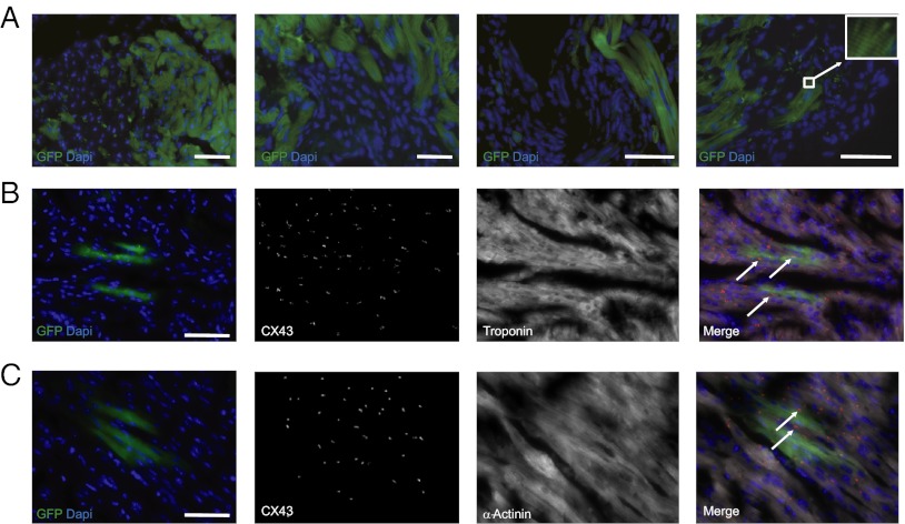
A goal of regenerative medicine is to identify cardiovascular progenitors from human ES cells (hESCs) that can functionally integrate into the human heart. Previous studies to evaluate the developmental potential of candidate hESC-derived progenitors have delivered these cells into murine and porcine cardiac tissue, with inconclusive evidence regarding the capacity of these human cells to physiologically engraft in xenotransplantation assays. Further, the potential of hESC-derived cardiovascular lineage cells to functionally couple to human myocardium remains untested and unknown. Here, we have prospectively identified a population of hESC-derived ROR2(+)/CD13(+)/KDR(+)/PDGFRα(+) cells that give rise to cardiomyocytes, endothelial cells, and vascular smooth muscle cells in vitro at a clonal level. We observed rare clusters of ROR2(+) cells and diffuse expression of KDR and PDGFRα in first-trimester human fetal hearts. We then developed an in vivo transplantation model by transplanting second-trimester human fetal heart tissues s.c. into the ear pinna of a SCID mouse. ROR2(+)/CD13(+)/KDR(+)/PDGFRα(+) cells were delivered into these functioning fetal heart tissues: in contrast to traditional murine heart models for cell transplantation, we show structural and functional integration of hESC-derived cardiovascular progenitors into human heart.


Conflict of interest statement

The authors declare no conflict of interest.

Figures

Fig. 1.
Fig. 1.
Identification of a cardiac mesoderm population marked by four surface markers—ROR2, CD13, KDR, and PDGFRα—and their expression in human fetal hearts. (A) Flow cytometric analysis of EBs at different time points of differentiation. On day 5, a distinct population defined by coexpression of ROR2 and CD13 (II) appeared, which was further analyzed for expression of KDR and PDGFRα. (B) Quantitative RT-PCR gene expression analysis of the QP (III), ROR2+CD13+ (II), and QN (I) cells isolated from day-5 EBs. The average expression is normalized to GAPDH. Data represent mean ± SD for three biologically independent experiments (P < 0.05, one-way ANOVA, populations III vs. I and II vs. I). (C) Presence of NKX2-5 (Left), MEF2C (Middle), and GATA-4 (Right) immunostaining in the QP population 24 h after sorting (Fig. S2). (Scale bar, 50 μm.) (D) Immunofluorescence staining of first trimester human hearts revealed pockets of ROR2-positive cells and diffuse KDR and PDGFRα staining in the left ventricle (arrows). (Scale bar, 120 μm.) (E) An area of the left ventricle with a cluster of ROR2+ cells that also costain with NKX2-5. (Scale bar, 120 μm.)
Fig. 2.
Fig. 2.
In vitro characterization of QP cells. (A) Immunofluorescence analysis of QP cells 6 d after sorting for markers of cardiomyocytes and smooth muscle and endothelial cells. (Scale bar, 25 μm.) (B) Quantitative RT-PCR analysis of QP cells grown in culture after 13 d after sorting for cardiac genes. Data represent mean ± SD for three biologically independent experiments. (C) Upon exposure to VEGF after sorting, QP cells (derived from hBCL2-hESC line and therefore expressing GFP) formed a lattice of tubular structures. (Scale bar, 100 μm.) (D) Endothelial phenotype was further confirmed by Dil-labeled acetylated LDL uptake. (Scale bar, 50 μm.)
Fig. 3.
Fig. 3.
Developmental potential of QP cells. (A) Whole-cell current-clamp recordings of spontaneous APs demonstrate ventricular-, atrial-, and nodal-like APs in the cultured QP population (Fig. S5). (B) Immunostaining of cells grown from a single GFP-QP cell indicates the presence of cardiomyocytes and endothelial and smooth muscle cells. (Top Left) Corresponding GFP cells (Fig. S6). (Scale bar, 50 μm.).
Fig. 4.
Fig. 4.
Engraftment of QP cells in mouse myocardium. (A) (Left) Whole mouse heart explanted 8 wk after injection of GFP+ QP cells. The dotted circle indicates the approximate location of the infarct zone. (Scale bar, 0.5 mm.) GFP-hESC–derived QP cells engraft into the periinfarct regions of mouse hearts (Middle and Right). (Scale bar, 120 μm.) (B and C) Immunofluorescence staining of mouse hearts 8 wk after QP transplantation reveals presence of GFP-hESC–derived QP cells that costain with troponin (B) and with human cardiomyocyte-specific β-myosin heavy chain (C). (Scale bar, 100 μm.) (D) Immunohistochemical evidence for teratoma formation after 8 wk upon transplantation of QN cells. The QN-derived cells gave rise to all three germ layers including columnar epithelium (Left), cartilage (Center), and neural rosette (Right). (Scale bar, 100 μm.)
Fig. 5.
Fig. 5.
Structural Integration of QP cells in a human fetal heart. (A) Myocardial sections from human fetal heart tissues 8 wk after transplantation s.c. into mouse ear and delivery of QP cells shows clusters of GFP+ cells spread throughout the ventricle. The site of injection of QP cells in the first micrograph is in the left upper corner. (Right, Inset) High-magnification image of sarcomeric structure. (Scale bar, 100 μm.) (B and C) Coexpression of GFP with cardiac-specific markers troponin (B) or α-actinin (C) and Connexin43 staining between host and transplanted GFP+ cells. (Right) Overlay with Connexin43 staining depicted in red and troponin or α-actinin in white. (Scale bar, 100 μm.)
Fig. 6.
Fig. 6.
QP cells integrate into fetal human myocardium. (A) Myocardial sections show evoked calcium signals when paced electrically ex vivo. Fluo-4 calcium dye was added to tissue, which was then electrically paced at 2 Hz. (Right) Same area after treatment with anti-GFP antibody reveals a GFP+ area. This region was analyzed for dye intensity changes (f) and results are plotted normalized to the intensity of the initial movie frame (f0). Real-time Ca2+ flux through the tissue indicate functional integration of GFP+ cells into the host tissue. (B) GFP+ cells, derived from XX H9 ESCs, were traced for FISH analyses to reveal XX karyotype (white arrowhead, Inset), whereas the host myocardium, from a male donor, expresses XY karyotype (white arrowhead, Inset). (Scale bar, 50 μm.)

References

    1. Bergmann O, et al. Evidence for cardiomyocyte renewal in humans. Science. 2009;324(5923):98–102. - PMC - PubMed
    1. Porrello ER, et al. Transient regenerative potential of the neonatal mouse heart. Science. 2011;331(6020):1078–1080. - PMC - PubMed
    1. Segers VF, Lee RT. Stem-cell therapy for cardiac disease. Nature. 2008;451(7181):937–942. - PubMed
    1. Laflamme MA, Murry CE. Heart regeneration. Nature. 2011;473(7347):326–335. - PMC - PubMed
    1. Soonpaa MH, Koh GY, Klug MG, Field LJ. Formation of nascent intercalated disks between grafted fetal cardiomyocytes and host myocardium. Science. 1994;264(5155):98–101. - PubMed

Publication types

MeSH terms

Substances