. 2013 Feb 19;110(8):3053-8.
doi: 10.1073/pnas.1220098110. Epub 2013 Feb 7.

STAT2 deficiency and susceptibility to viral illness in humans

Affiliations

STAT2 deficiency and susceptibility to viral illness in humans

Sophie Hambleton et al. Proc Natl Acad Sci U S A. .

Abstract

Severe infectious disease in children may be a manifestation of primary immunodeficiency. These genetic disorders represent important experiments of nature with the capacity to elucidate nonredundant mechanisms of human immunity. We hypothesized that a primary defect of innate antiviral immunity was responsible for unusually severe viral illness in two siblings; the proband developed disseminated vaccine strain measles following routine immunization, whereas an infant brother died after a 2-d febrile illness from an unknown viral infection. Patient fibroblasts were indeed abnormally permissive for viral replication in vitro, associated with profound failure of type I IFN signaling and absence of STAT2 protein. Sequencing of genomic DNA and RNA revealed a homozygous mutation in intron 4 of STAT2 that prevented correct splicing in patient cells. Subsequently, other family members were identified with the same genetic lesion. Despite documented infection by known viral pathogens, some of which have been more severe than normal, surviving STAT2-deficient individuals have remained generally healthy, with no obvious defects in their adaptive immunity or developmental abnormalities. These findings imply that type I IFN signaling [through interferon-stimulated gene factor 3 (ISGF3)] is surprisingly not essential for host defense against the majority of common childhood viral infections.


Conflict of interest statement

The authors declare no conflict of interest.

Figures

Fig. 1.
Fig. 1.
Patient dermal fibroblasts have a major defect in their IFN system. (A) Relative plaque sizes of a panel of negative-strand RNA viruses [vaccine strain measles virus (MeV), parainfluenza virus types 3 and 5 (PIV3, PIV5), influenza A virus (FLUAV), respiratory syncytial virus (RSV), and Bunyamwera virus (BUNV)] in patient and control cells. Monolayers of cells were virally infected, and fixed at 4 d postinfection (p.i.); plaques were visualized by immunostaining. (B) Relative plaque sizes of IFN-sensitive viruses PIV5VΔC and BUNΔNSs in control cells, those rendered IFN-unresponsive by forced expression of PIV5-V (control/PIV5-V, see text), or patient cells. (C) Activation (phosphorylation) of IRF3 in patient and control cells. Fibroblast monolayers were or were not infected with the vM2 preparation of PIV5VΔC (19) in the presence or absence of cycloheximide (CHX; 50 µg/mL). At 10 h p.i., cell lysates were made and the presence of p-IRF3 detected by immunoblot analysis. Note that more p-IRF3 is detected in the presence of CHX than in its absence because activated IRF3 induces a ubiquitin ligase that targets p-IRF3 for proteasome-mediated degradation (9). (D) Monolayers of control and patient cells were, or were not (Unt.), pretreated with IFN-α for 15 h before infection with a panel of negative-sense RNA viruses at a multiplicity of infection of 0.2 to 10 pfu/cell. At 48 h p.i., virus-infected cells were visualized by immunofluorescence.
Fig. 2.
Fig. 2.
Patient cells fail to up-regulate ISGF3-dependent genes in response to IFN-α. (A) Control and patient skin fibroblasts were treated for 18 h with IFN-α or IFN-γ, infected with the vM2 preparation of PIV5VΔC for 18 h, or left untreated (Unt.). Total cell lysates were immunoblotted for STAT1, MxA, IFIT1/ISG56, and actin. (BD) Whole genome microarray analysis of the transcriptional response to IFN-α in control and patient cells. (B) Volcano plots show the effect of IFN-α treatment of control and patient fibroblasts for 10 h. Data are plotted for all 29,298 probes used (log2 fold change on the x axis, −log10-adjusted P value on the y axis) to show significance of expression change after treatment. Probes with at least twofold change (positively or negatively regulated) and adjusted P ≤ 0.001 are shown in red; those not significantly changed (i.e., less than twofold) are in black. (C) Venn diagram shows genes up-regulated in control and patient cells following IFN-α treatment. Numbers of genes showing at least twofold change and adjusted P ≤ 0.001 cutoff are shown. Genes in control cells are shown in gray, genes in patient cells are shown in red. (D) Venn diagrams to show numbers of differentially expressed genes with predicted binding sites for the indicated transcription factors within 10,000 bp of their promoter. ISRE-containing genes are shown in red, IRF1 consensus binding site-containing genes are shown in gray, and GAS-containing genes are shown in blue.
Fig. 3.
Fig. 3.
Loss of ISGF3 activity reflects the absence of STAT2, caused by a homozygous splicing mutation in intron 4 of STAT2. (A) Total cell lysates of patient and control cells that had, or had not (Unt.), been treated with IFN-α or IFN-γ for 18 h were immunoblotted for STAT1, STAT2, IRF9, and actin. (B) Capillary sequencing traces from healthy control (Upper) and affected child V.3 (Lower) demonstrate homozygous G>C variant at position c.381+5. (C) Pedigree from a consanguineous family (roman numerals refer to the generation, numbers the individuals) with susceptibility to severe viral illness. Letters beneath family member represent the genotype [i.e., homozygous (CC; filled boxes) or heterozygous (GC; half-filled boxes) for the G>C variant at position c.381+5; unfilled boxes indicate unknown zygosity]. Proband is indicated by an arrow, line through V:4 indicates deceased. (D) Schematic showing exon structure of genomic DNA of STAT2 between exons 3 and 6. (E) Agarose gel electrophoresis of products obtained from RT-PCR by using primers anchored in exons 3 and 6 of STAT2 (indicated in D) and template cDNA from the indicated family members or control, together with schematic showing mutant and natively spliced mRNA species.
Fig. 4.
Fig. 4.
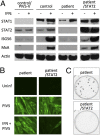
Lentiviral transduction of STAT2 into patient cells restores their ability to respond to IFN-α and induce an antiviral state. (A) Control and V:3 patient skin fibroblasts, patient cells that had been genetically engineered to express STAT2 (patient/STAT2), and control cells that expressed the V protein of PIV5 (control/PIV5-V) that targets STAT1 for proteasome-mediated degradation (Fig. S7) were treated for 18 h with IFN-α or left untreated. Total cell lysates were immunoblotted for STAT1, STAT2, ISG56, MxA, and actin. (B) Monolayers of patient and patient/STAT2 cells were, or were not, pretreated with IFN-α for 15 h before mock infection (Uninf) or infection with PIV5 at 10 pfu/cell. At 24 h p.i., virus-infected cells were visualized by immunofluorescence. (C) Relative plaque size of PIV5VΔC in unmodified patient cells and patient cells expressing STAT2. Monolayers of cells were infected with PIV5VΔC, and fixed at 4 d p.i.; plaques were visualized by immunostaining.

References

    1. Randall RE, Goodbourn S. Interferons and viruses: An interplay between induction, signalling, antiviral responses and virus countermeasures. J Gen Virol. 2008;89(pt 1):1–47. - PubMed
    1. Müller U, et al. Functional role of type I and type II interferons in antiviral defense. Science. 1994;264(5167):1918–1921. - PubMed
    1. Durbin JE, Hackenmiller R, Simon MC, Levy DE. Targeted disruption of the mouse Stat1 gene results in compromised innate immunity to viral disease. Cell. 1996;84(3):443–450. - PubMed
    1. Meraz MA, et al. Targeted disruption of the Stat1 gene in mice reveals unexpected physiologic specificity in the JAK-STAT signaling pathway. Cell. 1996;84(3):431–442. - PubMed
    1. Park C, Li S, Cha E, Schindler C. Immune response in Stat2 knockout mice. Immunity. 2000;13(6):795–804. - PubMed

Publication types

Associated data