. 2013 Apr;20(4):525-34.
doi: 10.1038/cdd.2013.3. Epub 2013 Feb 8.

ASPP1 and ASPP2 bind active RAS, potentiate RAS signalling and enhance p53 activity in cancer cells

Affiliations

ASPP1 and ASPP2 bind active RAS, potentiate RAS signalling and enhance p53 activity in cancer cells

Y Wang et al. Cell Death Differ. 2013 Apr.

Abstract

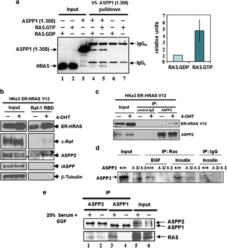
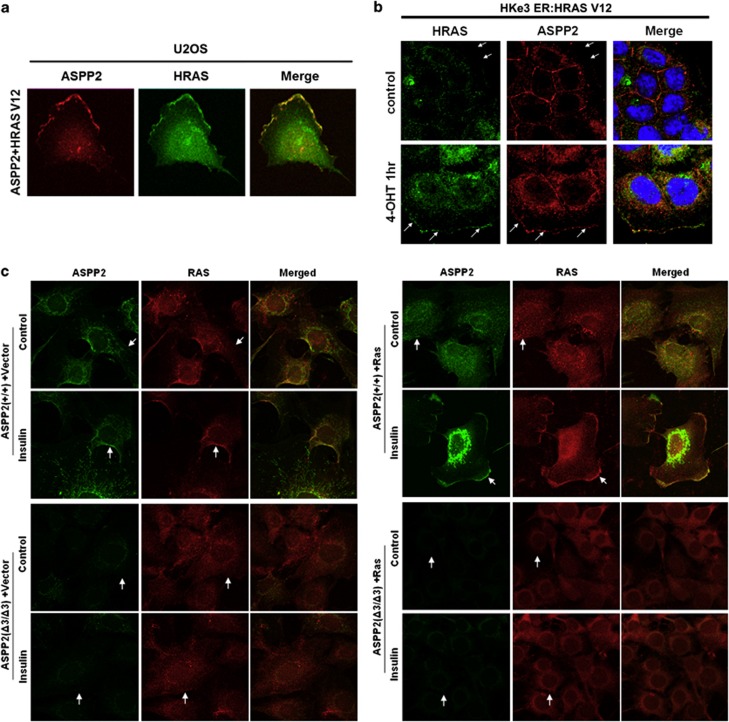
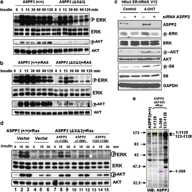
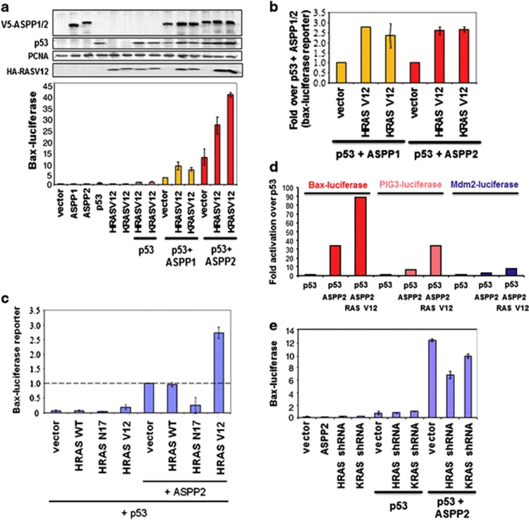
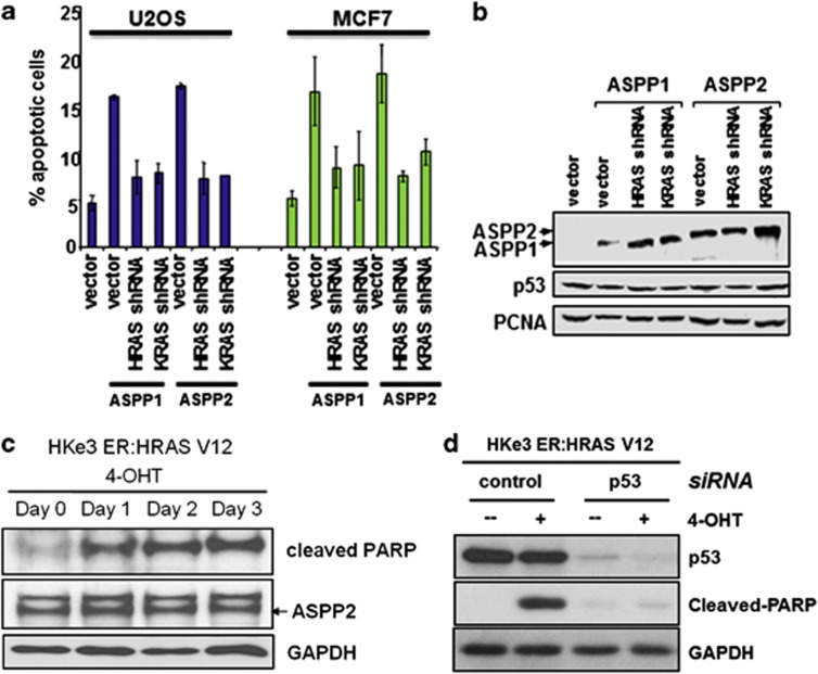
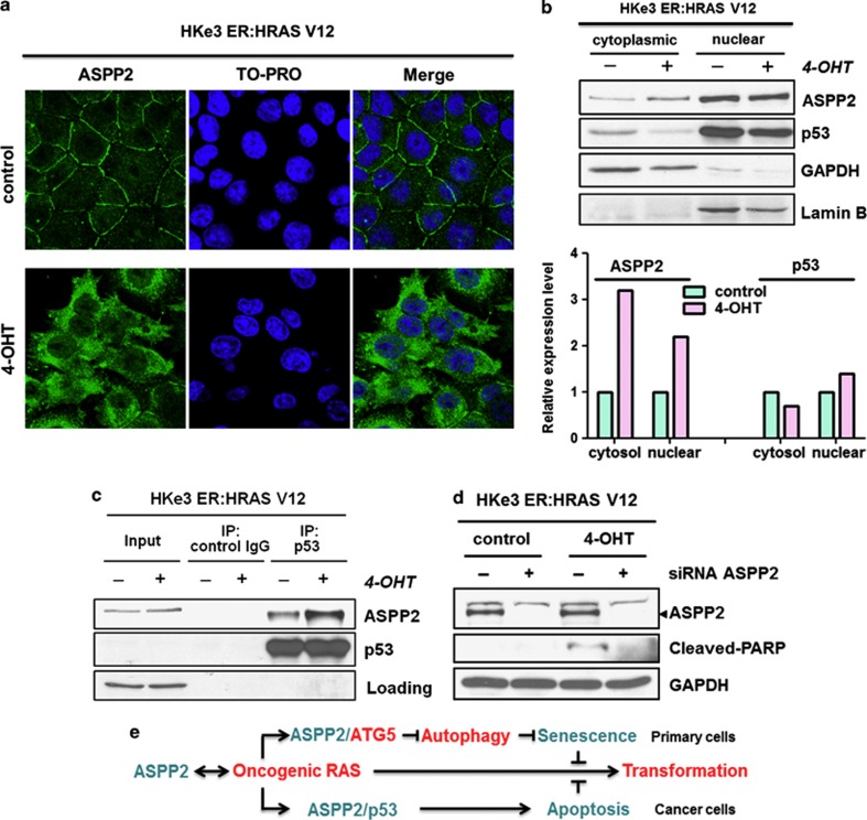
RAS mutations occur frequently in human cancer and activated RAS signalling contributes to tumour development and progression. Apart from its oncogenic effects on cell growth, active RAS has tumour-suppressive functions via its ability to induce cellular senescence and apoptosis. RAS is known to induce p53-dependent cell cycle arrest, yet its effect on p53-dependent apoptosis remains unclear. We report here that apoptosis-stimulating protein of p53 (ASPP) 1 and 2, two activators of p53, preferentially bind active RAS via their N-terminal RAS-association domains (RAD). Additionally, ASPP2 colocalises with and contributes to RAS cellular membrane localisation and potentiates RAS signalling. In cancer cells, ASPP1 and ASPP2 cooperate with oncogenic RAS to enhance the transcription and apoptotic function of p53. Thus, loss of ASPP1 and ASPP2 in human cancer cells may contribute to the full transforming property of RAS oncogene.


Figures

Figure 1
Figure 1
ASPP1 and ASPP2 preferentially bind active RAS. (a) ASPP1's N-terminus preferentially binds RAS–GTP in vitro. RAS protein, loaded with either tritium-labelled GDP or GTP, was added to V5-tagged recombinant ASPP1 (1–310) and immunoprecipitated with V5 antibody. As a negative control, RAS.GDP and RAS–GTP were immunoprecipitated with V5 antibody in the absence of ASPP1 (1–310). Co-immunoprecipitated RAS was quantified according to the presence of 3H-GTP or 3H-GDP. Values are shown as a bar graph. Standard deviation represents the mean of three independent experiments. (b) ASPP2 binds activated RAS in HKe3 cells. HKe3 ER:HRASV12 cells were treated with 100 nM 4-OHT for 2 days and Raf-1–RBD agarose pull-down assays were performed to pull down GTP-bound active RAS. (c) ASPP2 binds activated ER:HRASV12. Total cell lysates from HKe3 ER:HRASV12 cells treated with or without 4-OHT were immunoprecipitated with an anti-ASPP2 antibody, or control IgG as indicated. (d) ASPP2 binds activated RAS in MEFs. Total cell lysates from ASPP2(+/+) or ASPP2(Δ3/Δ3) MEFs treated with EGF (20 ng/ml) or insulin (1 μg/ml) for 15 min were immunoprecipitated with an anti-HRAS antibody or control IgG. (e) Saos2 cells were either starved in 0.5%, or stimulated with 20%, FCS plus 20 ng/ml EGF overnight. Lysates were collected and immunoprecipitated with rabbit anti-ASPP1 (ASPP1.88) or anti-ASPP2 (ASPP2/77) antibodies, respectively. The ASPP proteins were detected with mAbASPP1.54.1, which is known to crossreact with both ASPP1 and ASPP2
Figure 2
Figure 2
ASPP2 colocalises with and contributes to RAS activation at the cell membrane. (a) U2OS cells co-transfected with ASPP2 (red) and HRASV12 (green) show colocalisation of both at the cell membrane (yellow). (b) Immunofluorescence staining of HRAS in HKe3 ER:HRASV12 cells transfected with control or ASPP2 siRNA for 3 days, followed by treatment without (control) or with 100 nM 4-OHT for 1 h. Arrows indicate cell membrane. (c) Immunofluorescence staining of ASPP2 or HRAS in ASPP2(+/+) or ASPP2(Δ3/Δ3) MEFs, with or without HRASV12 expression, before or after treatment as indicated. Cells were treated with 1 μg/ml insulin for 15 min. Arrows indicate cell membrane
Figure 3
Figure 3
ASPP2's N-terminus is required and is sufficient to potentiate RAS signalling. (a and b) ASPP2(+/+) or ASPP2(Δ3/Δ3) MEFs, in the absence (a), or presence of HRASV12 (b) were serum-starved overnight, followed by stimulation with 1 μg/ml insulin for the indicated times. The activation status of ERK or AKT was determined by western blots. (c) Western blots showing the expression levels of endogenous ASPP2, phosphorylated ERK, ERK, phosphorylated AKT, AKT, phosphorylated S6 and S6 in HKe3 ER:HRASV12 cells with indicated treatments. GAPDH was used as a loading control. (d) Oncogenic HRASV12-expressing ASPP2(+/+) or ASPP2(Δ3/Δ3) MEFs infected with retroviruses expressing full-length ASPP2 or truncated mutants were serum-starved overnight before stimulation with 1 μg/ml insulin for the indicated times. The activation status of ERK or AKT was determined by western blot analysis. (e) Western blots showing expression levels of full-length ASPP2 and its mutants in HRASV12-expressing ASPP2(Δ3/Δ3) MEFs as indicated
Figure 4
Figure 4
ASPP1 and ASPP2 cooperates with RAS to enhance the transcriptional activity of p53. (a) Saos2 cells were transfected as indicated with a Bax–luciferase reporter and the luciferase activity shown (lower left panel). The expression of the proteins was verified by western blot (upper left panel). (b) The value of ASPP1+p53 or ASPP2+p53 were taken as 1.0 to reflect the fold increase of ASPP1/ASPP2 and p53 in the presence of mutant RAS. The mean values were derived from three independent experiments. (c) Saos2 cells were transfected with a Bax–luciferase reporter, p53, wild-type or mutant RAS, in the presence or absence of ASPP2 as indicated. The value of ASPP2+p53 was taken as 1.0 to reflect the fold increase. The mean values were derived from three independent experiments. (d) Saos2 cells were transfected as indicated with a Bax–luciferase or PIG3–luciferase or Mdm2–luciferase reporter; luciferase activity is reported here. (e) Saos2 cells were co-transfected with the p53 reporter Bax–luciferase, p53, ASPP2, HRAS shRNA or KRAS shRNA, as indicated
Figure 5
Figure 5
RAS oncogene enhances the apoptotic function of p53 via ASPP1 and ASPP2. (a) ASPP1 and ASPP2 work with endogenous RAS to stimulate apoptosis. shRNA against HRAS or KRAS was co-transfected with ASPP1 or ASPP2 in U2OS or MCF7 cells. FACS analysis was used to determine apoptotic cells. (b) Western blots showing the expression levels of p53 and transfected ASPP1 and ASPP2. PCNA was used as a loading control. (c) Apoptotic activity is detected following oncogenic RAS activation. HKe3 ER:HRASV12 cells were treated with 4-OHT (100 nM) for the indicated time. Western blot showing expression levels of cleaved PARP and ASPP2. (d) p53 depletion reduces oncogenic RAS-induced apoptosis. HKe3 ER:HRASV12 cells transfected with control siRNA or siRNA against p53 for 3 days were treated with or without 4-OHT (100 nM) for another day. Western blots showing expression levels of cleaved PARP and p53
Figure 6
Figure 6
RAS activation induces ASPP2 translocation and enhances p53 binding in colon cancer cells. (a) RAS activation induces ASPP2 translocation. Immunofluorescence staining of ASPP2 in HKe3 ER:HRASV12 cells treated without (control) or with 100 nM 4-OHT for 3 days. (b) RAS activation induces cytoplasmic and nuclear accumulation of ASPP2. Cytoplasmic and nuclear fractions in HKe3 ER:HRASV12 cells with indicated treatments were isolated. GAPDH was used as a loading control for the cytoplasmic fraction, whereas Lamin B was used as a loading control for the nuclear fraction. The levels of ASPP2 and p53 were calculated by densitometry. Fold increase was calculated by normalising control group. (c) RAS activation enhances the binding between ASPP2 and p53. Total cell lysates from HKe3 ER:HRASV12 cells treated with or without 4-OHT were immunoprecipitated with an anti-p53 antibody or control IgG as indicated. (d) ASPP2 depletion reduces oncogenic RAS-induced apoptosis. HKe3 ER:HRASV12 cells transfected with control siRNA or siRNA against ASPP2 for 3 days were treated with or without 4-OHT (100 nM) for another day. Western blots showing expression levels of cleaved PARP and ASPP2. (e) Diagram summarises the interactions between ASPP2 and RAS for their regulation and functions (details are provided in Discussion)

References

    1. Downward J. Targeting RAS signalling pathways in cancer therapy. Nat Rev. 2003;3:11–22. - PubMed
    1. Buday L, Downward J. Many faces of Ras activation. Biochim Biophys Acta. 2008;1786:178–187. - PubMed
    1. Okines A, Cunningham D, Chau I. Targeting the human EGFR family in esophagogastric cancer. Nat Rev Clin Oncol. 2011;8:492–503. - PubMed
    1. Wheeler DL, Dunn EF, Harari PM. Understanding resistance to EGFR inhibitors-impact on future treatment strategies. Nat Rev Clin Oncol. 2010;7:493–507. - PMC - PubMed
    1. Cox AD, Der CJ. The dark side of Ras: regulation of apoptosis. Oncogene. 2003;22:8999–9006. - PubMed

Publication types

MeSH terms