. 2013 Mar 1;14(3):284-91.
doi: 10.1038/embor.2013.6. Epub 2013 Feb 8.

FIP200 regulates targeting of Atg16L1 to the isolation membrane

Affiliations

FIP200 regulates targeting of Atg16L1 to the isolation membrane

Taki Nishimura et al. EMBO Rep. .

Abstract

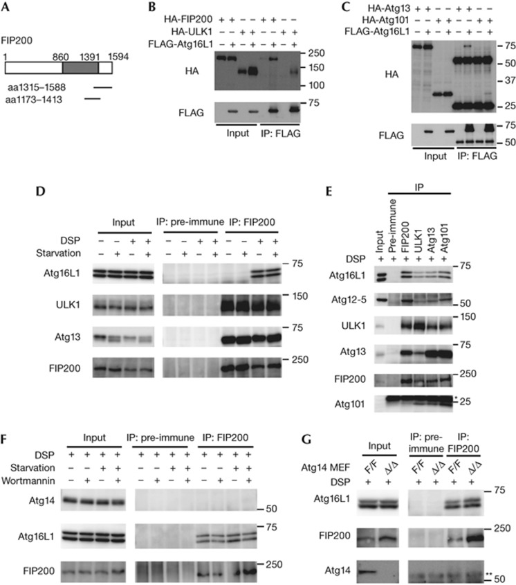
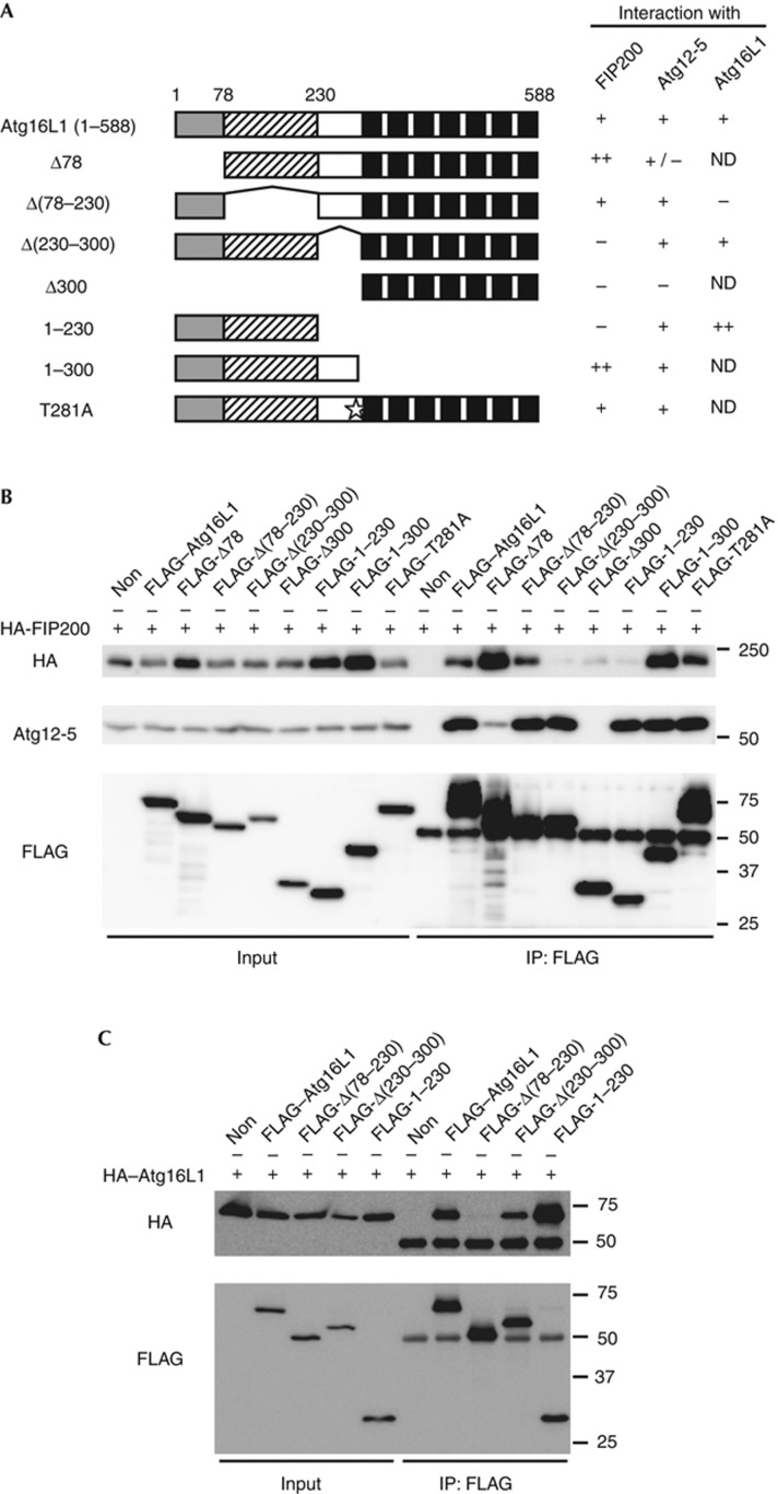
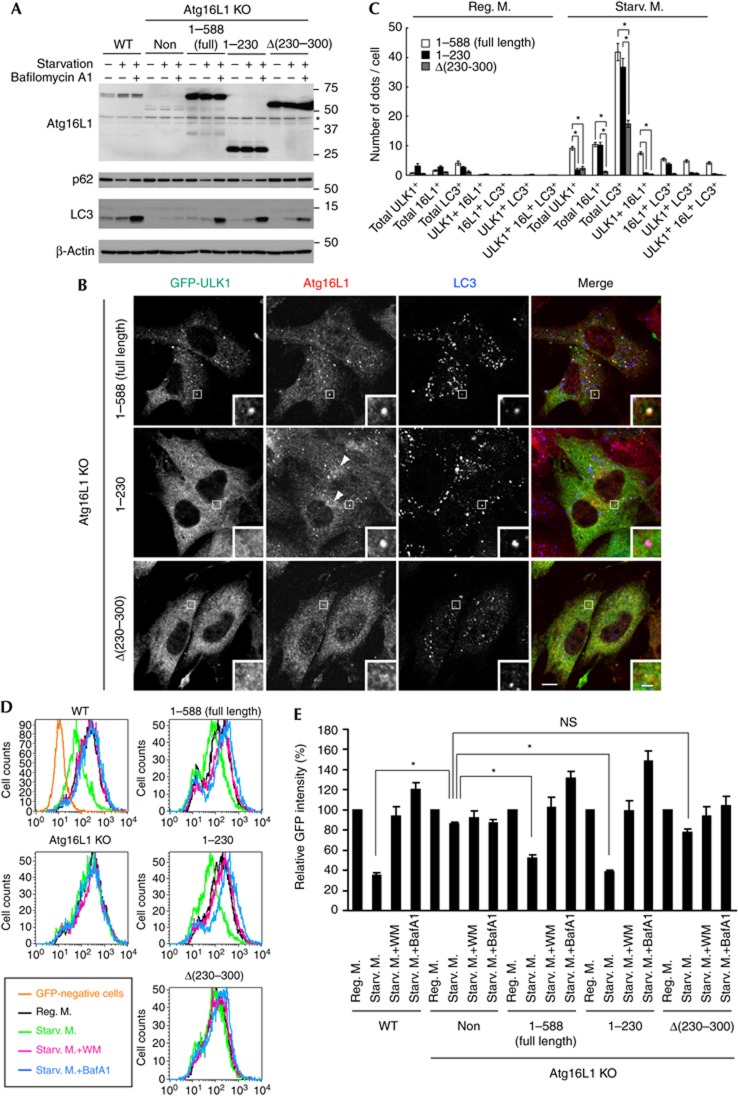
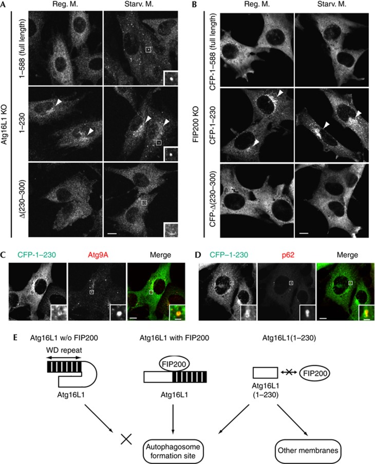
Autophagosome formation is a dynamic process that is strictly controlled by autophagy-related (Atg) proteins. However, how these Atg proteins are recruited to the autophagosome formation site or autophagic membranes remains poorly understood. Here, we found that FIP200, which is involved in proximal events, directly interacts with Atg16L1, one of the downstream Atg factors, in an Atg14- and phosphatidylinositol 3-kinase-independent manner. Atg16L1 deletion mutants, which lack the FIP200-interacting domain, are defective in proper membrane targeting. Thus, FIP200 regulates not only early events but also late events of autophagosome formation through direct interaction with Atg16L1.


Conflict of interest statement

The authors declare that they have no conflict of interest.

Figures

Figure 1
Figure 1
Atg16L1 interacts with the ULK1–Atg13–FIP200–Atg101 complex. (A) Yeast two-hybrid screening for Atg16L1-interacting proteins. The Atg16L1-interacting FIP200 clones are indicated. (B, C) HEK293T cells were co-transfected with the indicated constructs. Cell lysates were subjected to IP using anti-FLAG antibodies. The resulting precipitates were examined by immunoblot analysis with anti-FLAG and anti-HA antibodies. (D) HEK293T cells were cultured in regular or starvation medium for 2 h. Cells were harvested and treated with DSP. (E) HEK293T cells were harvested and treated with DSP. (F) HEK293T cells were cultured in regular DMEM or starvation medium in the presence or absence of 200 nM wortmannin for 2 h. After treatment with DSP, cell lysates were subjected to IP analysis. (G) Atg14F/F (undeleted) or Atg14Δ/Δ (deleted) MEFs were harvested and treated with DSP. * (E) and ** (G) indicate the positions of the immunoglobulin light and heavy chains, respectively. aa, amino acid; DSP, dithiobis(succinimidyl propionate); IP, immunoprecipitation; MEFs, mouse embryonic fibroblasts.
Figure 2
Figure 2
The Atg12—Atg5–Atg16L1 complex regulates the stability of the ULK1–Atg13–FIP200 complex. (A) Immunoblot analysis of Atg proteins in the indicated MEFs. (B) m5-7 cells were cultured in regular medium containing 10 ng/ml Dox for 8 days, and then cultured in the absence of Dox for a further 2 days. (C,D) m5-7 cells were cultured in regular medium with or without 10 ng/ml Dox for 4 days, and then cultured in the presence of 50 μg/ml CHX for the indicated time periods (C). mRNA levels of the indicated genes were measured by quantitative PCR following the 4-day Dox treatment. Data represent mean±s.e. (*P<0.05) (D). CHX, cycloheximide; Dox, doxycycline; MEFs, mouse embryonic fibroblasts; mRNA, messenger RNA; WT, wild-type.
Figure 3
Figure 3
The middle region of Atg16L1 is required for interaction with FIP200. (A) Schematic representations of the wild-type Atg16L1 and its mutants. Human Atg16L1α (NP_060444) has an amino-terminal Atg5-interacting domain (grey box), a coiled–coil domain (shaded box) and seven WD repeats (black boxes). Star indicates the position of the Crohn’s disease-associated T281A mutation. (B,C) HEK293T cells were co-transfected with the indicated constructs. Cell lysates were analysed by IP. IP, immunoprecipitation; ND, not determined.
Figure 4
Figure 4
Atg16L1Δ(230–300) mutant is impaired in isolation membrane targeting and partially restores the autophagic defects of Atg16L1 KO MEFs. (A) WT MEFs, Atg16L1 KO MEFs or Atg16L1 KO MEFs stably expressing either full-length Atg16L1(1–588), Atg16L1(1–230) or Atg16L1Δ(230–300) were cultured in regular DMEM or starvation medium in the presence or absence of 100 nM BafA1 for 2 h. * indicates nonspecific immunoreactive bands. (B,C) Atg16L1 KO MEFs stably expressing GFP–ULK1 and either full-length Atg16L1(1–588), Atg16L1(1–230) or Atg16L1Δ(230–300) were cultured in regular DMEM (C) or starvation medium (B,C) for 1 h. Cells were fixed and analysed by immunofluorescence microscopy using anti-GFP, anti-Atg16L1 and anti-LC3 antibodies. Arrowheads indicate the perinuclear localization of Atg16L1(1–230). The number of dots was quantified from more than 30 randomly selected cells from three independent samples as described in the Methods. Data represent mean±s.e. (*P<0.001, analysis of variance followed by Bonferroni/Dunn post hoc test). Scale bar, 10 μm, and 2 μm in inset. (D, E) Cells stably expressing GFP–LC3 were cultured in regular DMEM, starvation medium or starvation medium in the presence of 0.2 μM WM or 100 nM BafA1 for 6 h. Total cellular GFP–LC3 signals were analysed by flow cytometry. Representative FACS data were shown (D). The geometric mean of fluorescence intensity was determined. Values are expressed as a percentage of the mean of control cells cultured in regular DMEM. Data represent mean±s.e. (*P<0.05) (E). BafA1, bafilomycin A1; GFP, green fluorescent protein; KO, knockout; MEFs, mouse embryonic fibroblasts; NS, not significant; WM, wortmannin; WT, wild-type.
Figure 5
Figure 5
Atg16L1(1–230) localizes to aberrant membranes in a FIP200-independent manner. (A) Atg16L1 KO MEFs stably expressing either full-length Atg16L1(1–588), Atg16L1(1–230) or Atg16L1Δ(230–300) were cultured in regular DMEM or starvation medium for 1 h. Cells were fixed and subjected to immunofluorescence microscopy using anti-Atg16L1 antibody. Note that full-length Atg16L1 and Atg16L1(1–230), but not Atg16L1Δ(230–300), showed punctate structures under starvation conditions (inset). (B) FIP200 KO MEFs stably expressing either CFP–Atg16L1(1–588), CFP–Atg16L1(1–230) or CFP–Atg16L1Δ(230–300) were cultured in regular DMEM or starvation medium for 1 h. Cells were fixed and analysed by immunofluorescence microscopy using anti-GFP antibody. Arrowheads indicate the perinuclear localization of Atg16L1(1–230). (C,D) FIP200 KO MEFs stably expressing CFP–Atg16L1(1–230) were cultured in regular DMEM. Scale bar, 10 μm, and 2 μm in inset. (E) Hypothetical model of membrane targeting of Atg16L1. Interaction of Atg16L1 with FIP200 leads to proper targeting of this complex to the autophagosome formation site (middle). Without FIP200, puncta formation of Atg16L1 is impaired probably owing to a self-inhibitory role of the C-terminal WD-repeat domain (left). If the WD-repeat domain is deleted, the N-terminal half of Atg16L1 localizes to aberrant membranes as well as the autophagosome formation site (right). KO, knockout; MEFs, mouse embryonic fibroblasts.

References

    1. Mizushima N, Yoshimori T, Ohsumi Y (2011) The role of Atg proteins in autophagosome formation. Annu Rev Cell Dev Biol 27: 107–132 - PubMed
    1. Xie Z, Klionsky DJ (2007) Autophagosome formation: core machinery and adaptations. Nat Cell Biol 9: 1102–1109 - PubMed
    1. Nakatogawa H, Suzuki K, Kamada Y, Ohsumi Y (2009) Dynamics and diversity in autophagy mechanisms: lessons from yeast. Nat Rev Mol Cell Biol 10: 458–467 - PubMed
    1. Suzuki K, Kubota Y, Sekito T, Ohsumi Y (2007) Hierarchy of Atg proteins in pre-autophagosomal structure organization. Genes Cells 12: 209–218 - PubMed
    1. Itakura E, Mizushima N (2010) Characterization of autophagosome formation site by a hierarchical analysis of mammalian Atg proteins. Autophagy 6: 764–776 - PMC - PubMed

Publication types

Substances