. 2013 May;27(5):2001-12.
doi: 10.1096/fj.12-221317. Epub 2013 Feb 7.

The kinase domains of obscurin interact with intercellular adhesion proteins

Affiliations

The kinase domains of obscurin interact with intercellular adhesion proteins

Li-Yen R Hu et al. FASEB J. 2013 May.

Erratum in

Abstract

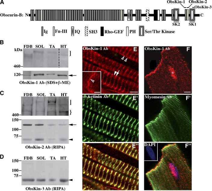
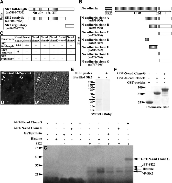
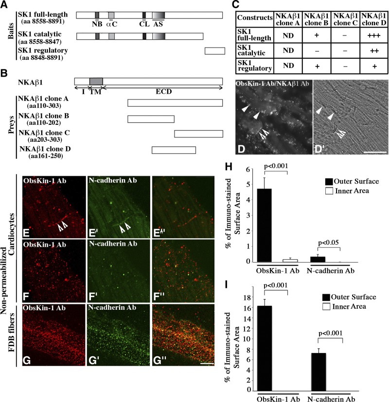
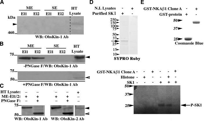
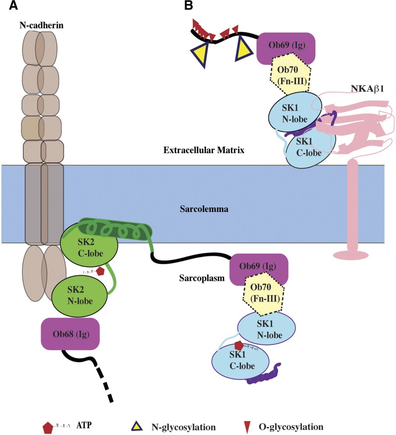
Obscurins comprise a family of giant (~870- to 600-kDa) and small (~250- to 55-kDa) proteins that play important roles in myofibrillogenesis, cytoskeletal organization, and cell adhesion and are implicated in hypertrophic cardiomyopathy and tumorigenesis. Giant obscurins are composed of tandem structural and signaling motifs, including 2 serine/threonine kinase domains, SK1 and SK2, present at the COOH terminus of giant obscurin-B. Using biochemical and cellular approaches, we show for the first time that both SK1 and SK2 possess enzymatic activities and undergo autophosphorylation. SK2 can phosphorylate the cytoplasmic domain of N-cadherin, a major component of adherens junctions, and SK1 can interact with the extracellular domain of the β1-subunit of the Na(+)/K(+)-ATPase, which also resides in adherens junctions. Immunostaining of nonpermeabilized myofibers and cardiocytes revealed that some obscurin kinase isoforms localize extracellularly. Quantification of the exofacial expression of obscurin kinase proteins indicated that they occupy ~16 and ~5% of the sarcolemmal surface in myofibers and cardiocytes, respectively. Treatment of heart lysates with peptide-N-glycosidase F revealed that while giant obscurin-B localizes intracellularly, possessing dual kinase activity, a small obscurin kinase isoform that contains SK1 localizes extracellularly, where it undergoes N-glycosylation. Collectively, our studies demonstrate that the obscurin kinase domains are enzymatically active and may be involved in the regulation of cell adhesion.


Figures

Figure 1.
Figure 1.
Expression profile of obscurin kinase proteins. A) Schematic representation of giant obscurin-B denoting the epitopes used for antibody production. B–D) Immunoblots using protein homogenates prepared from skeletal [FDB, soleus (SOL), and tibialis anterior (TA)] and heart (HT) muscles. Note that the lane including heart lysates was exposed for a longer time period than those including skeletal muscle lysates. Obscurin-B (square bracket) is expressed in higher amounts in skeletal than in cardiac muscle. An immunoreactive band of ∼600 kDa (curly bracket) was consistently detected in skeletal and cardiac muscle lysates with the ObsKin-2 antibody and may represent a novel obscurin isoform or a degradation product of obscurin B. Moreover, 2 small obscurin isoforms of ∼120 kDa (arrow) and ∼70 kDa (arrowhead) were also present in all striated muscles examined. E–E″) Mouse cardiac tissue sections stained with ObsKin-1 (red; E) and α-actinin (green; E′) antibodies and merged image (E″). F–F″) Immunolabeling of adult rat cardiocytes with ObsKin-1 (red; F) and myomesin (green; F″) antibodies counterstained with DAPI (blue) and merged image (E″). Scale bars = 5 μm.
Figure 2.
Figure 2.
N-cadherin is a substrate of SK2. A, B) Schematic diagrams illustrating the SK2 bait (A) and N-cadherin prey (B) constructs. αC, α-helix C; AS, activation segment; CDR, extracellular cadherin repeats; CL, catalytic loop; I, intracellular domain; NB, nucleotide binding domain; PRO, prodomain; TM, transmembrane region. C) Identification of minimal interacting domains using the Y2H system; relative strength of the identified interactions is indicated by + and − symbols. D, D′) Fluorescent (D) and bright field (D′) images obtained from the PLA assay. Scale bar = 30 μm. E) Purified His-tagged SK2 protein analyzed by SDS-PAGE and stained with SYPRO Ruby. F) Purified control GST-protein and recombinant N-cadherin peptides fused to GST separated by SDS-PAGE and stained with Coomassie Brilliant Blue-R250. G) Autoradiogram of an in vitro kinase assay using His-tagged SK2 and different substrates; bands denoted as P-SK2 and PP-SK2 represent SK2 species autophosphorylated to different extents.
Figure 3.
Figure 3.
Extracellular localization of obscurin kinase proteins in striated muscle cells. A, B) Schematic representations of SK1 bait (A) and NKAβ1 prey (B) constructs. αC, α-helix C; AS, activation segment; CL, catalytic loop; ECD, extracellular domain; I, intracellular domain; NB, nucleotide binding domain; TM, transmembrane domain. C) Identification of minimal interacting domains using the Y2H system; relative strength of the identified interactions is indicated by + and − symbols. ND, not determined due to autoactivation. D, D′) Fluorescent (D) and bright field (D′) images obtained from the PLA assay. Scale bar =30 μm. E–G″) Colabeling of nonpermeabilized cardiocytes (E–F″) and FDB fibers (G–G″) with ObsKin-1 (red; E, F, G) and N-cadherin (green; E′, F′, G′) antibodies that recognize epitopes in its extracellular domain and merged images (E″, F″, G″). Scale bar = 8 μm. H, I) Quantification of the percentage of the outer sarcolemmal surface and the inner sarcoplasmic area of nonpermeabilized cardiocytes (H) and FDB myofibers (I) immunostained with the ObsKin-1 and N-cadherin antibodies.
Figure 4.
Figure 4.
A small obscurin kinase isoform undergoes glycosylation. A–C) Adult mouse heart lysates were incubated with lectin resins. Eluted fractions were immunoprobed for the presence of giant obscurin-B (A) or smaller obscurin kinase isoforms prior to (B, top panel) and after (C) treatment with PNGase F glycosidase. Protein lysates were also treated with PNGase F before they were applied to the lectin resin (B, bottom panel); ME and SE, elution buffers (see Materials and Methods), El1 and El2, eluted fractions 1 and 2. D) Purified His-tagged SK1 protein analyzed by SDS-PAGE and stained with SYPRO Ruby. E) Purified control GST-protein and recombinant NKAβ1 peptide fused to GST, separated by SDS-PAGE, and stained with Coomassie Brilliant Blue-R250. F) Autoradiogram of an in vitro kinase assay using His-tagged SK1 and different substrates; bands marked as P-SK1 represent autophosphorylated species of SK1. Notably, the autophosphorylation activity of SK1 is significantly diminished in the presence of recombinant NKAβ1, possibly due to direct binding of the latter to the catalytic part of the former, as our Y2H results indicated.
Figure 5.
Figure 5.
Topology of obscurin kinase proteins. A) SK2 domain of obscurin-B (or of other intracellular obscurin kinase isoforms) may phosphorylate the cytoplasmic domain of N-cadherin at ICD. B) A small obscurin kinase isoform (∼70/55 kDa) containing SK1 localizes extracellularly, where it can undergo glycosylation and interact with the β1 subunit of NKA. N- and O-glycosylation sites were predicted using NetNGlyc1.0 and NetOGlyc3.1 software, respectively.

References

    1. Kontrogianni-Konstantopoulos A., Ackermann M. A., Bowman A. L., Yap S. V., Bloch R. J. (2009) Muscle giants: molecular scaffolds in sarcomerogenesis. Physiol. Rev. 89, 1217–1267 - PMC - PubMed
    1. Fukuzawa A., Idowu S., Gautel M. (2005) Complete human gene structure of obscurin: implications for isoform generation by differential splicing. J. Muscle Res. Cell Motil. 26, 427–434 - PubMed
    1. Russell M. W., Raeker M. O., Korytkowski K. A., Sonneman K. J. (2002) Identification, tissue expression and chromosomal localization of human obscurin-MLCK, a member of the titin and Dbl families of myosin light chain kinases. Gene 282, 237–246 - PubMed
    1. Sutter S. B., Raeker M. O., Borisov A. B., Russell M. W. (2004) Orthologous relationship of obscurin and Unc-89: phylogeny of a novel family of tandem myosin light chain kinases. Dev. Genes Evol. 214, 352–359 - PubMed
    1. Borisov A. B., Raeker M. O., Russell M. W. (2008) Developmental expression and differential cellular localization of obscurin and obscurin-associated kinase in cardiac muscle cells. J. Cell. Biochem. 103, 1621–1635 - PMC - PubMed

Publication types

MeSH terms