Extracorporeal membrane oxygenation and cerebral blood flow velocity in children
- PMID: 23392359
- PMCID: PMC3873647
- DOI: 10.1097/PCC.0b013e3182712d62
Extracorporeal membrane oxygenation and cerebral blood flow velocity in children
Abstract
Objective: To determine how extracorporeal membrane oxygenation affects cerebral blood flow velocity and to determine whether specific changes in cerebral blood flow velocity may be associated with neurologic injury.
Design: Prospective, observational study.
Setting: PICU in a tertiary care academic center.
Patients: Children (age less than or equal to 18 yr) requiring extracorporeal membrane oxygenation support.
Interventions: None.
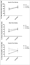
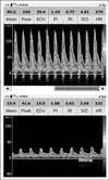
Measurements and main results: Eighteen patients (age 3.8 ± 7.2 years; venovenous neck, n = 5; venoarterial neck, n = 8; venoarterial chest, n = 5) requiring extracorporeal membrane oxygenation underwent daily transcranial Doppler ultrasound measurements of cerebral blood flow velocity in bilateral middle cerebral arteries. Cerebral blood flow velocity measurements were recorded as a percentage of age and gender normal value. On extracorporeal membrane oxygenation, cerebral blood flow velocities in patients not suffering clinically evident neurologic injury were decreased with systolic flow velocity (Vs) 54% ± 3% predicted and mean flow velocity (Vm) 52% ± 4% predicted. After decannulation, Vs and Vm were higher than while on extracorporeal membrane oxygenation at 73% ± 3% predicted (p = 0.0007 vs. value on extracorporeal membrane oxygenation) and 64% ± 4% predicted (p = 0.01 vs. value on extracorporeal membrane oxygenation).Patients who developed clinically evident cerebral hemorrhage had higher Vs, diastolic flow velocity (Vd), and Vm compared with those who did not: 123% ± 8% predicted, 130% ± 18% predicted, 127% ± 9% predicted (p < compared to values in children not suffering neurological injury). Supranormal flow velocities were noted 2-6 days before clinical recognition of cerebral hemorrhage in all four patients. There were no significant differences in mean arterial blood pressure, circuit flow, or hematocrit between the children who suffered cerebral hemorrhage and those who did not. Partial pressure of carbon dioxide was lower in the group of patients who experienced cerebral hemorrhage than in those who did not (38 ± 2 vs. 44 ± 1 mm Hg, p = 0.03).
Conclusion: In children who did not suffer clinically apparent neurologic injury, cerebral blood flow velocities were lower than normal while on extracorporeal membrane oxygenation support and increased after decannulation. However, children who developed cerebral hemorrhage had higher than normal cerebral blood flow velocities noted for days prior to clinical recognition of bleeding. Cerebral blood flow velocity measurement may represent a portable, noninvasive way to predict cerebral complications of extracorporeal membrane oxygenation and deserves further study.
Conflict of interest statement
The authors have not disclosed any potential conflicts of interest.
Figures


Similar articles
-
Cerebrovascular Physiology During Pediatric Extracorporeal Membrane Oxygenation: A Multicenter Study Using Transcranial Doppler Ultrasonography.Pediatr Crit Care Med. 2019 Feb;20(2):178-186. doi: 10.1097/PCC.0000000000001778. Pediatr Crit Care Med. 2019. PMID: 30395027
-
Transcranial Doppler Identification of Neurologic Injury during Pediatric Extracorporeal Membrane Oxygenation Therapy.J Stroke Cerebrovasc Dis. 2017 Oct;26(10):2336-2345. doi: 10.1016/j.jstrokecerebrovasdis.2017.05.022. Epub 2017 Jun 2. J Stroke Cerebrovasc Dis. 2017. PMID: 28583819
-
The association of carotid artery cannulation and neurologic injury in pediatric patients supported with venoarterial extracorporeal membrane oxygenation*.Pediatr Crit Care Med. 2014 May;15(4):355-61. doi: 10.1097/PCC.0000000000000103. Pediatr Crit Care Med. 2014. PMID: 24622166
-
Brain Injury Is More Common in Venoarterial Extracorporeal Membrane Oxygenation Than Venovenous Extracorporeal Membrane Oxygenation: A Systematic Review and Meta-Analysis.Crit Care Med. 2020 Dec;48(12):1799-1808. doi: 10.1097/CCM.0000000000004618. Crit Care Med. 2020. PMID: 33031150
-
Cerebrovascular complications and neurodevelopmental sequelae of neonatal ECMO.Clin Perinatol. 1997 Sep;24(3):655-75. Clin Perinatol. 1997. PMID: 9394865 Review.
Cited by
-
Subtypes and Mechanistic Advances of Extracorporeal Membrane Oxygenation-Related Acute Brain Injury.Brain Sci. 2023 Aug 4;13(8):1165. doi: 10.3390/brainsci13081165. Brain Sci. 2023. PMID: 37626521 Free PMC article. Review.
-
Incidence, Outcome, and Predictors of Intracranial Hemorrhage in Adult Patients on Extracorporeal Membrane Oxygenation: A Systematic and Narrative Review.Front Neurol. 2018 Jul 6;9:548. doi: 10.3389/fneur.2018.00548. eCollection 2018. Front Neurol. 2018. PMID: 30034364 Free PMC article. Review.
-
Multimodal Neurologic Monitoring in Patients Undergoing Extracorporeal Membrane Oxygenation.Cureus. 2024 May 1;16(5):e59476. doi: 10.7759/cureus.59476. eCollection 2024 May. Cureus. 2024. PMID: 38826870 Free PMC article.
-
Neurological complications during veno-venous extracorporeal membrane oxygenation: Does the configuration matter? A retrospective analysis of the ELSO database.Crit Care. 2021 Mar 17;25(1):107. doi: 10.1186/s13054-021-03533-5. Crit Care. 2021. PMID: 33731186 Free PMC article.
-
Multimodal Neurologic Monitoring in Children With Acute Brain Injury.Pediatr Neurol. 2022 Apr;129:62-71. doi: 10.1016/j.pediatrneurol.2022.01.006. Epub 2022 Feb 2. Pediatr Neurol. 2022. PMID: 35240364 Free PMC article. Review.
References
Publication types
MeSH terms
Grants and funding
LinkOut - more resources
Full Text Sources
Other Literature Sources

