

Diffuse optical tomography in the presence of a chest wall
- PMID: 23392384
- PMCID: PMC3566530
- DOI: 10.1117/1.JBO.18.2.026016
Diffuse optical tomography in the presence of a chest wall
Abstract
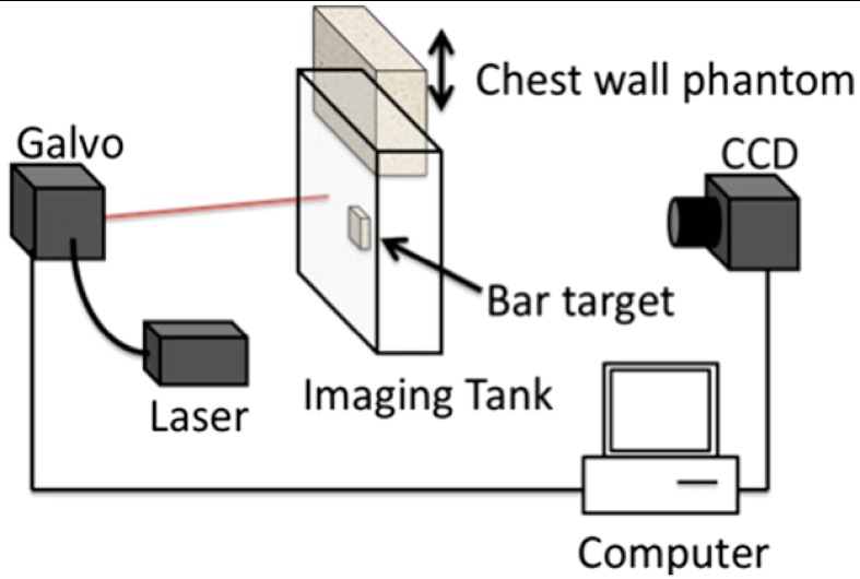
Diffuse optical tomography (DOT) has been employed to derive spatial maps of physiologically important chromophores in the human breast, but the fidelity of these images is often compromised by boundary effects such as those due to the chest wall. We explore the image quality in fast, data-intensive analytic and algebraic linear DOT reconstructions of phantoms with subcentimeter target features and large absorptive regions mimicking the chest wall. Experiments demonstrate that the chest wall phantom can introduce severe image artifacts. We then show how these artifacts can be mitigated by exclusion of data affected by the chest wall. We also introduce and demonstrate a linear algebraic reconstruction method well suited for very large data sets in the presence of a chest wall.
Figures


Similar articles
-
Flux density calibration in diffuse optical tomographic systems.J Biomed Opt. 2013 Feb;18(2):26023. doi: 10.1117/1.JBO.18.2.026023. J Biomed Opt. 2013. PMID: 23423331
-
Study of the effect of mechanical pressure on determination of position and size of tumor in biological phantoms.Appl Opt. 2013 Apr 20;52(12):2739-49. doi: 10.1364/AO.52.002739. Appl Opt. 2013. PMID: 23669685
-
Image reconstruction method for a two-layer tissue structure accounts for chest-wall effects in breast imaging.J Biomed Opt. 2008 Nov-Dec;13(6):064029. doi: 10.1117/1.3041497. J Biomed Opt. 2008. PMID: 19123675 Free PMC article.
-
Optical tomography method that accounts for tilted chest wall in breast imaging.J Biomed Opt. 2010 Jul-Aug;15(4):041515. doi: 10.1117/1.3449570. J Biomed Opt. 2010. PMID: 20799793 Free PMC article.
-
Time-resolved diffuse optical tomography and its application to in vitro and in vivo imaging.J Biomed Opt. 2007 Nov-Dec;12(6):062107. doi: 10.1117/1.2815724. J Biomed Opt. 2007. PMID: 18163810 Review.
References
-
- Arridge S. R., “Optical tomography in medical imaging,” Inverse Probl. 15(2), R41–R93 (1999).INPEEY10.1088/0266-5611/15/2/022 - DOI
-
- Boas D. A., et al. , “Imaging the body with diffuse optical tomography,” IEEE Signal Proc. Mag. 18(6), 57–75 (2001).ISPRE610.1109/79.962278 - DOI
-
- Arridge S. R., Schotland J. C., “Optical tomography: forward and inverse problems,” Inverse Probl. 25(12), 123010 (2009).INPEEY10.1088/0266-5611/25/12/123010 - DOI
-
- Colak S. B., et al. , “Clinical optical tomography and NIR spectroscopy for breast cancer detection,” IEEE J. Sel. Top. Quantum Electron. 5(4), 1143–1158 (1999).IJSQEN10.1109/2944.796341 - DOI
Publication types
MeSH terms
Grants and funding
LinkOut - more resources
Full Text Sources
Other Literature Sources
