. 2013 May;22(5):1037-44.
doi: 10.1007/s00586-013-2701-0. Epub 2013 Feb 8.

Irrigation endoscopic discectomy: a novel percutaneous approach for lumbar disc prolapse

Affiliations

Irrigation endoscopic discectomy: a novel percutaneous approach for lumbar disc prolapse

Hesham Magdi Soliman. Eur Spine J. 2013 May.

Abstract

Purpose: The purpose of this study is to present a new endoscopic procedure, aiming to achieve the success rate equivalent to microsurgical discectomy, while addressing the drawbacks and limitations of other percutaneous techniques.

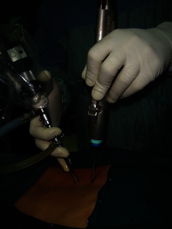
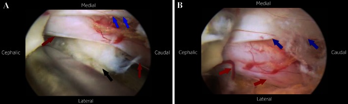
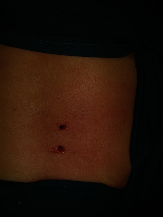
Methods: A series of 43 patients with uncontained lumbar disc herniation underwent surgery with irrigation endoscopic discectomy (IED). The endoscope and instruments are placed directly over the surface of the lamina through two posterior skin portals 5 mm each without any muscle retraction or dilatation. Pump irrigation is used for the opening of a potential working space. The rest of the procedure is performed endoscopically like the standard microsurgical discectomy.

Results: Outcome according to modified Macnab criteria was excellent in 78%, good in 17%, and poor in 5% of patients. VAS for leg pain dropped from 78 preoperatively to 7, and the Oswestry Low-Back Pain Disability Questionnaire dropped from 76 to 19. The mean time for postoperative ambulation was 4 h, hospital stay was 8 h, and for return to work was 7 days.

Conclusions: Preliminary clinical experience with IED shows it to be as effective as microsurgical discectomy, and in comparison to other percutaneous procedures addressing noncontained herniations, a reduction in the cost, technical difficulty and surgical invasiveness has been demonstrated.


Figures

Fig. 1
Fig. 1
Standard arthroscopic facilities: A video monitor, B xenon light source device, C video integrator device, D arthroscopic pump, E arthroscopic shaver device
Fig. 2
Fig. 2
Standard arthroscopic facilities: A arthroscope cannula 5 mm in diameter with a port for pump irrigation, B arthroscope with a view angle 30°; length 18 cm; outer diameter 4 mm, C abrader extension for arthroscopic shaver; size 4.5, D burr extension for arthroscopic shaver; size 5.5, E camera head, F arthroscopic shaver handle
Fig. 3
Fig. 3
Intraoperative image showing the endoscope and arthroscopic shaver introduced through two separate portals
Fig. 4
Fig. 4
Arthroscopic abrader cleaning the soft tissue; white arrows point to the superior lamina and base of spinous process, and red arrows to the interlaminar space
Fig. 5
Fig. 5
Kerrison removing the inferior edge of superior lamina and medial half of the facet; red arrow points to the superior lamina and the white arrow to the medial facetectomy site
Fig. 6
Fig. 6
Laminotomy site; blue arrows point to the superior free edge of the ligamentum flavum, yellow arrow to the epidural space, black arrow to the superior lamina and green arrow to the medial facetectomy site
Fig. 7
Fig. 7
aBlack arrow point to the kerrison rongeour removing the ligamentum flavum, blue arrows to the ligamentum flavum, red arrows to the dural sac and green arrow to the shoulder of the nerve root. b After removal of the ligamentum flavum with the red arrows pointing to the dural sac and the green arrow to the nerve root
Fig. 8
Fig. 8
a Removal of the herniated fragment; black arrow points to the disc fragment, red arrows point to the nerve root and the blue arrows to the lateral edge of the dural sac. b Nerve root and dural sac after disc fragment removal; red arrows show the nerve root after relief of compression and the blue arrows point to the dura
Fig. 9
Fig. 9
Picture showing postoperative wound before closure
Fig. 10
Fig. 10
a Picture of a morbidly obese patient with L4–L5 lumbar disc herniation. b Picture showing the 18 cm long endoscope and pituitary rongeur placed entirely in the patients back during discectomy

References

    1. Perez-Cruet MJ, Fessler RG, Perin NI. Review: complications of minimally invasive spinal surgery. Neurosurgery. 2002;51(Suppl 5):S26–S36. - PubMed
    1. Smith L. Enzyme dissolution of the nucleus pulposus in humans. JAMA. 1964;265:137–140. doi: 10.1001/jama.1964.03060150061016. - DOI - PubMed
    1. Hijikata S, Yamgishi M, Nakayama T, Oomon K. Percutaneous discectomy: a new treatment method for lumbar disc herniation. J Toden Hosp. 1975;5:5–13.
    1. Onik G, Helms CA, Ginsberg L. Percutaneous lumbar discectomy using a new aspiration probe. AJR Am J Roentgenol. 1985;144:1137–1140. doi: 10.2214/ajr.144.6.1137. - DOI - PubMed
    1. Schreiber A, Suezawa Y. Transdiscoscopic percutaneous nucleotomy in disc herniation. Orthop Rev. 1986;15:35–38. - PubMed

LinkOut - more resources