. 2013 Feb 6;33(6):2408-18.
doi: 10.1523/JNEUROSCI.3406-12.2013.

A mutant prion protein sensitizes neurons to glutamate-induced excitotoxicity

Affiliations

A mutant prion protein sensitizes neurons to glutamate-induced excitotoxicity

Emiliano Biasini et al. J Neurosci. .

Abstract

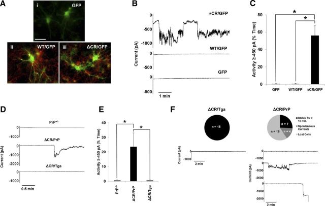
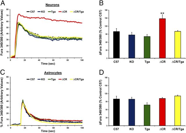
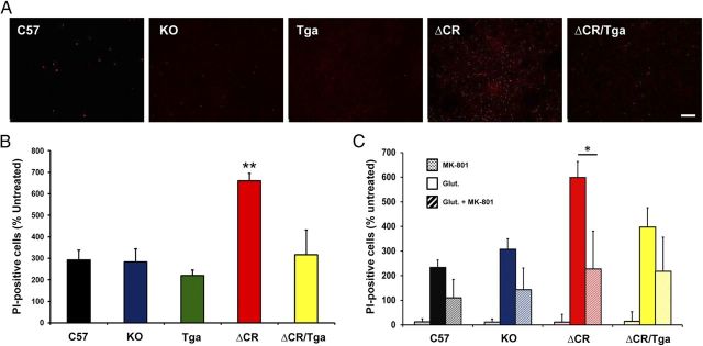
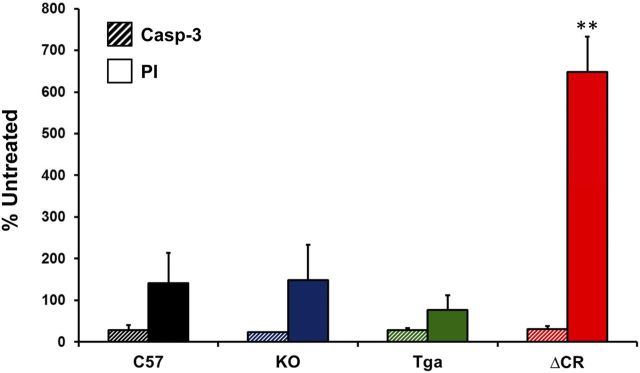
Growing evidence suggests that a physiological activity of the cellular prion protein (PrP(C)) plays a crucial role in several neurodegenerative disorders, including prion and Alzheimer's diseases. However, how the functional activity of PrP(C) is subverted to deliver neurotoxic signals remains uncertain. Transgenic (Tg) mice expressing PrP with a deletion of residues 105-125 in the central region (referred to as ΔCR PrP) provide important insights into this problem. Tg(ΔCR) mice exhibit neonatal lethality and massive degeneration of cerebellar granule neurons, a phenotype that is dose dependently suppressed by the presence of wild-type PrP. When expressed in cultured cells, ΔCR PrP induces large, ionic currents that can be detected by patch-clamping techniques. Here, we tested the hypothesis that abnormal ion channel activity underlies the neuronal death seen in Tg(ΔCR) mice. We find that ΔCR PrP induces abnormal ionic currents in neurons in culture and in cerebellar slices and that this activity sensitizes the neurons to glutamate-induced, calcium-mediated death. In combination with ultrastructural and biochemical analyses, these results demonstrate a role for glutamate-induced excitotoxicity in PrP-mediated neurodegeneration. A similar mechanism may operate in other neurodegenerative disorders attributable to toxic, β-rich oligomers that bind to PrP(C).


Figures

Figure 1.
Figure 1.
ΔCR PrP induces spontaneous ionic currents in CGNs. A–C, CGNs isolated from P5 PrP−/− mice were transduced with recombinant lentiviruses encoding GFP alone (i) or GFP plus either WT (ii) or ΔCR PrP (iii). A, Surface immunofluorescence staining shows PrP expression (red) in GFP (green) containing neurons (ii, iii). Coincidence of PrP fluorescence (on the cell membrane) and EGFP fluorescence (in the cytoplasm) is most evident in neuronal processes, in which PrP is preferentially expressed. Scale bar, 50 μm. B, Whole-cell patch-clamp recordings were made at a holding potential of −80 mV from GFP-positive cells 48 h after transduction. C, Quantitation of the currents recorded in B, plotted as the percentage of total time the cells exhibited inward current at 450 pA (mean ± SEM, n = 5 cells). *p < 0.05, statistically significant differences in spontaneous channel activity, one-tailed Student's t test. D, CGNs were cultured from Tg mice with the following genotypes: PrP+/−; ΔCR+/−/PrP+/− (ΔCR/PrP); or ΔCR+/−/Tga20+/−/PrP+/− (ΔCR/Tga). Whole-cell patch-clamp recordings were made at a potential of −80 mV. E, Quantitation of the currents recorded in D, plotted as the percentage of total time the cells exhibited inward current of 450 pA (mean ± SEM, n = 5 cells). *p < 0.05, statistically significant differences in spontaneous channel activity, one-tailed Student's t test. F, Expression of ΔCR PrP induces spontaneous currents and increases fragility of CGNs in acute cerebellar slices. Cerebellar slices dissected from P10 mice of the indicated genotypes were allowed to equilibrate in external recording buffer for 1 h before whole-cell patch-clamp recordings at −80 mV. Unlike CGNs from ΔCR/Tga mice, which remain stable for >10 min of recording without current activity, the majority of CGNs in ΔCR/PrP slices exhibited spontaneous inward currents that returned to baseline or were unable to be observed for 10 min because of cell death or instability of the patch. The pie charts indicate the number and proportion cells in each category.
Figure 2.
Figure 2.
ΔCR PrP causes abnormal, glutamate-induced Ca2+ influx in NSC-derived neurons. NSCs dissected from mouse embryos at 13.5 d were cultured and propagated as neurospheres, differentiated for 7 d, and then incubated with the Ca2+-sensitive dye fura-2 AM before imaging analysis. Ca2+ influx in response to stimulation with 0.5 mm glutamate was detected by alternating excitation at 340 and 380 nm and monitoring emission at 510 nm. Raw data were collected as the ratio F340/F380, which is directly proportional to the amount of Ca2+ in the cytosol. Neuronal cells (A, B) were discriminated from non-neuronal cells (C, D) on the basis of the expression of the MAP2, which was detected by immunostaining after acquiring Ca2+ influx recordings. Examples of recordings from individual neuronal (A) and non-neuronal (C) cells from the indicated genotypes are shown. B, D, Bar graphs show quantitation of the Ca2+ burst in the different cells, calculated as the difference between F340/F380 before and after glutamate stimulation. Data are reported as percentage of the C57 control. **p < 0.01, one-tailed Student's t test.
Figure 3.
Figure 3.
Differentiated NSCs expressing ΔCR PrP are hypersensitive to glutamate-induced excitotoxicity. A, Differentiated NSCs of the indicated genotypes were treated for 24 h with 0.5 mm glutamate and then stained by PI (red) to reveal cell death. Scale bar, 10 μm. B, The bar graph shows the number of PI-positive cells after treatment with 0.5 mm glutamate, expressed as a percentage of the untreated cells, which was determined in five fields for each sample group. Error bars show means ± SEM (n = 5 independent experiments). The number of PI-positive cells was significantly higher in ΔCR cells than in controls. **p < 0.01, one-tailed Student's t test. C, Differentiated NSCs of the indicated genotypes were treated for 24 h with 0.1 mm MK-801, 0.5 mm glutamate, or a mixture of the two, and then stained with PI to reveal cell death. The graph shows the number of PI-positive cells, expressed as a percentage of the untreated cells, determined in three fields for each sample group. Error bars show means ± SEM (n = 3 independent experiments). The number of PI-positive cells was significantly higher in ΔCR cultures treated with glutamate (red bar) compared with those cotreated with glutamate and MK-801 (red striped bar). *p < 0.01, one-tailed Student's t test.
Figure 4.
Figure 4.
Cell death in glutamate-treated, ΔCR-expressing NSCs is not caspase-3 dependent. Differentiated NSCs of the indicated genotypes were treated for 24 h with 0.5 mm glutamate and then stained for activated caspase-3 (Casp-3) (striped bars) or PI (solid bars). The bar graph shows the number of positively stained cells, expressed as a percentage of the untreated cells, determined in three fields for each sample group. Error bars show means ± SEM (n = 3 independent experiments). The number of PI-positive cells, but not the number of activated caspase 3-positive cells, was significantly higher in ΔCR cultures than in control cultures. **p < 0.01, one-tailed Student's t test.
Figure 5.
Figure 5.
Morphology of organotypic slice cultures of mouse cerebellum. WT cerebellar slices were kept in culture for 15 d before assessment of slice morphology and integrity (slices were dissected and cultured on P10). A, H&E staining of a formalin-fixed, paraffin-embedded slice cut at 4 μm. B, C, Immunofluorescence staining for NeuN (neurons) in Formalin-fixed slices. Scale bars: A, C, 20 μm; B, 800 μm. ML, Molecular layer; PCL, Purkinje cell layer; GCL, granule cell layer.
Figure 6.
Figure 6.
Granule neurons expressing ΔCR PrP in cerebellar slices are hypersensitive to Zeocin. A, PI labeling of slices obtained from ΔCR/PrP+/−, ΔCR/Tga20+/−/PrP+/−, and PrP+/− animals was evaluated 15 d after slices were placed in culture. B, Quantitation of labeled cells (as a percentage of DAPI-positive nuclei) revealed a significant difference between untreated slices from ΔCR/PrP+/− and control animals. **p < 0.01. C, PI labeling of slices treated with 500 μg/ml Zeocin for 24 h beginning on day 15 in culture. D, Quantitation of the proportion of labeled cells in Zeocin-treated slices (as a percentage of those in untreated slices) revealed a significant difference between ΔCR/PrP+/− and control animals in Zeocin-induced toxicity. **p < 0.01. Scale bars, 10 μm.
Figure 7.
Figure 7.
ΔCR neurons in cerebellar slices show ultrastructural features reminiscent of excitotoxic cell death. Electron micrographs of the cerebellar granule layer in slices cultures kept in culture for 15 d before fixing and embedding. Note the heterogeneously condensed chromatin in granule cell nuclei of ΔCR/PrP+/− mice (B) that is not present in granule cell nuclei of PrP+/− (A) or ΔCR/Tga+/−/PrP+/− (C) animals (see insets). Scale bar, 2 μm.
Figure 8.
Figure 8.
ΔCR granule neurons in cerebellar slices are hypersensitive to glutamate, NMDA, and kainate. A, PI labeling of slices after treatment with 500 μm glutamate for 24 h shows that cells within the cerebellar granule cell layer of ΔCR/PrP+/− animals are significantly more susceptible to glutamate-induced toxicity than controls. Overlay of bright-field images and PI fluorescence images confirms that the dying cells localize to the granule cell layer. Scale bar, 20 μm. B, Quantitation of labeled cells revealed significant differences in glutamate-induced toxicity between ΔCR/PrP+/− animals and controls. **p < 0.01. C, Quantitation of PI-positive cells after treatment with receptor subtype agonists reveals that ΔCR/PrP+/− slices contain significantly more labeled cells compared with controls after treatment with NMDA and kainate but not AMPA. *p < 0.05, **p < 0.01. ML, Molecular layer; GCL, granule cell layer.

References

    1. Aguzzi A, Heikenwalder M, Polymenidou M. Insights into prion strains and neurotoxicity. Nat Rev Mol Cell Biol. 2007;8:552–561. - PubMed
    1. Balducci C, Beeg M, Stravalaci M, Bastone A, Sclip A, Biasini E, Tapella L, Colombo L, Manzoni C, Borsello T, Chiesa R, Gobbi M, Salmona M, Forloni G. Synthetic amyloid-beta oligomers impair long-term memory independently of cellular prion protein. Proc Natl Acad Sci U S A. 2010;107:2295–2300. - PMC - PubMed
    1. Beraldo FH, Arantes CP, Santos TG, Queiroz NG, Young K, Rylett RJ, Markus RP, Prado MA, Martins VR. Role of alpha7 nicotinic acetylcholine receptor in calcium signaling induced by prion protein interaction with stress-inducible protein 1. J Biol Chem. 2010;285:36542–36550. - PMC - PubMed
    1. Biasini E, Harris DA. Targeting the cellular prion protein to treat neurodegeneration. Future Med Chem. 2012;4:1655–1658. - PubMed
    1. Biasini E, Turnbaugh JA, Unterberger U, Harris DA. Prion protein at the crossroads of physiology and disease. Trends Neurosci. 2012a;35:92–103. - PMC - PubMed

Publication types

LinkOut - more resources