. 2013 Feb 6;33(6):2626-38.
doi: 10.1523/JNEUROSCI.2906-12.2013.

Arborization of dendrites by developing neocortical neurons is dependent on primary cilia and type 3 adenylyl cyclase

Affiliations

Arborization of dendrites by developing neocortical neurons is dependent on primary cilia and type 3 adenylyl cyclase

Sarah M Guadiana et al. J Neurosci. .

Abstract

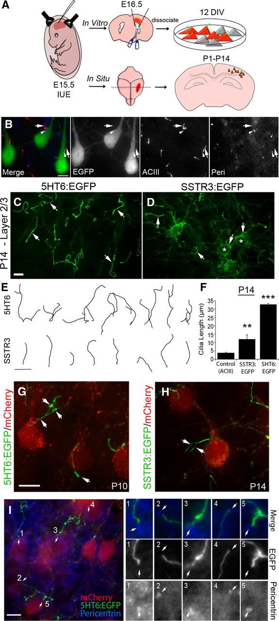
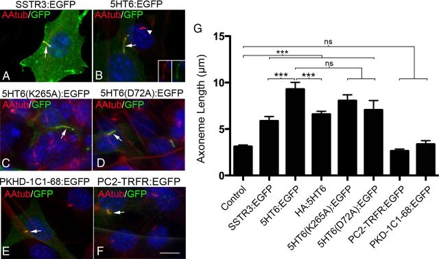
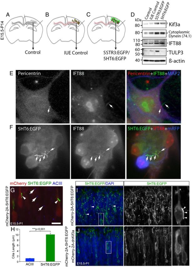
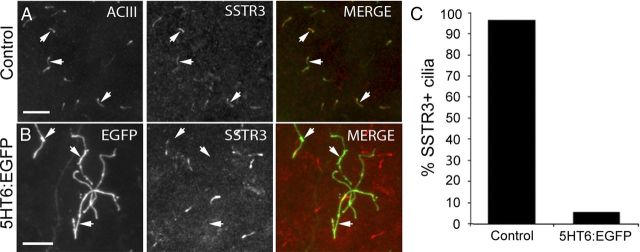
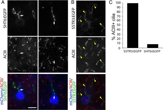
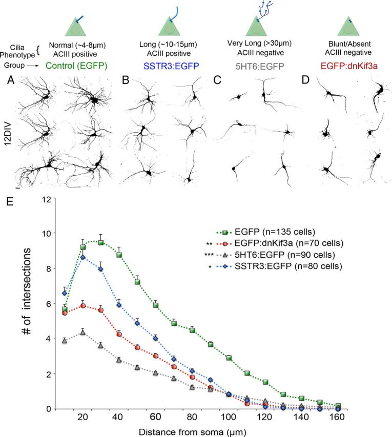
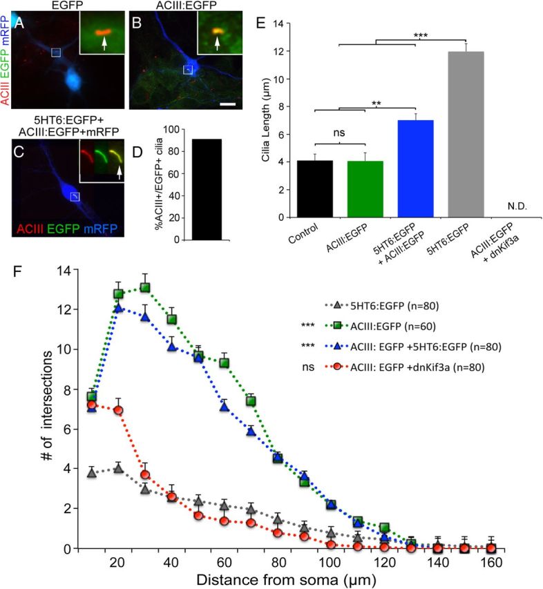
The formation of primary cilia is a highly choreographed process that can be disrupted in developing neurons by overexpressing neuromodulatory G-protein-coupled receptors GPCRs or by blocking intraflagellar transport. Here, we examined the effects of overexpressing the ciliary GPCRs, 5HT6 and SSTR3, on cilia structure and the differentiation of neocortical neurons. Neuronal overexpression of 5HT6 and SSTR3 was achieved by electroporating mouse embryo cortex in utero with vectors encoding these receptors. We found that overexpression of ciliary GPCRs in cortical neurons, especially 5HT6, induced the formation of long (>30 μm) and often forked cilia. These changes were associated with increased levels of intraflagellar transport proteins and accelerated ciliogenesis in neonatal neocortex, the induction of which required Kif3a, an anterograde motor critical for cilia protein trafficking and growth. GPCR overexpression also altered the complement of signaling molecules within the cilia. We found that SSTR3 and type III adenylyl cyclase (ACIII), proteins normally enriched in neuronal cilia, were rarely detected in 5HT6-elongated cilia. Intriguingly, the changes in cilia structure were accompanied by changes in neuronal morphology. Specifically, disruption of normal ciliogenesis in developing neocortical neurons, either by overexpressing cilia GPCRs or a dominant-negative form of Kif3a, significantly impaired dendrite outgrowth. Remarkably, coexpression of ACIII with 5HT6 restored ACIII to cilia, normalized cilia structure, and restored dendrite outgrowth, effects that were not observed in neurons coexpressing ACIII and dominant-negative form of Kif3a. Collectively, our data suggest the formation of neuronal dendrites in developing neocortex requires structurally normal cilia enriched with ACIII.


Figures

Figure 1.
Figure 1.
Overexpression of 5HT6 and SSTR3 in mouse neocortical neurons induces abnormal growth of their primary cilia. A, Flow diagram of electroporation and experimental procedures used to induce and analyze the effects of overexpression of ciliary GPCRs on ciliogenesis in cultured and in situ neocortical neurons. Neurons analyzed in culture were dissected from electroporated cortex at E16.5, dissociated, and cultured up to 12 DIV. Neurons in situ were analyzed in sections of brains between P1 and P40 electroporated mice. B, Examples of layer 2/3 neurons electroporated at E15.5 with a vector encoding EGFP and labeled at P14. EGFP+ pyramidal neurons show normal positioning and morphology of ACIII+ cilia (red, arrows) with pericentrin+ basal bodies (blue, arrowheads) near the base of the apical dendrite, and are comparable to neighboring nonelectroporated neurons. EGFP does not colocalize with ACIII in the EGFP+ neurons. C, D, E15.5 cortices were electroporated with vectors encoding either 5HT6:EGFP (C) or SSTR3:EGFP (D) under control of the EF1α promoter and were analyzed in situ at P14. C, Example of layer 2/3 neurons overexpressing 5HT6:EGFP, which is enriched in cilia (arrows). D, Example of layer 2/3 neurons overexpressing SSTR3:EGFP, which is detected in cilia (arrows) and throughout the cell bodies (*). E, Tracings of EGFP+ cilia elongated by neurons expressing either 5HT6:EGFP and SSTR3:EGFP. Scale bar, 10 μm. F, Comparison of cilia lengths from control (ACIII+), SSTR3:EGFP+, and 5HT6:EGFP+ neurons in layers 2/3 of P14 neocortex. **p < 0.01 (ANOVA). ***p < 0.001 (ANOVA). G, H, Higher-magnification confocal z-stacks of branching cilia with varicosities (arrows) synthesized by mCherry+ pyramidal neurons expressing either 5HT6:EGFP (E) or SSTR3:EGFP (F). Scale bar, 10 μm. I, Sections of P14 neocortex containing mCherry+ neurons (red) with abnormally long and malformed 5HT6:EGFP+ cilia (green) were immunostained for pericentrin (blue). Five EGFP+ cilia with basal bodies were numbered, and their magnified images are displayed as separate channels (3 images) grouped vertically by cilium on the right. Pericentrin+ basal bodies are present at the base of each EGFP+ cilium (I, arrows). Scale bar, 10 μm.
Figure 2.
Figure 2.
Cilia growth induced by GPCR overexpression is not significantly affected by loss of GPCR function or the presence of protein tags. (A–F) Representative confocal images of NIH3T3 cells expressing the proteins indicated above each panel whose expression was driven by the CMV promoter. All cells were immunostained with antibodies against the axoneme-enriched protein, acetylated α tubulin (AAtub) (red) and GFP (green). Nuclei were labeled with DAPI (blue). A, Cilium of a cell transfected with a vector encoding SSTR3:EGFP (arrow). B, Cilium of cell overexpressing 5HT6:EGFP (arrow) adjacent to a cilium of a nontransfected control cell (arrowhead). Insets, Single-channel EGFP and AAtub staining of the cilium of the transfected cell. C, D, Cilia elaborated by cells overexpressing the signaling defective 5HT6 receptors, 5HT6(K265A):EGFP (C) or 5HT6(D72A):EGFP (D) (arrows). E, F, Overexpression of EGFP fused to fibrocystin (PKHD-1C1–68:EGFP) (E) or human transferrin receptor (PC2-TRFR:EGFP) (F), two noncilia proteins fused to a cilia localization signal. Scale bar (in F), 10 μm. G, Mean axoneme lengths of cilia produced by cells expressing the experimental vectors shown in panels A–F and HA:5HT6. From left to right, n = 50, 34, 44, 56, 38, 22, 26, and 30 cilia analyzed/group, respectively. Error bar indicates mean ± SEM. ***p < 0.001 (one-way ANOVA). ns, Not significant.
Figure 3.
Figure 3.
GPCR overexpression induces upregulation of IFT proteins and premature cilia lengthening. A–D, Comparisons were made between protein expression in nonelectroporated control cortex (A), fetal cortex that was electroporated at E15.5 with either a vector encoding EGFP and mCherry:AU1 (in utero electroporation control) (B), or mCherry:AU1 and either SSTR3:EGFP or 5HT6:EGFP (C). Expression of all transgenes was under the control of the EF1α promoter. Hyphenated lines indicate cortical regions of P14 brains that were used to prepare the protein lysates analyzed by Western blot. D, Western blots (10 μg of total protein/group) were probed for proteins associated with either anterograde (Kif3a) or retrograde (cytoplasmic dynein, D1 IC74) IFT complex B protein (IFT88), or GPCR trafficking into cilia (TULP3). β-Actin was used as a loading control. E, Cultured, nonelectroporated control cortical neuron immunostained for pericentrin (basal body, red), IFT88 (green), and the neuronal marker, MAP2 (blue). The arrow in the middle panel points to an IFT88+ cilium extending from a pericentrin+ basal body (arrow left panel). F, Example of an abnormally long, branched 5HT6:EGFP+ cilium synthesized by a cultured neuron expressing 5HT6:EGFP (green) under the control of the CMV promoter and mRFP (pseudocolored blue). IFT88 (red) and EGFP were colocalized along the length and branches of the cilium (white arrows). Scale bar, 5 μm. G, E15.5 brains were electroporated with a vector encoding mCherry(AU1)-2a-5HT6:EGFP. At P1, electroporated brains were sectioned and stained with an antibody against ACIII. Examination of the upper layers of the cortical plate revealed mCherry+ neurons (red) that possessed longer 5HT6:EGFP+ cilia (arrowheads) than their neighboring nonelectroporated cells whose ACIII-stained cilia appear punctate (blue, arrows). Scale bar, 10 μm. H, Comparison of the lengths of the cilia of neurons overexpressing 5HT6:EGFP and control neurons: ***Student's t test. (I) Section of brain electroporated and processed as described for G, but not including the red channel used to visualize mCherry. Numerous, often long cilia (arrows) were present in the upper layers of the cortical plate. J, P1 neurons in the upper cortical plate that were coelectroporated at E15.5 with vectors encoding mCherry and 5HT6:EGFP (mCherry(AU1)-2a-5HT6:EGFP) and mCherry and dnKif3a (mCherry(AU1)-2a-dnKif3a). The elongated 5HT6:EGFP+ cilia of neurons expressing 5HT6:EGFP alone (I) are not observed in cells coexpressing 5HT6:EGFP and dnKif3a.
Figure 4.
Figure 4.
Cilia of neurons overexpressing 5HT6:EGFP do not contain detectable levels of SSTR3. A, Control P14 neocortex stained with antibodies against ACIII (green) and SSTR3 (red). The majority of ACIII+ cilia are also SSTR3+ (arrows). Scale bar, 10 μm. B, Cilia synthesized by neurons expressing 5HT6:EGFP (arrows) are not correspondingly SSTR3+. Scale bar, 10 μm. C, The percentage of ACIII+ control (n = 1056) or 5HT6:EGFP+ (n = 688) cilia that are also SSTR3+.
Figure 5.
Figure 5.
Cilia of neurons overexpressing 5HT6:EGFP do not contain detectable levels of ACIII. The brains of E15.5 embryos were electroporated with vectors encoding either 5HT6:EGFP or SSTR3:EGFP under control of the EF1α promoter and were immunostained for ACIII at P14. A, Pyramidal neurons in layers 2/3 of neocortex expressing mCherry:AU1 (blue) and 5HT6:EGFP (green). Sections were immunostained for ACIII (red), which normally is enriched in cilia of virtually all neocortical neurons. White arrows point to 5HT6:EGFP+ cilia projecting from mCherry:AU1+ neurons that lack detectable ACIII staining. Scale bar, 10 μm. B, Pyramidal neurons in layers 2/3 of neocortex expressing mCherry:AU1 (blue) and SSTR3:EGFP (green). SSTR3:EGFP+ cilia also stain for ACIII (yellow arrowheads). C, The percentage of SSTR3:EGFP+ (n = 123) or 5HT6:EGFP+ (n = 89) cilia that are also ACIII+.
Figure 6.
Figure 6.
5HT6, SSTR3, and dnKif3a overexpression reduces dendrite outgrowth of cultured cortical neurons. Neurons were electroporated at E15.5 with a vector encoding EGFP (Control) or the vector encoding EGFP plus vectors encoding SSTR3:EGFP, 5HT6:EGFP, or EGFP:dnKif3a under the CMV promoter. Typical cilia phenotypes associated with each group are indicated. At E16.5, cells were harvested, dissociated, and fixed after 12 DIV. Confocal images of EGFP+ cells were converted to black-and-white images. A–D, Examples of neurons at 12 DIV expressing (A) EGFP alone, (B) SSTR3:EGFP, (C) 5HT6:EGFP, and (D) EGFP:dnKif3a. E, Sholl analyses reveal the extent of arborization of EGFP control (green line), SSTR3:EGFP (blue), dnKif3a (red), and 5HT6:EGFP (gray) neurons. N = total number of cells analyzed. ***p < 0.001 versus EGFP (two-way ANOVA). **p < 0.01 versus EGFP (two-way ANOVA). *p < 0.05, versus EGFP (two-way ANOVA).
Figure 7.
Figure 7.
Coexpression of ACIII:EGFP with 5HT6:EGFP, but not dnKif3a, restores ciliary ACIII, cilia structure, and dendrite outgrowth. A, B, Neurons electroporated at E15.5 with vectors encoding either (A) mRFP and EGFP or (B) mRFP and ACIII:EGFP. Electroporated neurons were cultured for 6 DIV, fixed, and immunostained for ACIII (red). Analyses of mRFP+ neurons (blue) showed that EGFP did not traffic into the cilia as evidenced by an absence of colocalization with ACIII (A; arrow in inset). When fused to ACIII, the cilia were positive for both ACIII staining and EGFP fluorescence (B; arrow in inset). Scale bar (in B), 10 μm. C, Example of a cultured neuron coelectroporated with 5HT6:EGFP and ACIII:EGFP possessing a cilium that is positive for both ACIII staining and EGFP (inset shows higher magnification of cilia in individual channels and merge). D, Quantification of the number of cells coelectroporated with 5HT6:EGFP and ACIII:EGFP whose cilia were both ACIII+ and EGFP+ (N = 139 of 153). E, Comparisons of the lengths of the cilia elaborated by neighboring nonelectroporated (control) neurons (n = 142) or neurons transfected with vectors encoding mRFP plus ACIII:EGFP (n = 60), ACIII:EGFP and 5HT6:EGFP (n = 120), 5HT6:EGFP (n = 120), or dnKif3a (n = 118) and cultured for 12 DIV. **p < 0.01. ***p < 0.001. ns, Not significant; N.D., not determinable. F, Sholl analyses of the complexity of the dendritic arbors elaborated by neurons transfected with mRFP plus either 5HT6:EGFP (gray), ACIII:EGFP (green), EGFP:dnKif3a + ACIII:EGFP (red), or ACIII:EGFP + 5HT6:EGFP (blue) and maintained in culture for 12 d (n = number of cells analyzed). The complexity of the arbors of neurons expressing 5HT6:EGFP were statistically compared with those of the other groups using two-way ANOVA: ***p < 0.001. ns, Not significant).

References

    1. Anastas SB, Mueller D, Semple-Rowland SL, Breunig JJ, Sarkisian MR. Failed cytokinesis of neural progenitors in citron kinase-deficient rats leads to multiciliated neurons. Cereb Cortex. 2011;21:338–344. - PubMed
    1. Arellano JI, Guadiana SM, Breunig JJ, Rakic P, Sarkisian MR. Development and distribution of neuronal cilia in mouse neocortex. J Comp Neurol. 2012;520:848–873. - PMC - PubMed
    1. Avasthi P, Marshall WF. Stages of ciliogenesis and regulation of ciliary length. Differentiation. 2012;83(Suppl):S30–S42. - PMC - PubMed
    1. Avasthi P, Marley A, Lin H, Gregori-Puigjane E, Shoichet BK, von Zastrow M, Marshall WF. A chemical screen identifies class a G-protein coupled receptors as regulators of cilia. ACS Chem Biol. 2012;7:911–919. - PMC - PubMed
    1. Bennouna-Greene V, Kremer S, Stoetzel C, Christmann D, Schuster C, Durand M, Verloes A, Sigaudy S, Holder-Espinasse M, Godet J, Brandt C, Marion V, Danion A, Dietemann JL, Dollfus H. Hippocampal dysgenesis and variable neuropsychiatric phenotypes in patients with Bardet-Biedl syndrome underline complex CNS impact of primary cilia. Clin Genet. 2011;80:523–531. - PubMed

Publication types