. 2013 Mar 29;339(6127):1621-5.
doi: 10.1126/science.1231677. Epub 2013 Feb 7.

(R)-2-hydroxyglutarate is sufficient to promote leukemogenesis and its effects are reversible

Affiliations

(R)-2-hydroxyglutarate is sufficient to promote leukemogenesis and its effects are reversible

Julie-Aurore Losman et al. Science. .

Abstract

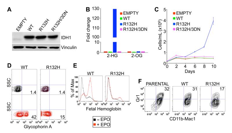
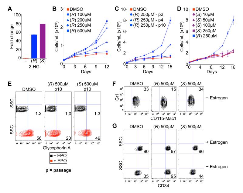
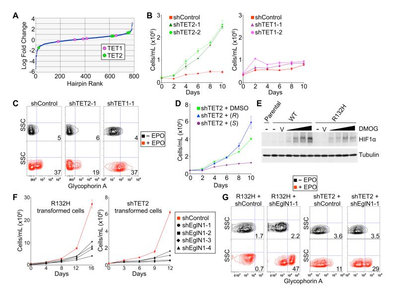
Mutations in IDH1 and IDH2, the genes coding for isocitrate dehydrogenases 1 and 2, are common in several human cancers, including leukemias, and result in overproduction of the (R)-enantiomer of 2-hydroxyglutarate [(R)-2HG]. Elucidation of the role of IDH mutations and (R)-2HG in leukemogenesis has been hampered by a lack of appropriate cell-based models. Here, we show that a canonical IDH1 mutant, IDH1 R132H, promotes cytokine independence and blocks differentiation in hematopoietic cells. These effects can be recapitulated by (R)-2HG, but not (S)-2HG, despite the fact that (S)-2HG more potently inhibits enzymes, such as the 5'-methylcytosine hydroxylase TET2, that have previously been linked to the pathogenesis of IDH mutant tumors. We provide evidence that this paradox relates to the ability of (S)-2HG, but not (R)-2HG, to inhibit the EglN prolyl hydroxylases. Additionally, we show that transformation by (R)-2HG is reversible.


Figures

Fig. 1
Fig. 1
IDH1 R132H leukemic transformation assays. (A and B) Immunoblot (A) and LC-MS (B) analysis of TF-1 cells infected with lentiviruses encoding GFP alone (empty) or GFP and the indicated IDH1 variants. Shown are mean LC-MS values of triplicate experiments. (C) Proliferation of TF-1 cells described in (A) and (B) under cytokine-poor conditions. Shown are mean values of duplicate experiments ± SD. (D and E) Erythroid differentiation of parental TF-1 cells and derivatives described in (A) and (B) as determined by fluorescence activated cell sorting (FACS) for Glycophorin A (D) or fetal hemoglobin (E) after 8 days of EPO treatment. (F) Differentiation of parental SCF ER-Hoxb8 cells and derivatives expressing the indicated IDH1 variants as determined by dual staining for CD11b/Mac1 and Gr1 3 days after withdrawal of β-estradiol. FACS plots shown are representative results of three independent experiments.
Fig. 2
Fig. 2
(R)-2-HG is sufficient to promote leukemogenesis. (A) LC-MS analysis of TF-1 cells after treatment for 3 hours with DMSO (-) or 250 μM of the indicated esterified (TFMB) 2-HG. Shown are mean values of triplicate experiments. (B-D) Proliferation of TF-1 cells under cytokine-poor conditions in the presence of the indicated amounts of TFMB-(R)-2-HG (B and C) or TFMB-(S)-2-HG (D). Cells in (B and D) were passaged 10 times prior to GM-CSF withdrawal. Cells treated with 250 μM TFMB-(R)-2-HG are also included in (D) for comparison. Shown are mean values of duplicate experiments ± SD. (E) Differentiation of TF-1 cells, as determined by Glycophorin A FACS, after 8 days of EPO treatment following pretreatment for 10 passages with DMSO or 500 μM TFMB-2-HG (R or S). Shown are representative results of three independent experiments. (F and G) Differentiation of SCF ER-Hoxb8 cells, as determined by dual staining for CD11b/Mac1 and Gr1 (F) or staining for CD34 (G), 3 days after of withdrawal of β-estradiol following pretreatment with DMSO or 500 μM TFMB-2-HG (R or S) for 20 passages. Shown are representative results of two independent experiments.
Fig. 3
Fig. 3
Opposing roles of TET2 and EglN1 in TF-1 cell transformation. (A) Relative enrichment/depletion of shRNAs targeting TET2 or TET1 in TF-1 cells infected with a pool of ~800 lentiviral shRNAs vectors targeting 2-OG-dependent dioxygenases (4-8 shRNAs/gene) and then grown under cytokine-poor conditions for 10 days. Shown are mean values of triplicate experiments. (B-C) Proliferation under cytokine-poor conditions (B) and differentiation after 8 days of EPO (C) of TF-1 cells expressing a non-targeting shRNA (shControl) or shRNAs targeting TET2 or TET1, as indicated. (D) Proliferation under cytokine-poor conditions of TF-1 cells expressing a TET2 shRNA after pretreatment for 6 days with DMSO, 250 μM TFMB-(R)-2-HG or TFMB-(S)-2-HG. (E) Immunoblot of TF-1 cells stably expressing the indicated IDH1 variants; untreated (-), treated with vehicle (V) or treated with 100 μM to 1 mM DMOG, as indicated by the triangles, for 3 hours prior to cell lysis. (F-G) Proliferation under cytokine-poor conditions (F) and differentiation after 8 days of EPO (G) of TF-1 cells expressing either IDH1 R132H or an shRNA targeting TET2, and either a non-targeting shRNA (shControl) or shRNAs targeting EglN1. Growth curves show mean values of duplicate experiments ± SD. FACS plots shown are representative results of three independent experiments.
Fig. 4
Fig. 4
Transformation by (R)-2-HG is reversible. (A) Proliferation of TF-1 cells under cytokine-poor conditions after being passaged 4 times in the presence of DMSO or 250 μM TFMB-(R)-2-HG. At the time of GM-CSF withdrawal TFMB-(R)-2-HG was either maintained [continuous (R)] or removed [out of (R) on day 0]. (B) Proliferation of TF-1 cells under cytokine-poor conditions after being passaged 20 times in the presence of DMSO or 250 μM TFMB-(R)-2-HG and then undergoing wash-out of TFMB-(R)-2-HG for the periods indicated. (C) Differentiation of TF-1 cells after being passaged 20 times in the presence of 500 μM TFMB-(R)-2-HG or DMSO. At the time of EPO administration TFMB-(R)-2-HG was either maintained [in (R)] or removed [out of (R) on day 0]. (D-F) Differentiation of TF-1 cells (D and E) and SCF ER-Hoxb8 cells (F) transformed with IDH1 R132H (D and F) or TFMB-(R)-2-HG (E) and then passaged 5 times in the presence of DMSO or a small molecule inhibitor of IDH1 R132H (1 μM). (G and H) Proliferation under cytokine-poor conditions of early-passage (p15) TF-1 cells transformed with IDH1 R132H (G) or TFMB-(R)-2-HG (H) and then passaged 5 times in the presence of DMSO or a small molecule inhibitor of IDH1 R132H (1 μM) prior to growth factor withdrawal. Growth curves show mean values of duplicate experiments ± SD. FACS plots shown are representative results of three independent experiments.

Comment in

References

    1. Falini B, et al. Blood. 2007;109:874. - PubMed
    1. Mardis ER, et al. N. Engl. J. Med. 2009;361:1058. - PMC - PubMed
    1. Kaelin WG., Jr Cold Spring Harb. Symp. Quant. Biol. 2011;76:335. - PMC - PubMed
    1. Dang L, et al. Nature. 2009;462:739. - PMC - PubMed
    1. Ward PS, et al. Cancer Cell. 2010;17:225. - PMC - PubMed

Publication types

MeSH terms