. 2013 Apr 1;41(6):3576-87.
doi: 10.1093/nar/gkt056. Epub 2013 Feb 7.

Single-molecule sorting reveals how ubiquitylation affects substrate recognition and activities of FBH1 helicase

Affiliations

Single-molecule sorting reveals how ubiquitylation affects substrate recognition and activities of FBH1 helicase

Tokiha Masuda-Ozawa et al. Nucleic Acids Res. .

Abstract

DNA repair helicases function in the cell to separate DNA duplexes or remodel nucleoprotein complexes. These functions are influenced by sensing and signaling; the cellular pool of a DNA helicase may contain subpopulations of enzymes carrying different post-translational modifications and performing distinct biochemical functions. Here, we report a novel experimental strategy, single-molecule sorting, which overcomes difficulties associated with comprehensive analysis of heterologously modified pool of proteins. This methodology was applied to visualize human DNA helicase F-box-containing DNA helicase (FBH1) acting on the DNA structures resembling a stalled or collapsed replication fork and its interactions with RAD51 nucleoprotein filament. Individual helicase molecules isolated from human cells with their native post-translational modifications were analyzed using total internal reflection fluorescence microscopy. Separation of the activity trajectories originated from ubiquitylated and non-ubiquitylated FBH1 molecules revealed that ubiquitylation affects FBH1 interaction with the RAD51 nucleoprotein filament, but not its translocase and helicase activities.


Figures
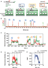
Figure 1.
Figure 1.
Surface-tethered FBH1 helicase displays a bona fide unwinding activity. (A) Schematic representation of TIRFM-based assay for analysis of DNA binding and unwinding by the individual surface-tethered helicases molecules. FBH1 is immobilized on the surface of the microscope flow cell illuminated with a 530-nm evanescent wave generated by TIR. DNA substrate molecules contain two fluorescence dyes (Cy3, donor; Cy5, acceptor). Both dyes are invisible unless found within the evanescent field because of association with the surface-tethered helicase (ON state). In the presence of ATP, FBH1-mediated unwinding of the duplex region of the DNA substrate results in gradual decrease in the FRET efficiency. Substrate dissociation results in a loss of the signal. (B) Activity of an individual surface-tethered FBH1 molecule was monitored >200 s in the presence of 1 mM ATP and PD1. Each spike in the fluorescence intensities of Cy3 (green) and Cy5 (red) marked by the blue arrow corresponds to binding and rearrangement of a new DNA substrate. Only events featuring fluorescence of both Cy3 and Cy5 dyes were analyzed. Each experiment yielded 200–600 trajectories originated from individual surface-tethered FBH1 molecules similar to the one depicted here. (C) A magnified fragment of a representative fluorescence trajectory depicting three consecutive events where FBH1 binds and unwinds PD1. Blue, orange and yellow arrows represent binding, dissociation and unwinding, respectively. (D) An individual unwinding event. (E) The Cy3 and Cy5 signals from the event depicted in the panel D and converted into a FRET trajectory, which can be divided into three sections: initial binding of the DNA substrate to FBH1 (flat portion), active unwinding of the duplex (linear decrease in FRET efficiency), dissociation of the displaced Cy5-labeled oligo, whereas the Cy3 (translocating) oligo remains bound to the FBH1.
Figure 2.
Figure 2.
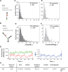
DNA structure influences FBH1 unwinding and translocation behaviors. (A and B) Schematic representation of the PD1 and RFL substrates, respectively. (C–F) Distributions of Δttotal (C and D) and Δtunwinding (E and F) for PD (upper panels) and RFL (lower panels). All dwell times were binned with the 0.25-s bin size. (G) A particular repetitive shuttling trajectory and FRET value for RFL. (H) Classification and percentage of each event over longer event (Δttotal >5 s) for RFL.
Figure 3.
Figure 3.
FBH1NTD interacts with ssDNA, dsDNA and RAD51. (A) SDS–PAGE of purified FBH1NTD visualized by Coomassie brilliant blue staining. N-terminal his-tagged and C-terminal FLAG-tagged FBH1NTD is expressed as 371 amino acids (42 kDa) polypeptide. Purification procedure outlined in the Supplementary Methods yielded a pure FBH1NTD protein of expected molecular weight. (B) To determine whether FBH1NTD contains secondary DNA-binding site, we tested its ability to interact with ssDNA and dsDNA by following changes in the intrinsic fluorescence of the aromatic residues. We observed the most profound difference between FBH1NTD fluorescence in the absence and in the presence of DNA by exciting it at 280 nm and following fluorescence at 320 nm. This fluorescence change indicates change in the hydrophobic environment of aromatic residues, and thereby it can be used as an indicator of the DNA binding. (C) Interaction between the 6× his-tagged FBH1NTD and RAD51 protein was investigated using pull-down on Ni-NTA beads. SDS–PAGE shows the proteins co-eluted from the beads.
Figure 4.
Figure 4.
Immobilized FBH1 interacts with RAD51 nucleoprotein filament. (A) Schematic representation of TIRFM-based assay for analysis of the RAD51 nucleoprotein filament binding by the individual surface-tethered helicases molecules. The experimental strategy is similar to that described for the DNA substrates, except the preformed nucleoprotein filaments assembled by RAD51 recombinase on the 60mer ssDNA were used as a substrate. Appearance of the fluorescence in both Cy3 and Cy5 channels corresponds to the nucleoprotein filament binding by the immobilized FBH1; the low FRET between the two dyes confirms that the nucleoprotein filament is fully formed and is in the extended (active) conformation. (B) Activity of an individual surface-tethered FBH1 molecule was monitored for >200 s. Blue arrow indicates RAD51 filament-binding event. (C) Representative FRET trajectories of two binding events. FRET values were calculated from the Cy3 and Cy5 intensities exactly as described for the DNA substrates (left) and binned in 0.05 U intervals for each event (right) using GraphPad Prism 4.0.
Figure 5.
Figure 5.
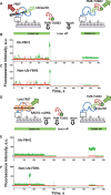
Sorting of ubiquitylated and non-ubiquitylated FBH1 molecules. (A) Schematic representation of the single-molecule sorting experiment. (B) Representative trajectory for ubiquitylated FBH1 in the presence of 1 mM ATP, 3 mM MgCl2 and 1 nM PD1. Time points at which the DNA substrate was exchanged with TMR-TUBE2, when unbound TMR-TUBE2 was washed out of the flow cell and when the laser was switched off and on are indicated over the trajectory. (C) Representative trajectory of unmodified FBH1 from the same experiment. (D) Schematic representation of the single-molecule sorting experiment with RAD51 nucleoprotein filament used as a substrate. (E) Representative trajectory of the ubiquitylated FBH1 in the presence of 1 mM ATP, 5 mM MgCl2, 5 mM CaCl2, 40 nM RAD51 and 1 nM Cy3/Cy5-labeled poly(dT)60. Time points at which the DNA substrate was exchanged with TMR-TUBE2, when unbound TMR-TUBE2 was washed out of the flow cell and when the laser was switched off and on are indicated over the trajectory. (F) Representative trajectory of unmodified FBH1 from the same experiment.

References

    1. Walsh CT, Garneau-Tsodikova S, Gatto GJ., Jr Protein posttranslational modifications: the chemistry of proteome diversifications. Angew Chem. Int. Ed. Engl. 2005;44:7342–7372. - PubMed
    1. Thomson TM, Guerra-Rebollo M. Ubiquitin and SUMO signalling in DNA repair. Biochem. Soc. Trans. 2010;38:116–131. - PubMed
    1. van Kasteren S. Synthesis of post-translationally modified proteins. Biochem. Soc. Trans. 2012;40:929–944. - PubMed
    1. Matos J, Blanco MG, Maslen S, Skehel JM, West SC. Regulatory control of the resolution of DNA recombination intermediates during meiosis and mitosis. Cell. 2011;147:158–172. - PMC - PubMed
    1. Honda M, Okuno Y, Yoo J, Ha T, Spies M. Tyrosine phosphorylation enhances RAD52-mediated annealing by modulating its DNA binding. EMBO J. 2011;30:3368–3382. - PMC - PubMed

Publication types