Review
. 2013 May 1;98(2):204-15.
doi: 10.1093/cvr/cvt020. Epub 2013 Feb 7.

Emerging mechanisms of T-tubule remodelling in heart failure

Affiliations
Review

Emerging mechanisms of T-tubule remodelling in heart failure

Ang Guo et al. Cardiovasc Res. .

Abstract

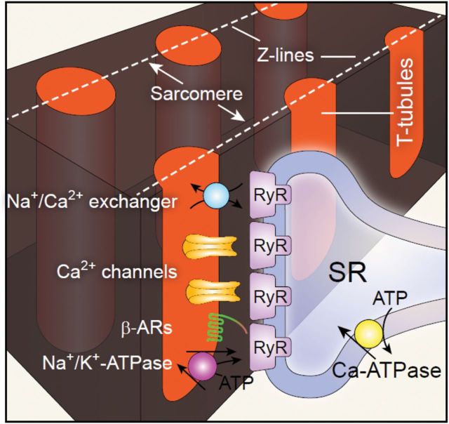
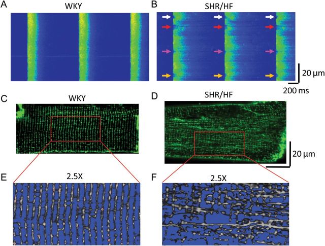
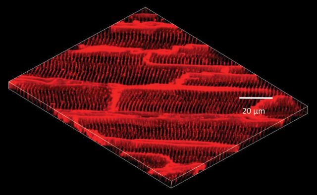
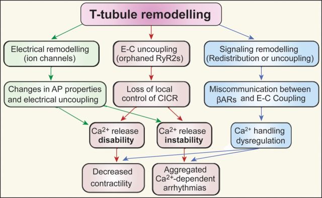
Cardiac excitation-contraction coupling occurs primarily at the sites of transverse (T)-tubule/sarcoplasmic reticulum junctions. The orderly T-tubule network guarantees the instantaneous excitation and synchronous activation of nearly all Ca(2+) release sites throughout the large ventricular myocyte. Because of the critical roles played by T-tubules and the array of channels and transporters localized to the T-tubule membrane network, T-tubule architecture has recently become an area of considerable research interest in the cardiovascular field. This review will focus on the current knowledge regarding normal T-tubule structure and function in the heart, T-tubule remodelling in the transition from compensated hypertrophy to heart failure, and the impact of T-tubule remodelling on myocyte Ca(2+) handling function. In the last section, we discuss the molecular mechanisms underlying T-tubule remodelling in heart disease.


Figures

Figure 1
Figure 1
Ultrastructure and organization of T-tubules in cardiac muscle (myocytes). (A) Electron micrograph of a transverse section of cat cardiac muscle showing four T-tubules extending inward from the periphery of the fibre (×32 000). (B) Electron micrograph of a longitudinal section of cat papillary muscle showing two T-tubules and multiple junctional couplings between T-tubules and terminal cisternae of SR, as indicated by the arrows (×40 000; reproduced with permission from Fawcett and McNutt). (C) A confocal fluorescence image of the T-tubules in an isolated living rat ventricular myocyte stained with lipophilic membrane marker Di-8 ANEPPS. (D) 3D projection of the T-tubule network from 30 sequential sections (at 0.2 μm per section) of confocal images from the same myocyte. (E) Schematic drawing of the T-tubule system in a ventricular myocyte, viewed from the transverse section.
Figure 2
Figure 2
Cartoon of local Ca2+ micro-domain and major proteins concentrated in the dyadic junction. The local Ca2+ micro-domain includes primarily (L-type) Ca2+ channels and opposing RyRs within a 12–15 nm distance between T-tubule and SR membrane, forming functional Ca2+ release units (CRU). Other important components such as Na+/Ca2+ exchanger, Na+/K+-ATPase, and β-adrenergic receptor (β-AR) are also condensed on the T-tubules.
Figure 3
Figure 3
Synchrony of local Ca2+ release during E–C coupling. (A) Ca2+ spikes recorded in ventricular myocyte under whole-cell voltage-clamp condition, and 4 mM EGTA and 1 mM Oregon Green 488 BAPTA 5N in patch pipette solution. The confocal scan line was placed along the longitudinal axis of myocyte. Discrete Ca2+ spikes were evoked synchronously from all T-tubule/SR junctions upon membrane depolarization to 0 mV from a holding potential of −70 mV. (B) Surface plot of Ca2+ spikes shown in (A). (C and D) Sparklet–spark coupling—a direct visualization of local control of CICR. (C) LTCC Ca2+ influx mediated sparklets (the low-amplitude events, pink arrows) and triggered sparks (the high-amplitude events) recorded under loose-seal patch-clamp conditions. Note that not every sparklet can trigger a spark. (D) Surface plot of sparklet–spark coupling. The arrow indicates a sparklet foot that triggered a spark (from Wang et al.).
Figure 4
Figure 4
Ca2+ handling defects and T-tubule remodelling in failing myocytes. (A and B) Field-stimulated Ca2+ transients (1 Hz) in isolated myocytes from a control Wistar-Kyoto (WKY) rat and failing spontaneously hypertension rat (SHR/HF), respectively. Control healthy myocyte exhibits uniform, synchronous and stable Ca2+ transients from beat to beat. The failing myocyte displays dyssynchronous Ca2+ release at different regions of the cell. As shown by these arrows, Ca2+ releases are delayed at fixed locations on every beat. (CF) T-tubule disorganization in a ventricular myocyte from a failing SHR heart (SHR/HF, D and F), compared with the organized T-tubule network from a WKY control myocyte (C and E). (From Song et al.)
Figure 5
Figure 5
3D reconstruction of epicardial myocyte T-tubule network in situ. Confocal images (25 confocal stacks at 0.2 μm interval) were acquired in situ from Langendorff-perfused intact healthy heart, demonstrating the periodically organized T-tubule structure in normal myocytes. (From Wei et al.)
Figure 6
Figure 6
Progressive T-tubule remodelling of left-ventricular myocytes in pressure overload rat cardiomyopathy. (AD) Representative T-tubule images from left ventricle (LV) of age-matched sham-operated heart (A), hypertrophy (B), early HF (C), and advanced HF (D). At hypertrophy stage, discrete T-tubule loss (green arrows) was often observed with slight T-tubule disorganization (B). In moderately decompensated heart, LV myocytes exhibited widely impaired T-tubule system (C). At advanced HF stage, myocytes lost majority of T-tubules with striated pattern almost vanished (D). Each yellow framed inset is a zoom-in view of an area 40×40 μm2 from associated images. (E) A gradual reduction in TTpower (an index of T-tubule regularity) with heart disease progression. (F) Cardiac global function (ejection fraction) correlates well with T-tubule integrity. LV, left ventricle; EF, ejection fraction. (From Wei et al.)
Figure 7
Figure 7
Schematic chart depicting the consequences of T-tubule remodelling and the relationship with heart failure and Ca2+-dependent arrhythmogenesis. Myocyte T-tubule remodelling leads to alterations at multiple levels, including ultrastructural, electrical, and signal transduction changes, collectively contributing to the progression of cardiac failure and the genesis of fatal arrhythmias.

Comment in

References

    1. Bers DM. Excitation–Contraction Coupling and Cardiac Contractile Force. Boston: Kluwer Academic Publishers; 2001.
    1. Stern MD. Theory of excitation–contraction coupling in cardiac muscle. Biophys J. 1992;63:497–517. doi:10.1016/S0006-3495(92)81615-6. - DOI - PMC - PubMed
    1. Wang SQ, Song LS, Lakatta EG, Cheng H. Ca2+ signalling between single L-type Ca2+ channels and ryanodine receptors in heart cells. Nature. 2001;410:592–596. doi:10.1038/35069083. - DOI - PubMed
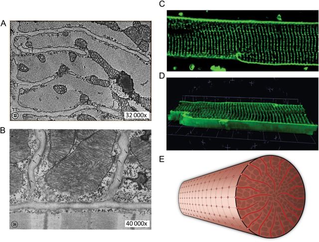
    1. Sun XH, Protasi F, Takahashi M, Takeshima H, Ferguson DG, Franzini-Armstrong C. Molecular architecture of membranes involved in excitation-contraction coupling of cardiac muscle. J Cell Biol. 1995;129:659–671. doi:10.1083/jcb.129.3.659. - DOI - PMC - PubMed
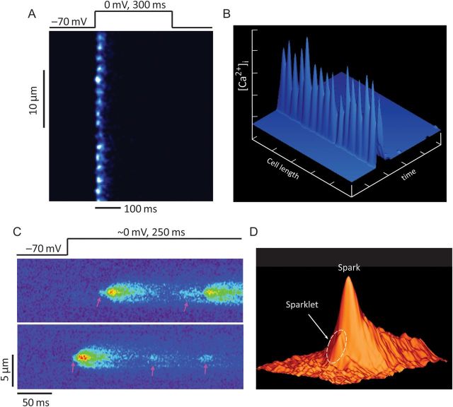
    1. Cheng H, Lederer WJ. Calcium sparks. Physiol Rev. 2008;88:1491–1545. doi:10.1152/physrev.00030.2007. - DOI - PubMed

Publication types