. 2013 Feb 8;339(6120):694-8.
doi: 10.1126/science.1229934.

Structural basis for hijacking of cellular LxxLL motifs by papillomavirus E6 oncoproteins

Affiliations

Structural basis for hijacking of cellular LxxLL motifs by papillomavirus E6 oncoproteins

Katia Zanier et al. Science. .

Abstract

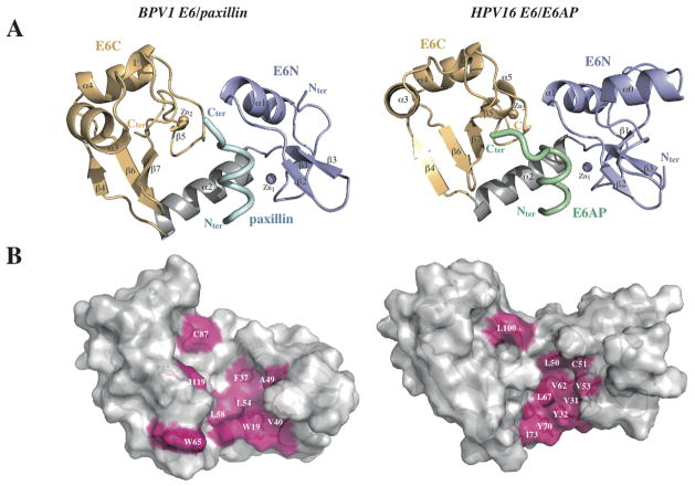
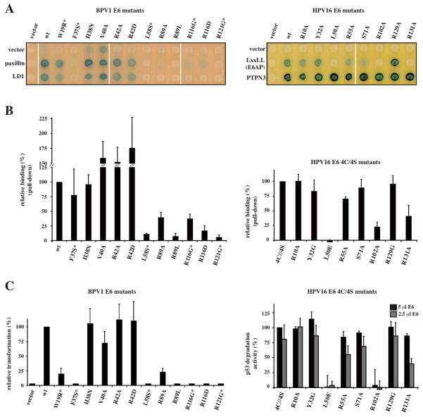
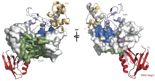
E6 viral oncoproteins are key players in epithelial tumors induced by papillomaviruses in vertebrates, including cervical cancer in humans. E6 proteins target many host proteins by specifically interacting with acidic LxxLL motifs. We solved the crystal structures of bovine (BPV1) and human (HPV16) papillomavirus E6 proteins bound to LxxLL peptides from the focal adhesion protein paxillin and the ubiquitin ligase E6AP, respectively. In both E6 proteins, two zinc domains and a linker helix form a basic-hydrophobic pocket, which captures helical LxxLL motifs in a way compatible with other interaction modes. Mutational inactivation of the LxxLL binding pocket disrupts the oncogenic activities of both E6 proteins. This work reveals the structural basis of both the multifunctionality and the oncogenicity of E6 proteins.


Figures

Fig. 1
Fig. 1
X-ray structures of E6/LxxLL complexes. (A) The structures of BPV1 E6 bound to residues 1–10 of paxillin (left panel) and of HPV16 E6 bound to residues 403–414 of E6AP (right panel). violet: E6N; grey: linker helix; gold: E6C; cyan/green: LxxLL peptides. (B) The hydrophobic pocket (pink) responsible for LxxLL motif recognition in BPV1 E6 (left panel) and HPV16 E6 (right panel). These structures show helical LxxLL peptides inserted in a deep pocket formed by the two domains, unlike other cellular domains (FAT, CH and LBD) interacting through shallow surfaces with the cognate LxxLL peptides. See also Supplementary Fig. 5.
Fig. 2
Fig. 2
Networks of E6/LxxLL peptide interactions. (A) Polar interactions between BPV1 E6 and paxillin. Red spheres: water molecules; black dashed lines: interactions mediated by “keystone” arginine R89; violet/orange dashed lines: intermolecular interactions mediated by E6N/E6C arginines; dotted lines: other polar interactions. (B) All contacts between BPV1 E6 and paxillin. Pink dashed lines: hydrophobic contacts; black lines: polar contacts mediated by side chain (continuous lines) or main chain (dotted lines); pink/blue boxed residues: E6 hydrophobic/polar contributors; E6 polar residues displaying favorable van der Waals terms (ΔE vdw< -1 kcal/mol, Supplemental Fig. 6A) are included as hydrophobic contributors. (C) Polar interactions between HPV16 E6 and E6AP. Black dashed lines: interactions mediated by “keystone” arginine R102; violet/orange dashed lines: intermolecular interactions mediated by E6N/E6C arginines. (D) All contacts between HPV16 E6 and E6AP. R129-E1 (dashed gray line) is deduced from MD simulations (Supplemental Fig. 7).
Fig. 3
Fig. 3
LxxLL binding and functional activities of E6 mutants. (A) Yeast two-hybrid analysis of BPV1 E6 (left panel) and HPV16 E6 (right panel) mutants binding to paxillin (full-length or LD1 peptide) and E6AP peptide respectively. PTPN3 (targeting the PDZ binding motif) is included as control of HPV16 E6 expression. (B) Pull-down assays of BPV1 E6 (left panel) and HPV16 E6 (right panel) mutants binding to GST-fused LxxLL peptides. The data (mean ± sd) were normalized to 100% for the reference protein. “*”: previously investigated mutants (15, 33, 34). HPV16 E6 mutations were introduced in the E6 4C/4S construct. (C) Oncogenic activities of E6 mutants. The data (mean ± sd) were normalized to 100% for the reference protein. (Left panel) Transformation phenotypes of BPV1 E6 mutants, quantified using numbers of anchorage-independent colonies. (Right panel) p53 degradation activities of HPV16 E6 mutants measured by incubating constant amounts of p53 with either 2.5 or 5 μl of E6 translation product. I0 and I correspond to, respectively, the p53 signal before and after incubation with E6. See also Supplemental Fig. 8.
Fig. 4
Fig. 4
Mapping HPV16 E6 functional regions. The different binding sites of HPV16 E6 map to distinct regions of the protein’s solvent accessible surface. LxxLL binding and E6 self-association residues are colored green and blue respectively. The position of the second E6 molecule shown in the ribbon representation was modeled based on the geometry of the E6N homodimer interface (2LJY.pdb). The C-terminal PDZ binding motif (pink) is disordered in the NMR structure of the isolated HPV16 E6C domain and adopts a β-strand conformation upon binding to the PDZ1 of Magi I (red) (2KPL.pdb). The relative orientation of E6 and the PDZ domain shown is arbitrary. Surfaces colored white are potentially available for binding to p53. See also Supplemental Fig. 9.

References

    1. Bernard HU, et al. Classification of papillomaviruses (PVs) based on 189 PV types and proposal of taxonomic amendments. Virology. 2010;401:70. - PMC - PubMed
    1. zur Hausen H. Immortalization of human cells and their malignant conversion by high risk human papillomavirus genotypes. Semin Cancer Biol. 1999;9:405. - PubMed
    1. Pfister H, et al. High prevalence of epidermodysplasia verruciformis-associated human papillomavirus DNA in actinic keratoses of the immunocompetent population. Arch Dermatol Res. 2003;295:273. - PubMed
    1. Nasir L, Campo MS. Bovine papillomaviruses: their role in the aetiology of cutaneous tumours of bovids and equids. Vet Dermatol. 2008;19:243. - PubMed
    1. Huibregtse JM, Scheffner M, Howley PM. A cellular protein mediates association of p53 with the E6 oncoprotein of human papillomavirus types 16 or 18. EMBO J. 1991;10:4129. - PMC - PubMed

Publication types

MeSH terms