. 2013 May 13;14(5):1299-310.
doi: 10.1021/bm301825q. Epub 2013 Apr 1.

Hyperbranched polyester hydrogels with controlled drug release and cell adhesion properties

Affiliations

Hyperbranched polyester hydrogels with controlled drug release and cell adhesion properties

Hongbin Zhang et al. Biomacromolecules. .

Abstract

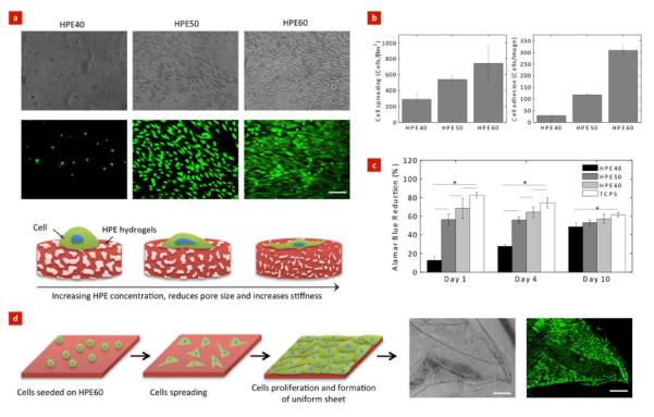
Hyperbranched polyesters (HPE) have a high efficiency to encapsulate bioactive agents, including drugs, genes, and proteins, due to their globe-like nanostructure. However, the use of these highly branched polymeric systems for tissue engineering applications has not been broadly investigated. Here, we report synthesis and characterization of photocrosslinkable HPE hydrogels with sustained drug release characteristics for cellular therapies. These HPE can encapsulate hydrophobic drug molecules within the HPE cavities due to the presence of a hydrophobic inner structure that is otherwise difficult to achieve in conventional hydrogels. The functionalization of HPE with photocrosslinkable acrylate moieties renders the formation of hydrogels with a highly porous interconnected structure and mechanically tough network. The compressive modulus of HPE hydrogels was tunable by changing the crosslinking density. The feasibility of using these HPE networks for cellular therapies was investigated by evaluating cell adhesion, spreading, and proliferation on hydrogel surface. Highly crosslinked and mechanically stiff HPE hydrogels have higher cell adhesion, spreading, and proliferation compared to soft and complaint HPE hydrogels. Overall, we showed that hydrogels made from HPE could be used for biomedical applications that require spatial control of cell adhesion and controlled release of hydrophobic clues.


Figures

Figure 1
Figure 1. Synthesis of HPE and HPE-A macromolecules
HPE (Generation 4) was synthesized y pseudo-one-step procedure using 1,1,1-trimethylolpropane (TMP) as a core and 2,2-bis(hydroxymethyl)propionic acid (bis-MPA) as a repeating unit.
Figure 2
Figure 2. Chemical characterization of HPE and HPE-A macromolecules
(a) 1H-NMR spectra of HPE and HPE-A with different degrees of acrylation are shown. Strong peaks related to acrylate groups at 6.36, 6.21 and 6.10 ppm were observed in HPE-A. (b) Strong peaks at 3400 cm−1, 1730 cm−1 and 1050 cm−1 are related to OH, C=O and C-O groups respectively; whereas a weak peak of methylene group is observed at 1635 cm−1 after acrylation of HPE macromolecules. (c) Size exclusion spectra of HPE and HPE-A dissolved in THF were obtained using GPC. The GPC curve demonstrated an increase in the hydrodynamic volume of HPE in THF after acylation.
Figure 3
Figure 3. Structural and physical characterization of HPE hydrogels made by crosslinking HPE macromers
Effect of HPE macromolecules concentration on microstructure of freeze dried hydrogel network was evaluated using scanning electron microscopy. At low HPE concentration (40% HPE), highly porous and interconnected structure was observed. With an increase in HPE concentration, pore size decreased due to increase in crosslinked density. The bars represent mean ± standard deviation (n=3), (*p < 0.05).
Figure 4
Figure 4. Mechanical properties of HPE hydrogels was evaluated using unconfined compression test
(a) Representative stress strain curves for different concentration of HPE hydrogels are shown. (b) Increase in HPE concentration resulted in an increase in compressive modulus indicating formation of highly crosslinked network. The error bars represent mean ± standard deviation (n=3), (*p < 0.05).
Figure 5
Figure 5. Stability of HPE hydrogels in physiological conditions
(a) Stability of HPE hydrogels was investigated by evaluating swelling behavior and in vitro degradation of 40%, 50% and 60% HPE hydrogels in physiological environment. All the hydrogels reached equilibrium hydration degree within 24 hours. A significant decrease in the swelling ratio of HPE hydrogels due to increase in HPE concentration was observed. (b) In degradation study, an initial mass loss (Phase I) observed was due to leaching out of sol content. A slow and linear degradation after initial mass loss was observed (Phase II). The bars represent mean ± standard deviation (n=3), (*p < 0.05).
Figure 6
Figure 6. Sustain release of hydrophobic drug from HPE hydrogels made by crosslinking HPE macromers
(a) Schematic showing encapsulation of dexamethasone acetate (DA) within hydrophobic cavities of HPE-A macromolecules. After photocrosslinking, DA loaded HPE-A macromers forms covalently crosslinked network. (b) Image showing PEG-DA and HPE-DA solution. Due to hydrophobic nature of DA, HPE macromolecule is able to uniformly entrap DA and result in a stable solution. Whereas, PEG-DA solution shows phase separation due to precipitation of DA aggregates. The release profile of DA was investigated from PEG and HPE hydrogels. PEG hydrogels reached a plateau phase after initial release of entrapped DA. (c) Whereas, HPE hydrogels made by crosslinking HPE macromers showed sustain release of entrapped DA. The sustain release of DA can also be attributed to linear degradation profile of HPE hydrogels.
Figure 7
Figure 7. Microfabricated photocrosslinkable HPE hydrogels made by crosslinking HPE macromers
(a) Precursor solution containing 50% HPE macromers showed low viscosity and can be injected using 22-gauge needle. When subjected to UV radiation the prepolymer solution quickly formed covalently crosslinked network. The optical images representing the flexibility of 150 μm thin methylene blue dyed HPE hydrogel sheet. The HPE hydrogel sheet can be easily handled without breaking. After folding, the hydrogel sheet easily unfolds and regains its original shape in the DPBS bath. (b) Schematic presentation for developing patterned HPE hydrogel structures using photolithography. Fluorescent images of different micropatterned structures obtained using photolithography (scale bar = 500 μm).
Figure 8
Figure 8. Control cells adhesion and proliferation on HPE hydrogels
(a) Representative phase contrast and fluorescent images of NIH-3T3 fibroblasts on the surface of the HPE hydrogels. Cells readily attached and spread on hydrogels with higher HPE concentration. (Scale bar = 100 μm). Schematic showing the increase in cells adhesion and spreading can be attributed to higher stiffness of hydrogels resulting from an increase in HPE concentration. (b) Cells adhesion and spreading on the hydrogels surface was quantified using ImageJ. Increase in HPE concentration results in an increase in cells adhesion and spreading. (c) The metabolic activity of cells strongly depends on the HPE concentration. Tissue culture polystyrene was used as a positive control. (d) Controlled cell adhesion properties of HPE60 can be utilized to fabricate uniform cell layer. We observed after 4 day, cells uniformly covered the surface of hydrogels and the cell layer can be easily handled using forceps (Scale bar = 500 μm). The bars represent mean ± standard deviation (n=3), (*p < 0.05).

References

    1. Khademhosseini A, Langer R. Biomaterials. 2007;28(34):5087–5092. - PubMed
    1. Khademhosseini A, Vacanti JP, Langer R. Sci. Am. 2009;300(5):64–71. - PubMed
    1. Langer R, Vacanti JP. Science. 1993;260(5110):920–926. - PubMed
    1. Zorlutuna P, Annabi N, Camci-Unal G, Nikkhah M, Cha JM, Nichol JW, Manbachi A, Bae H, Chen S, Khademhosseini A. Adv. Mater. 2012;24(14):1782–804. - PMC - PubMed
    1. Lutolf MP. Nat. Mater. 2009;8(6):451–453. - PubMed

Publication types

MeSH terms