

Characterization of microaneurysm closure after focal laser photocoagulation in diabetic macular edema
- PMID: 23394906
- PMCID: PMC3627831
- DOI: 10.1016/j.ajo.2012.12.005
Characterization of microaneurysm closure after focal laser photocoagulation in diabetic macular edema
Abstract
Purpose: To characterize microaneurysm closure following focal laser photocoagulation in diabetic macular edema (DME) using simultaneous fluorescein angiography (FA) and spectral-domain optical coherence tomography (SD-OCT).
Design: Retrospective observational case series.
Methods: Leaking microaneurysms (n = 123) were analyzed in eyes (n = 29) with nonproliferative diabetic retinopathy (NPDR) that underwent navigated focal laser photocoagulation in DME and were followed at 3, 6, and 12 months. Closure of diabetic microaneurysms was characterized in detail following focal laser using SD-OCT.
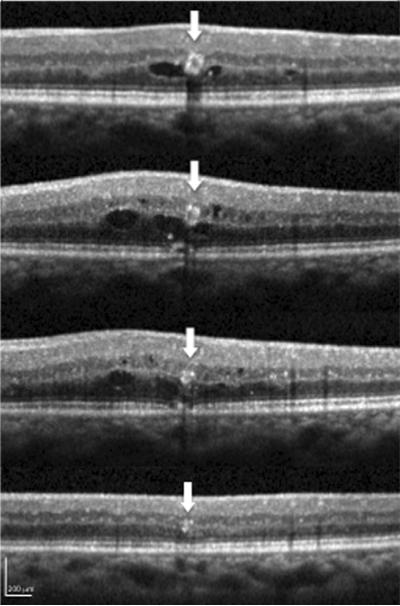
Results: Closure rate of microaneurysms by both FA and SD-OCT was 69.9% (84/123), 79.7% (98/123), and 82.9% (102/123) at 3, 6, and 12 months, respectively. Microaneurysm closure rate increased at 6 and 12 months compared to 3 months (P < .003, P < .001). Over half of closed microaneurysms (45/86, 52.3%) left hyperreflective spots while the remaining half (41/86, 47.7%) disappeared without any hyperreflectivity by SD-OCT at 3 months. Hyperreflective spots decreased at 6 (36/99, 36.4%) and 12 months (17/102, 16.7%) with a concomitant increase in complete loss of reflectivity at 6 (63/99, 63.6%) and 12 months (85/102, 83.3%). Smaller outer and inner diameters and heterogeneous lumen reflectivity were positively associated with microaneurysm closure at 12 months (P < .0001, P < .001, P < .03).
Conclusions: Characterization of microaneurysms following focal laser photocoagulation resulted in hyperreflective spots and complete resolution of all reflectivity using SD-OCT. Smaller microaneurysms and those with heterogeneous lumen were positively associated with microaneurysm closure. These findings provide greater understanding of localized retinal changes following focal laser photocoagulation in DME treatment.
Copyright © 2013 Elsevier Inc. All rights reserved.
Figures


Similar articles
-
Indocyanine green angiography-guided focal navigated laser photocoagulation for diabetic macular edema.Jpn J Ophthalmol. 2019 May;63(3):243-254. doi: 10.1007/s10384-019-00662-x. Epub 2019 Feb 26. Jpn J Ophthalmol. 2019. PMID: 30806869
-
Imaging of laser-photocoagulated diabetic microaneurysm with spectral domain optical coherence tomography.Retina. 2013 Apr;33(4):726-31. doi: 10.1097/IAE.0b013e3182753960. Retina. 2013. PMID: 23478291
-
Structural en face optical coherence tomography imaging for identification of leaky microaneurysms in diabetic macular edema.Int Ophthalmol. 2020 Apr;40(4):787-794. doi: 10.1007/s10792-019-01239-w. Epub 2019 Dec 3. Int Ophthalmol. 2020. PMID: 31797175
-
The Role of Laser Photocoagulation in Treating Diabetic Macular Edema in the Era of Intravitreal Drug Administration: A Descriptive Review.Medicina (Kaunas). 2023 Jul 17;59(7):1319. doi: 10.3390/medicina59071319. Medicina (Kaunas). 2023. PMID: 37512130 Free PMC article. Review.
-
[Angiopathy and the eye].Vnitr Lek. 2010 Apr;56(4):333-9. Vnitr Lek. 2010. PMID: 20465107 Review. Czech.
Cited by
-
Photocoagulation or sham laser in addition to conventional anti-VEGF therapy in macular edema associated with TelCaps due to diabetic macular edema or retinal vein occlusion (TalaDME): a study protocol for a multicentric, French, two-group, non-commercial, active-control, observer-masked, non-inferiority, randomized controlled clinical trial.Trials. 2024 Apr 22;25(1):273. doi: 10.1186/s13063-024-07994-1. Trials. 2024. PMID: 38649937 Free PMC article.
-
Navigated direct photocoagulation with a 30-ms short-pulse laser for treating microaneurysms in diabetic macular edema exhibits a high closure rate.Sci Rep. 2023 Apr 13;13(1):6092. doi: 10.1038/s41598-023-33260-6. Sci Rep. 2023. PMID: 37055549 Free PMC article.
-
Effect of focal laser photocoagulation in eyes with mild to moderate non-proliferative diabetic retinopathy.Int J Ophthalmol. 2016 Oct 18;9(10):1439-1443. doi: 10.18240/ijo.2016.10.12. eCollection 2016. Int J Ophthalmol. 2016. PMID: 27803861 Free PMC article.
-
Structural changes in individual retinal layers in diabetic macular edema.J Diabetes Res. 2013;2013:920713. doi: 10.1155/2013/920713. Epub 2013 Aug 29. J Diabetes Res. 2013. PMID: 24073417 Free PMC article. Review.
-
Bridging the resources gap: deep learning for fluorescein angiography and optical coherence tomography macular thickness map image translation.BMC Ophthalmol. 2022 Sep 1;22(1):355. doi: 10.1186/s12886-022-02577-7. BMC Ophthalmol. 2022. PMID: 36050661 Free PMC article. Clinical Trial.
References
-
- Moss SE, Klein R, Klein BE. The 14-year incidence of visual loss in a diabetic population. Ophthalmology. 1998;105(6):998–1003. - PubMed
-
- Murakami T, Nishijima K, Sakamoto A, Ota M, Horii T, Yoshimura N. Foveal cystoid spaces are associated with enlarged foveal avascular zone and microaneurysms in diabetic macular edema. Ophthalmology. 2011;118(2):359–367. - PubMed
-
- Early Treatment Diabetic Retinopathy Study Research group Photocoagulation for diabetic macular edema. Early Treatment Diabetic Retinopathy Study report number 1. Arch Ophthalmol. 1985;103(12):1796–1806. - PubMed
-
- Shah AM, Bressler NM, Jampol LM. Does laser still have a role in the management of retinal vascular and neovascular disease? Am J Ophthalmol. 2011;152(3):332–339. - PubMed
Publication types
MeSH terms
Grants and funding
LinkOut - more resources
Full Text Sources
Other Literature Sources
Medical
