. 2013 Feb 5;17(2):210-24.
doi: 10.1016/j.cmet.2013.01.004.

MicroRNA-133 controls brown adipose determination in skeletal muscle satellite cells by targeting Prdm16

Affiliations

MicroRNA-133 controls brown adipose determination in skeletal muscle satellite cells by targeting Prdm16

Hang Yin et al. Cell Metab. .

Abstract

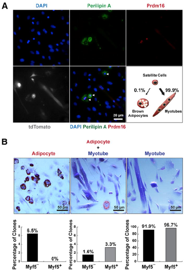
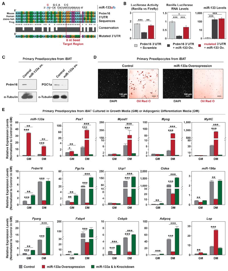
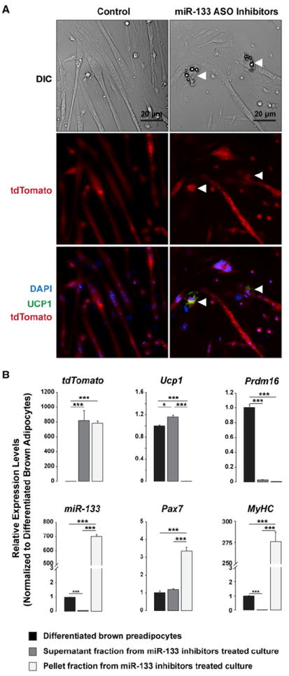
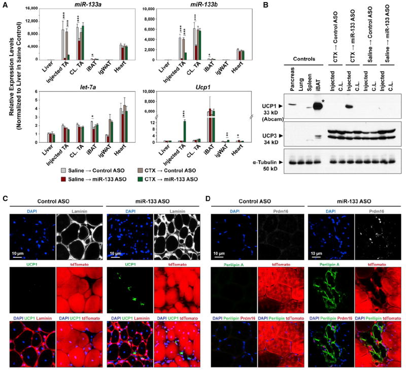
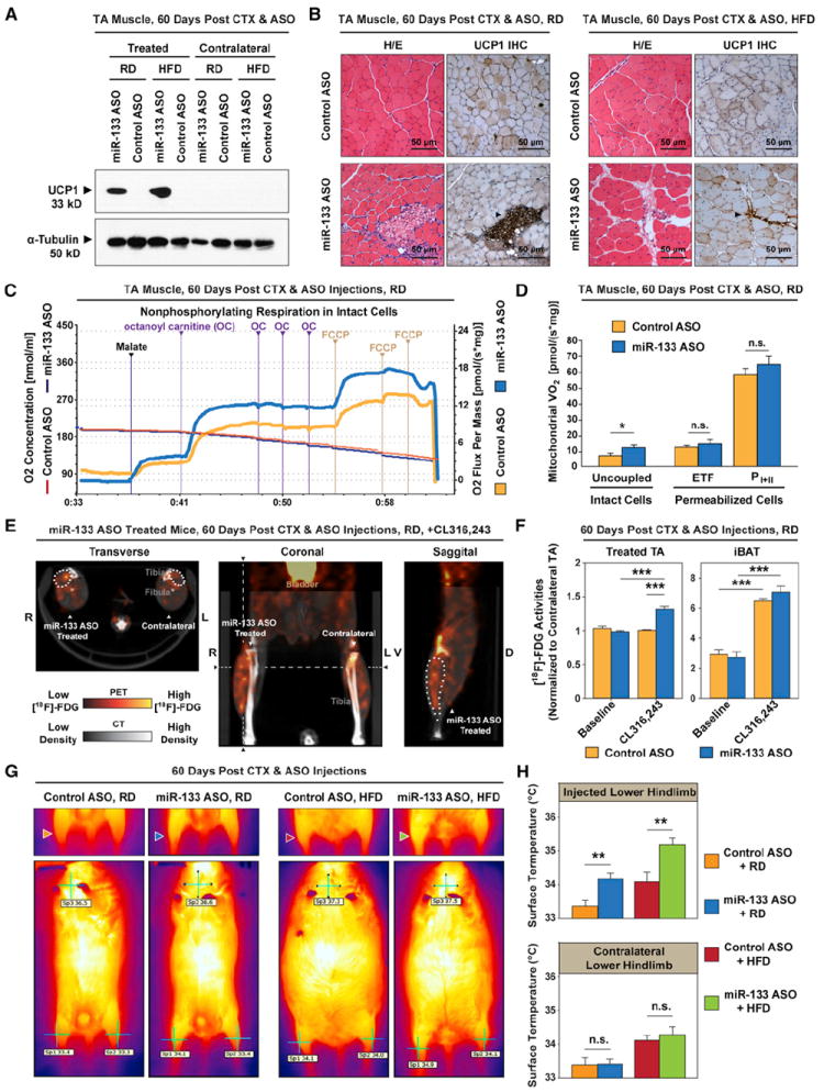
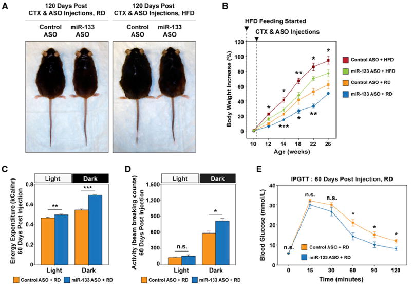
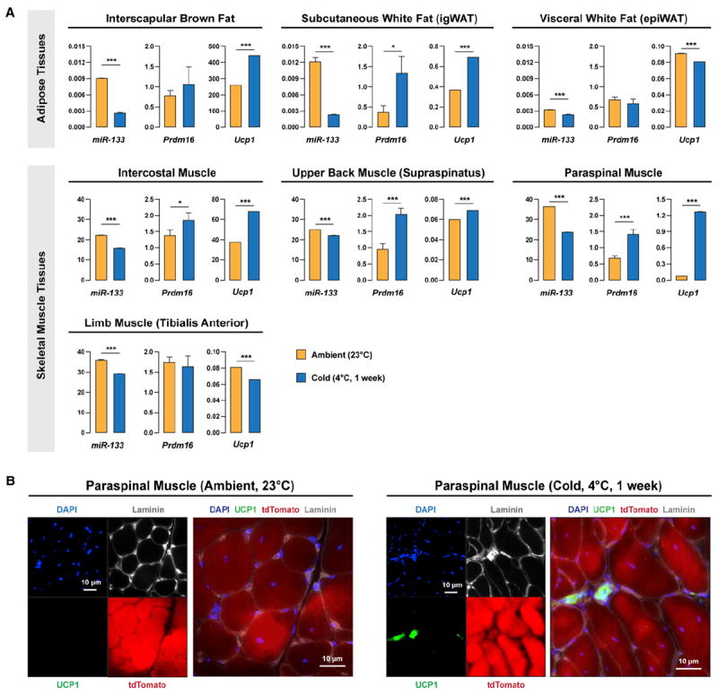
Brown adipose tissue (BAT) is an energy-dispensing thermogenic tissue that plays an important role in balancing energy metabolism. Lineage-tracing experiments indicate that brown adipocytes are derived from myogenic progenitors during embryonic development. However, adult skeletal muscle stem cells (satellite cells) have long been considered uniformly determined toward the myogenic lineage. Here, we report that adult satellite cells give rise to brown adipocytes and that microRNA-133 regulates the choice between myogenic and brown adipose determination by targeting the 3'UTR of Prdm16. Antagonism of microRNA-133 during muscle regeneration increases uncoupled respiration, glucose uptake, and thermogenesis in local treated muscle and augments whole-body energy expenditure, improves glucose tolerance, and impedes the development of diet-induced obesity. Finally, we demonstrate that miR-133 levels are downregulated in mice exposed to cold, resulting in de novo generation of satellite cell-derived brown adipocytes. Therefore, microRNA-133 represents an important therapeutic target for the treatment of obesity.


Figures

Figure 1
Figure 1. Satellite Cells Differentiate into Brown Adipocytes
(A) Satellite cells differentiated into brown adipocytes (arrowheads) in myofiber cultures under proadipogenic conditions. Myofibers (n > 600) with resident satellite cells were isolated from Pax7-CreER/R26R-tdTomato EDL muscles and cultured for 12 days in proadipogenic medium. Lineage-marked satellite cell-derived brown adipocytes expressed tdTomato and Prdm16, Perilipin A. (B) Clonal analysis of FACS-isolated single satellite stem cells and satellite myogenic progenitors (n > 2,000 for each cell type) indicates some satellite cells are bipotential. Approximately 1.6% of satellite stem cells and 3.3% satellite myogenic progenitors gave rise to mixed muscle and adipocyte-containing colonies. In addition, 6.5% of satellite stem cells clones but none of satellite myogenic progenitors gave rise to colonies uniformly composed of adipocytes. Shown are representative images of three types of clones derived from clonal satellite cell cultures stained with ORO, and the corresponding percentages from satellite stem cells (Myf5) and satellite myogenic progenitors (Myf5+) clones. See also Figure S1.
Figure 2
Figure 2. Prdm16 Is Targeted by miR-133
(A) Prdm16 3′UTR contains a conserved target site for both miR-133a and miR-133b. The absolutely conserved 8 nt seed sequences in multiple genomes and a mutated seed sequence used in this study (Prdm16_mutUTR) were enclosed in a red frame. (B) Luciferase assays and RT-qPCR indicate miR-133 targets Prdm16 3′UTR and this repression depends on the predicted 8 nt seed sequence. Ectopic miR-133 was overexpressed in HEK293T cells together with Renilla luciferase reporter constructs containing either intact or mutated Prdm16 3′UTR. The repression of luciferase activity and expression by miR-133 was abolished by mutating the predicted 8 nt seed sequence. (C) Immunoblots reveals that lentiviral miR-133 overexpression (ov.) in primary brown preadipocytes repressed Prdm16 and Pgc1-α protein levels. (D) Representative images depict that adipogenic differentiation from primary brown preadipocytes was severely impaired by miR-133 overexpression. ORO staining revealed drastically reduced number of differentiated adipocytes with oil droplets in the miR-133 ov. culture, while DAPI staining of the same fields indicated that same near-confluent density of cells were present for both cultures. (E) RT-qPCR shows lentiviral overexpression of miR-133 or inhibition of miR-133 by antisense oligos impaired or enhanced brown adipogenic commitment in primary brown preadipocytes, respectively. Notably, impaired brown adipogenic commitment in the miR-133 ov. culture was companied with the emergence of myogenic differentiation, as evidenced by increased expression of myogenic markers (Pax7, MyoD, Myog, MyHC), and white adipogenic differentiation, as evidenced by increased expression of Leptin. Error bars, SEM. See also Figure S2.
Figure 3
Figure 3. miR-133 Prevents Brown Adipose Determination in Satellite Cells
(A) Inhibition of miR-133 induced satellite cells to differentiate into brown adipocytes (arrowheads) in myofiber cultures under proadipogenic conditions. Myofibers (n > 300) with resident satellite cells were isolated from Pax7-CreER/R26R-tdTomato EDL muscles and transfected with mixed inhibitors for miR-133a and miR-133b or a control scramble inhibitor. Lineage-marked satellite cell-derived brown adipocytes (SC_BA) expressed tdTomato and Ucp1. (B) RT-qPCR reveals gene expression signatures of SC_BAs as compared to those of myotubes and differentiated primary brown adipocytes. miR-133 inhibitor-treated myofiber cultures were centrifuged to separate the supernatant faction enriched for SC_BAs and the pellet fraction which contains mostly myotubes. Error bars, SEM. See also Figure S3.
Figure 4
Figure 4. Antagonism of miR-133 Induces Brown Adipose Determination of Satellite Cells during Muscle Regeneration
(A) High efficacy and specificity of miR-133 antagomiR (ASO) in vivo. RT-qPCR indicates reduced expression of both miR-133a and miR-133b, but not let-7a in response to intramuscular miR-133 ASO administration. Ucp1 mRNA was drastically induced in regenerating TA muscles by miR-133 ASO. (B) Immunoblots reveal the evident induction of Ucp1, but not Ucp3, in regenerating TA muscles in response to miR-133 ASO treatment. Asterisk denotes that the loading of the iBAT lane on the Ucp1 immunoblot was 1/100 of other lanes to avoid overloading. (C and D) miR-133 antagonism during muscle regeneration induced satellite cells to differentiate into brown adipocytes located within the muscle interstitium. Representative images of TA muscle cross-sections stained with Ucp1 and Laminin (C) or Prdm16 and Perilipin A (D) together with tdTomato native fluorescence revealed SC_BAs within the muscle interstitium. Error bars, SEM. See also Figure S4.
Figure 5
Figure 5. miR-133 Antagonism Induces Metabolically Active Brown Adipocytes in Muscle
(A) Immunoblots reveal the induction of Ucp1 in regenerating TA muscles by miR-133 antagonism in C57BL/6 mice fed with either a regular diet (RD) or a high-fat diet (HFD). The contralateral TA muscles were included as control. (B) Hematoxylin and eosin (H/E) staining and immunohistochemistry (IHC) of Ucp1 protein reveal that miR-133 antagonism-induced brown adipocytes located within the muscle interstitium (arrowheads) under both diet conditions. (C) Representative oxygraph tracings from high-resolution respirometry depict markedly increased uncoupled (nonphosphorylating) respiration in intact (non-permeabilized) miR-133 ASO-treated TA muscles compared to control. Notably, octanoyl carnitine (OC) markedly increased the O2 consumption in miR-133 ASO-treated TA muscles compared to control. Responses before reoxygenation (which showed no O2 diffusion limitation) and titration of antimycin A are shown. (D) High-resolution respirometry reveals a marked increase of uncoupled respiration in miR-133 ASO-treated TA muscles (intact cells) as well as comparable levels of fatty acid β-oxidation-mediated electron transport through electron-transferring flavoprotein (ETF) and maximal oxidative phosphorylation capacity (PI+II) in control and miR-133 ASO-treated, permeabilized TA muscles (n = 6 per group). Notably, the respiration rate in miR-133 ASO-treated permabilized muscle was comparable to that of control muscle after titration of ADP (ETF). (E) Representative 18F-FDG microPET/CT images of miR-133 ASO-treated mice depict increased FDG uptake in the ASO-treated TA muscle compared to the contralateral nontreated TA muscle in response to acute CL316,243 treatment. Notably, hot spots with extremely high 18F-FDG activities, presumably representing clusters of active brown adipocytes, were only present within miR-133 ASO-treated TA muscles. Dashed lines denote the levels for transverse and sagittal cross-sections. The position of miR-133 ASO-treated TA muscle was demarcated by dots on these cross-sections. (F) Quantitative 18F-FDG activities within regions of interest (ROIs) reveal marked increases of FDG uptake in miR-133 ASO-treated TA muscles after acute CL316,243 treatment (n = 5 for miR-133 ASO-treated group, n = 4 for control ASO-treated group). We normalized the 18F-FDG activities within ROIs of treated TA muscles to those of contralateral TA muscles in order to cancel out potential effect of CL316,243 or physical activity on FDG uptake in differentiated muscle cells. 18 F-FDG uptake by interscapular BAT (iBAT) was also dramatically induced by CL316,243. (G) Representative thermographic images depict evident increase of surface temperatures in the miR-133 ASO-treated hindlimbs of mice fed with either a regular diet (RD, blue arrowhead) or a high-fat diet (HFD, green arrowhead) compared to the contralateral hindlimbs or both hindlimbs in control ASO-treated mice. Arrowheads denote ASO treated right hindlimbs. Whole-body thermographic images (lower panels) are shown to denote the representative temperatures at both hindlimbs and neck areas. (H) Quantitative temperature measurement by thermographic imaging reveals the marked increase of surface temperatures in hindlimbs that received miR-133 ASO treatment (n = 5 per treatment group per diet group). Error bars, SEM. See also Figure S5.
Figure 6
Figure 6. Antagonism of miR-133 during Muscle Regeneration Reduced Adiposity, Increased Total Energy Expenditure, and Improved Glucose Tolerance
(A) Representative images of direct comparisons of C57BL/6 mice fed the RD or HFD and received control or miR-133 ASO treatment during TA muscle regeneration depict that the miR-133 ASO-treated mice displayed leaner phenotype. (B) Retarded body weight increase over a 16 week body weight monitoring time course in mice that received miR-133 ASO treatment during TA muscle regeneration (n = 6 per treatment group per diet group). (C) miR-133 ASO treatment increased total energy expenditure. Indirect calorimetry reveals that mice receiving miR-133 ASO treatment during TA muscle regeneration had a recorded higher energy expenditure than the control mice (n = 5 per group), measured at 22°C during light and dark cycles (fed the RD). Values of total energy expenditure were plotted without normalization to lean body mass. (D) Physical activities measured within light and dark cycles during indirect calorimetry (C) reveal increased physical activities within the dark cycle for miR-133 ASO-treated mice (fed the RD). Physical activities are presented as arithmetic means of beam-breaking events at X, Y, and Z dimensions. (E) IPGTT tests for C57BL/6 mice received either control or miR-133 ASO treatment (fed the RD; n = 6 per group). Error bars, SEM. See also Figure S6.
Figure 7
Figure 7. Cold Exposure Downregulates miR-133 Expression in Skeletal Muscles
(A) Cold exposure induced downregulation of miR-133 (miR-133a and miR-133b) in multiple adipose and muscle tissues and increased expression of brown adipocyte markers, Prdm16 and Ucp1, in subcutaneous WAT and trunk muscles. C57BL/6 mice were raised under ambient temperature (23°C) or exposed to cold (4°C) for 1 week. Multiple tissues were dissected and investigated by RT-qPCR for the expressions of miR-133, Prdm16, and Ucp1. (B) Cold exposure induced satellite cells to differentiate into brown adipocytes located within the muscle interstitium in paraspinal muscles. Pax7-CreER;R26R-tdTomato mice were raised under ambient temperature (23°C) or exposed to cold (4°C) for 1 week. Representative images of paraspinal muscle cross-sections stained with DAPI, Ucp1, and Laminin together with tdTomato native fluorescence revealed SC_BAs within the muscle interstitium. Error bars, SEM.

References

    1. Almind K, Manieri M, Sivitz WI, Cinti S, Kahn CR. Ectopic brown adipose tissue in muscle provides a mechanism for differences in risk of metabolic syndrome in mice. Proc Natl Acad Sci USA. 2007;104:2366–2371. - PMC - PubMed
    1. Atit R, Sgaier SK, Mohamed OA, Taketo MM, Dufort D, Joyner AL, Niswander L, Conlon RA. Beta-catenin activation is necessary and sufficient to specify the dorsal dermal fate in the mouse. Dev Biol. 2006;296:164–176. - PubMed
    1. Bosnakovski D, Xu Z, Li W, Thet S, Cleaver O, Perlingeiro RC, Kyba M. Prospective isolation of skeletal muscle stem cells with a Pax7 reporter. Stem Cells. 2008;26:3194–3204. - PMC - PubMed
    1. Bowman WC, Nott MW. Actions of sympathomimetic amines and their antagonists on skeletal muscle. Pharmacol Rev. 1969;21:27–72. - PubMed
    1. Cannon B, Nedergaard J. Respiratory and thermogenic capacities of cells and mitochondria from brown and white adipose tissue. Methods Mol Biol. 2001;155:295–303. - PubMed

Publication types

MeSH terms