. 2013 Jun;144(7):1466-77, 1477.e1-9.
doi: 10.1053/j.gastro.2013.01.057. Epub 2013 Feb 7.

ZBP-89 regulates expression of tryptophan hydroxylase I and mucosal defense against Salmonella typhimurium in mice

Affiliations

ZBP-89 regulates expression of tryptophan hydroxylase I and mucosal defense against Salmonella typhimurium in mice

Bryan E Essien et al. Gastroenterology. 2013 Jun.

Abstract

Background & aims: ZBP-89 (also ZNF148 or Zfp148) is a butyrate-inducible zinc finger transcription factor that binds to GC-rich DNA elements. Deletion of the N-terminal domain is sufficient to increase mucosal susceptibility to chemical injury and inflammation. We investigated whether conditional deletion of ZBP-89 from the intestinal and colonic epithelium of mice increases their susceptibility to pathogens such as Salmonella typhimurium.

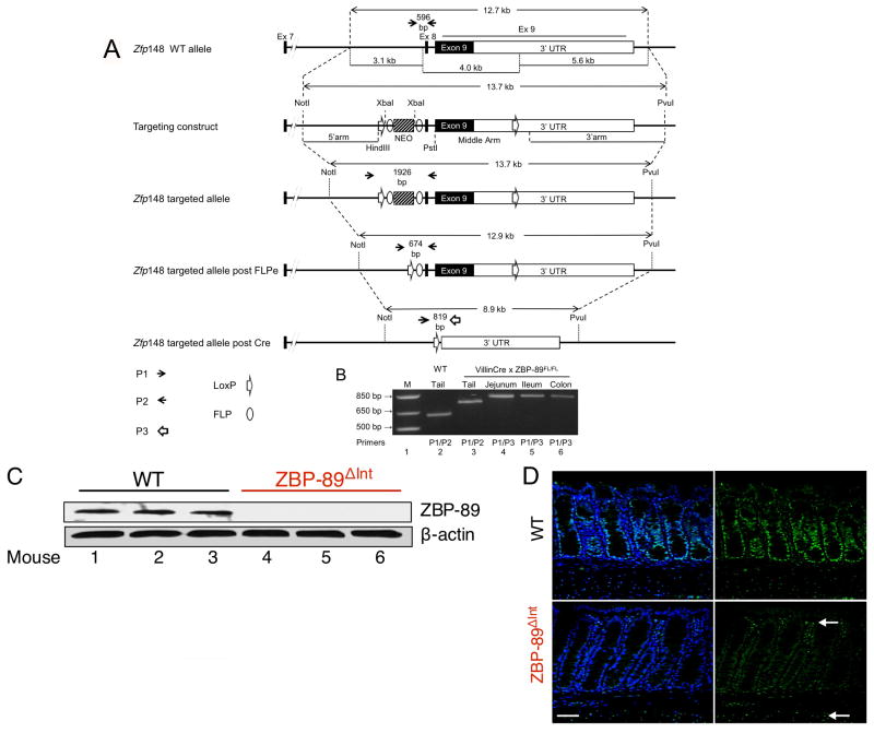
Methods: We generated mice with a conditional null allele of Zfp148 (ZBP-89(FL/FL)) using homologous recombination to flank Zfp148 with LoxP sites (ZBP-89(FL/FL)), and then bred the resulting mice with those that express VillinCre. We used microarray analysis to compare gene expression patterns in colonic mucosa between ZBP-89(ΔInt) and C57BL/6 wild-type mice (controls). Mice were gavaged with 2 isogenic strains of S. typhimurium after administration of streptomycin.

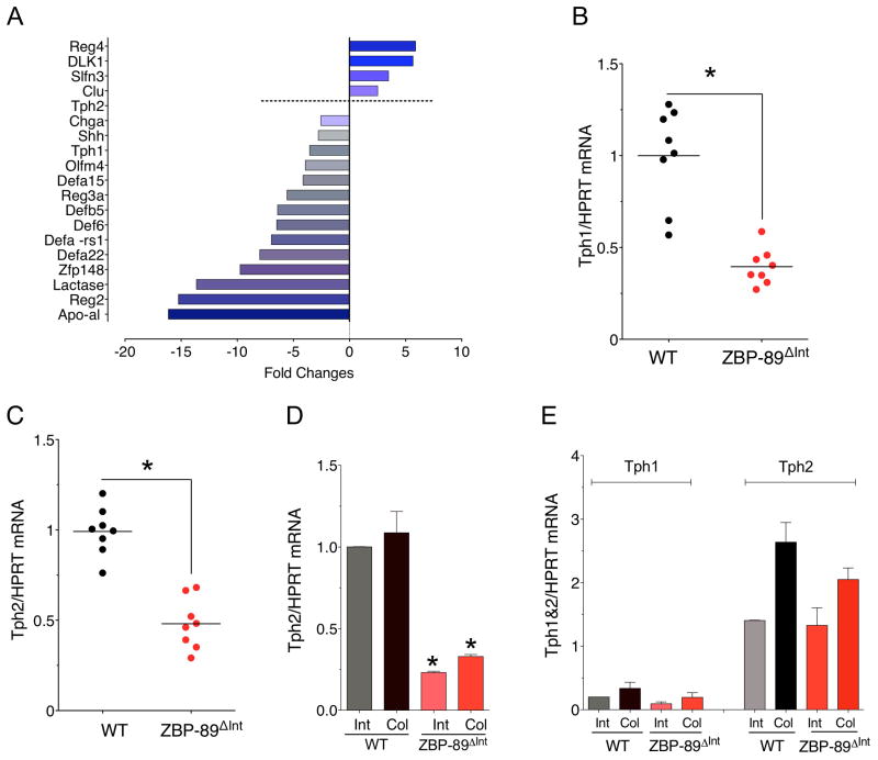
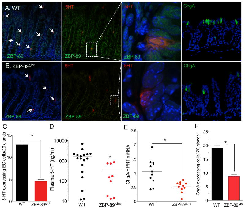
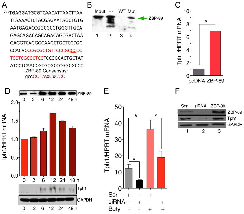
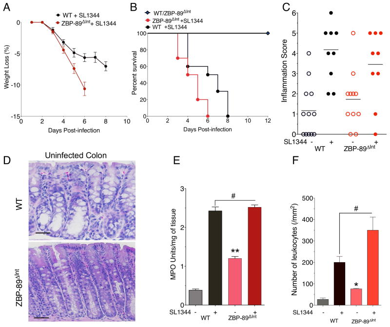
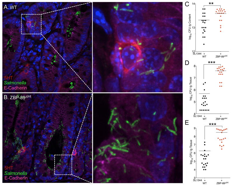
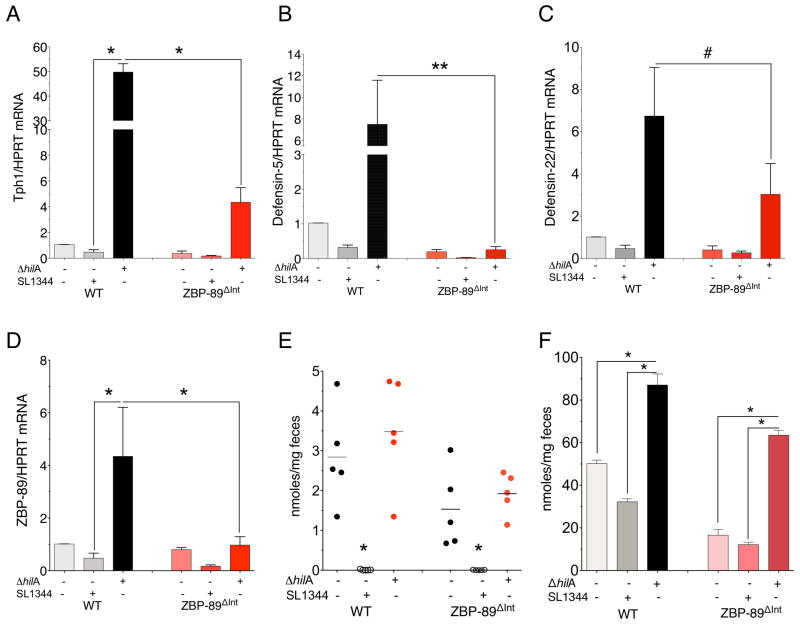
Results: Microarray analysis revealed that the colonic mucosa of ZBP-89(ΔInt) mice had reduced levels of tryptophan hydroxylase 1 (Tph1) messenger RNA, encoding the rate-limiting enzyme in enterochromaffin cell serotonin (5-hydroxytryptamine [5HT]) biosynthesis. DNA affinity precipitation demonstrated direct binding of ZBP-89 to the mouse Tph1 promoter, which was required for its basal and butyrate-inducible expression. ZBP-89(ΔInt) mice did not increase mucosal levels of 5HT in response to S. typhimurium infection, and succumbed to the infection 2 days before control mice. The ΔhilA isogenic mutant of S. typhimurium lacks this butyrate-regulated locus and stimulated, rather than suppressed, expression of Tph1 approximately 50-fold in control, but not ZBP-89(ΔInt), mice, correlating with fecal levels of butyrate.

Conclusions: ZBP-89 is required for butyrate-induced expression of the Tph1 gene and subsequent production of 5HT in response to bacterial infection in mice. Reductions in epithelial ZBP-89 increase susceptibility to colitis and sepsis after infection with S. typhimurium, partly because of reduced induction of 5HT production in response to butyrate and decreased secretion of antimicrobial peptides.


Conflict of interest statement

The authors have all declared that no conflict of interest exists.

Figures

Figure 1
Figure 1. Conditional deletion of locus encoding transcription factor Zfp148
(A) Schematic representation of mouse ZBP-89 locus (Zfp148) and strategy for gene targeting. Intestine specific deletion was achieved by breeding to the VillinCre line (ZBP-89ΔInt). (B) PCR Genotyping. Lane 1= DNA markers, lane 2 = wild-type (+/+) tail DNA, lane 3 = targeted locus, lanes 4–6, intestinal mucosa where Cre is active. ZBP-89 expression in the colon of WT and ZBP-89ΔInt mice detected by (C) Western blot of ZBP-89 normalized to β-actin from after colonic epithelium separated by chelation. (D) Immunofluorescence. FITC label = ZBP-89, DAPI = nuclei. Arrows indicate residual ZBP-89 nuclear staining in the lamina propria/smooth muscle layer.
Figure 2
Figure 2. Microarray analysis of ZBP-89ΔInt mice
(A) Mean relative expression (N=2 mice) of several genes in the ZBP-89ΔInt versus WT mice. (B) RT-qPCR for Tph1 mRNA and (C) for Tph2 mRNA. (D) Tph2 mRNA in isolated epithelium. (E) Tph1 versus Tph2 in isolated mesenchymal/smooth muscle layer. Shown is the mean ± SEM for N = 5. *P < 0.05.
Figure 3
Figure 3. Reduced 5HT+ cells in ZBP-89ΔInt mice
(A) Immunofluorescent staining for 5HT (red), and ZBP-89 (FITC, confocal) or ChgA (FITC) with DAPI (blue) in WT and (B) ZBP-89ΔInt mice. Low power merged views on left, (Scale bar, 100 μm). High power view of ZBP-89 with 5HT (600x) (confocal image) or ChgA with DAPI (400x). (C) Morphometric analysis of 5HT-expressing cells, N= 12 mice; (D) Plasma 5HT (ng/ml); (E) ChgA mRNA and (F) Morphometric analysis of ChgA-expressing cells in WT and ZBP-89ΔInt mouse colons, N = 8 mice. *P < 0.05.
Figure 4
Figure 4. ZBP-89 binds and regulates mouse Tph1
(A) mTph1 promoter from −255 to −55 bp. ZBP-89 DNA element highlighted (red); two bp mutation underlined. (B) DAPA using a 30 bp biotinylated DNA element at −123 bp. Lane 1: input, lane 2: probe alone with 50 μg of STC-1 nuclear extract, lane 3: 20x unlabeled probe competitor, lane 4: 20x 2 bp mutation competitor. (C) RT-qPCR analysis of mTph1 mRNA after ZBP-89 expression in STC-1 cells. (D) Butyrate (2.5 mM) induction of ZBP-89 protein, Tph1 mRNA and protein in STC-1 cells, N=5. Both ZBP-89 and Tph1 western blots normalized to same GAPDH. (E) ZBP-89 siRNA versus scrambled siRNA oligos transfected into STC-1 cells ± 2.5 mM butyrate for 6h. Tph1 mRNA levels determined by RT-qPCR. N = 3, *P < 0.05. (F) Western blot of ZBP-89 and mTph1 protein levels after transfection of scrambled (Scr) (lane 1) or siRNA oligos for mZBP-89 (lane 2) or a ZBP-89 expression vector (lane 3) into STC-1 cells.
Figure 5
Figure 5. Increased susceptibility to S. typhimurium in ZBP-89ΔInt mice
(A) Mouse body weight at euthanization. Percent survival of WT, N = 20 (black) versus ZBP-89ΔInt N = 20 SL1344 infected mice (red) from 4 independent experiments. Log-rank test demonstrated a statistically significant difference between the 2 groups (P = 0.0165) (C) Colitis scores (D) H&E stains of colon from WT and ZBP-89ΔInt mice prior to infection; (E) Myeloperoxidase (MPO) activity; (F) Leukocyte cell count from colons of WT versus ZBP-89ΔInt mice before and after SL1344 infection for 6 days. *P < 0.05.
Figure 6
Figure 6. Salmonella-infected colonic glands in WT and ZBP-89ΔInt mice
(A) Confocal image of Salmonella with 5HT-expressing EC cells 4 day p.i. in wild type mice. (B) versus ZBP-89ΔInt. Quantitative cultures of Salmonella SL1344 in (C) colon, (D) liver, (E) spleen for in WT versus ZBP-89ΔInt mice at euthanization (CFU/g content, Log10) for two separate experiments. * P < 0.05. ** P < 0.01; *** P < 0.001.
Figure 7
Figure 7. S. typhimurium ΔhilA induces Tph1 and defensin mRNA levels
mRNA levels 4 days after gavage with ΔhilA mutant for (A)Tph1; (B) defensin 5; (C) defensin 22; (D) ZBP-89. N = 6 mice per WT versus ZBP-89ΔInt for two independent experiments. Mean ± SEM. #P = 0.42; *P < 0.05; **P < 0.01. (E) Fecal butyrate and (F) Fecal SCFA levels in the colon. *P < 0.05.

References

    1. Bai L, Merchant JL. Transcription factor ZBP-89 cooperates with histone acetyltransferase p300 during butyrate activation of p21waf1 transcription in human cells. J Biol Chem. 2000;275:30725–33. - PubMed
    1. Merchant JL, Bai L, Okada M. ZBP-89 mediates butyrate regulation of gene expression. J Nutr. 2003;133:2456S–2460S. - PubMed
    1. Merchant JL, Iyer GR, Taylor BR, et al. ZBP-89, a Krüppel-type zinc finger protein, inhibits EGF induction of the gastrin promoter. Mol Cell Biol. 1996;16:6644–6653. - PMC - PubMed
    1. Bai L, Kao JY, Law DJ, et al. Recruitment of Ataxia-Telangiectasia Mutated to the p21(waf1) Promoter by ZBP-89 Plays a Role in Mucosal Protection. Gastroenterology. 2006;131:841–52. - PubMed
    1. Topping DL, Clifton PM. Short-chain fatty acids and human colonic function: roles of resistant starch and nonstarch polysaccharides. Physiol Rev. 2001;81:1031–64. - PubMed

Publication types