. 2013 Mar 18;587(6):717-23.
doi: 10.1016/j.febslet.2013.01.051. Epub 2013 Feb 5.

Neurofibrillary tangle-like tau pathology induced by synthetic tau fibrils in primary neurons over-expressing mutant tau

Affiliations

Neurofibrillary tangle-like tau pathology induced by synthetic tau fibrils in primary neurons over-expressing mutant tau

Jing L Guo et al. FEBS Lett. .

Erratum in

  • FEBS Lett. 2013 Aug 2;587(15):2484

Abstract

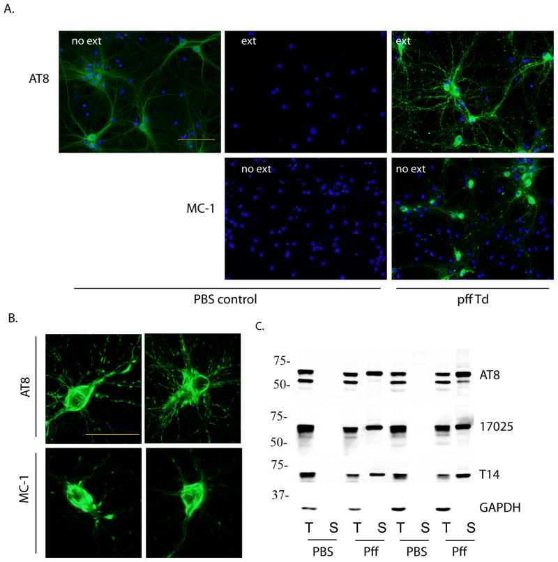
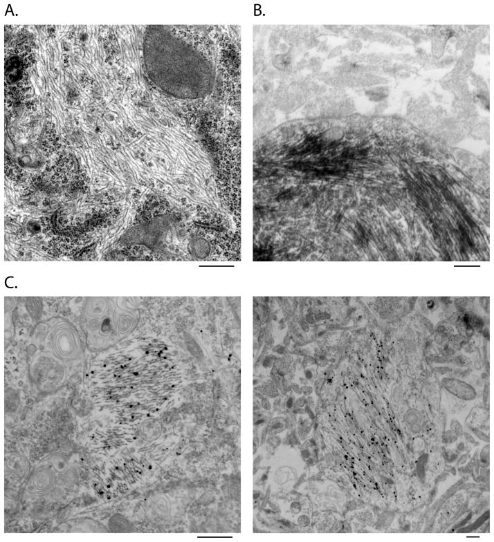
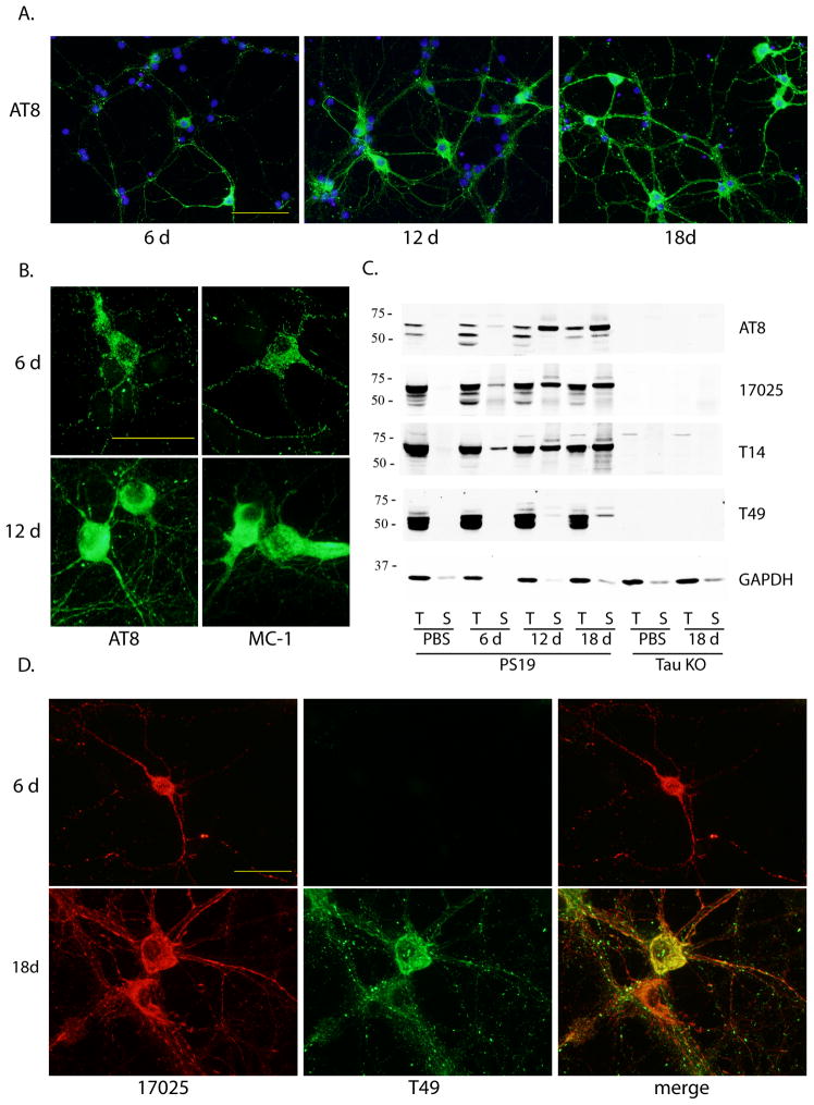
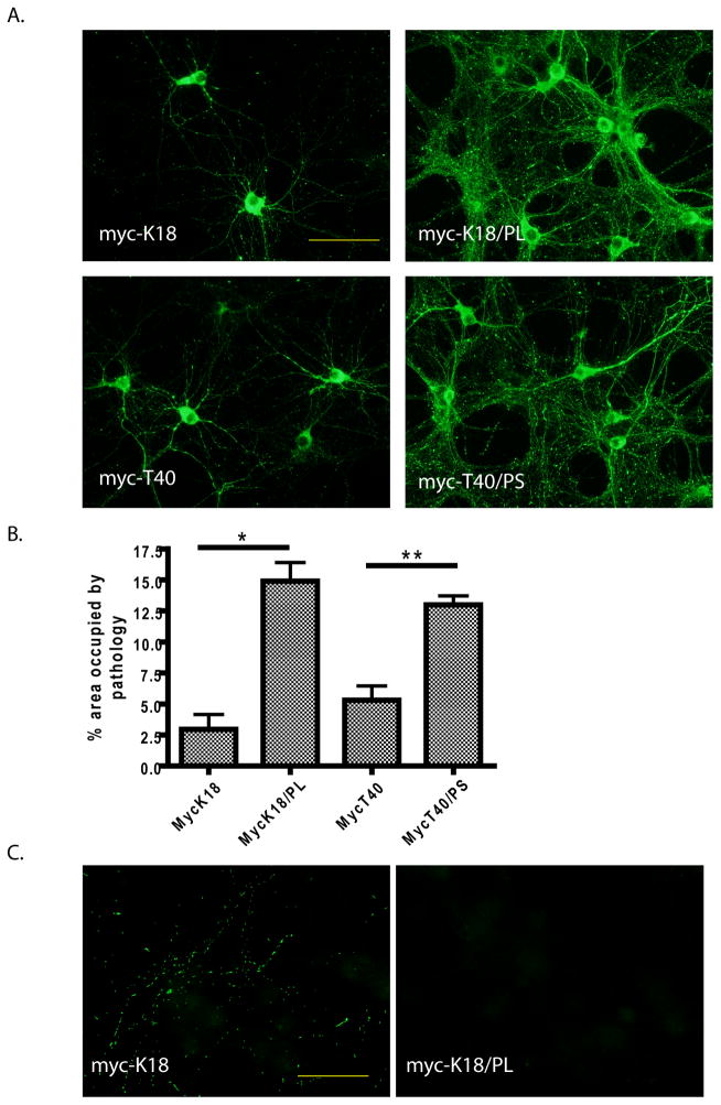
Increasing evidence demonstrates the transmissibility of fibrillar species of tau protein, but this has never been directly tested in neurons, the cell type most affected by formation of tau inclusions in neurodegenerative tauopathies. Here we show that synthetic tau fibrils made from recombinant protein not only time-dependently recruit normal tau into neurofibrillary tangle-like insoluble aggregates in primary hippocampal neurons over-expressing human tau, but also induce neuritic tau pathology in non-transgenic neurons. This study provides highly compelling support for the protein-only hypothesis of pathological tau transmission in primary neurons and describes a useful neuronal model for studying the pathogenesis of tauopathies.


Figures

Figure 1
Figure 1. Insoluble tau accumulated in PS19 primary hippocampal neurons after incubation with myc-K18/P301L pffs
(A) PS19 neurons treated with PBS (PBS control) or myc-K18/P301L fibrils (pff Td) were immunostained with phospho-tau mAb AT8 and conformational mAb MC-1 with or without 1% Triton-X100 extraction during fixing (ext, no ext, respectively). DAPI staining was used to visualize nuclei (blue). Scale bar: 100 μm. (B) Perikaryal tau aggregates recognized by AT8 and MC-1. Scale bar: 50 μm. (C) Neurons treated with PBS or myc-K18/P301L fibrils (pff) were sequentially extracted with 1% Triton-X100 lysis buffer (T) followed by 1% SDS lysis buffer (S) and immunoblotted with AT8, polyclonal antibody against total tau (17025), and mAb against human tau (T14). GAPDH served as loading control. Results from two independent sets of neurons were shown.
Figure 2
Figure 2. Ultrastructural analysis of tau pff-induced aggregates by EM and immune-EM
Routine EM (A) revealed filamentous structures in the cytoplasm of tau pff transduced neurons, which were recognized by MC-1 in immuno-EM using both HRP-labeling (B) and nanogold-amplification (C) detecting methods. Scale bar: 500 nm.
Figure 3
Figure 3. Time-dependent increase in pff-induced tau pathology and recruitment of mouse tau into aggregates
(A) Increasing population of cells developed tau pathology from 6 d to 18 d incubation after pff addition. Green: AT8. Blue: DAPI. Scale bar: 100 μm. (B) Morphological difference between 6 d and 12 d post-transduction aggregates immunostained with AT8 and MC-1. Scale bar: 50 μm. (C) Sequentially extracted PS19 neuron lysates at different time points after pff addition (6 d, 12 d, 18 d) or at 18 d after PBS treatment (PBS) and tau knock-out neuron lysates (tau KO) at 18 d post-addition of PBS or pffs were immunoblotted with AT8, 17025, T14 as well as mAb specific for mouse tau (T49). (D) Double-labeling of aggregates by T49 and 17025 at different time points after pff addition. Scale bar: 50 μm. For (A), (B) and (D), soluble proteins were removed by 1% Triton-X100 during fixing.
Figure 4
Figure 4. Differential seeding capacity of distinct forms of tau pffs on PS19 and non-Tg primary neurons
(A) Tau pathology (green: AT8) induced in PS19 neurons by myc-K18, myc-K18/P301L (myc-K18/PL), myc-T40 and myc-T40/P301S (myc-T40/PS) fibrils. Scale bar: 100 μm. (B) Quantification of percentage area occupied by AT8-positive pathology induced by the different pffs (mean + SEM). Number of independent sets of PS19 neurons tested: n = 2 for myc-K18, n = 3 for the other three. *: < 0.05. **: < 0.005. (C) Transduction of myc-K18 but not myc-K18/P301L pffs on non-Tg neurons resulted in neuritic tau pathology (green: AT8). Scale bar: 50 μm. For both (A) and (C), soluble proteins were removed by 1% Triton-X100 during fixing.

References

    1. Ballatore C, Lee VM, Trojanowski JQ. Tau-mediated neurodegeneration in Alzheimer’s disease and related disorders. Nature reviews Neuroscience. 2007;8:663–72. - PubMed
    1. Frost B, Jacks RL, Diamond MI. Propagation of tau misfolding from the outside to the inside of a cell. The Journal of biological chemistry. 2009;284:12845–52. - PMC - PubMed
    1. Nonaka T, Watanabe ST, Iwatsubo T, Hasegawa M. Seeded aggregation and toxicity of {alpha}-synuclein and tau: cellular models of neurodegenerative diseases. The Journal of biological chemistry. 2010;285:34885–98. - PMC - PubMed
    1. Guo JL, Lee VM. Seeding of normal Tau by pathological Tau conformers drives pathogenesis of Alzheimer-like tangles. The Journal of biological chemistry. 2011;286:15317–31. - PMC - PubMed
    1. Clavaguera F, Bolmont T, Crowther RA, Abramowski D, Frank S, Probst A, Fraser G, Stalder AK, Beibel M, Staufenbiel M, Jucker M, Goedert M, Tolnay M. Transmission and spreading of tauopathy in transgenic mouse brain. Nature cell biology. 2009;11:909–13. - PMC - PubMed

Publication types

MeSH terms