

Defining a direction: electron transfer and catalysis in Escherichia coli complex II enzymes
- PMID: 23396003
- PMCID: PMC3615059
- DOI: 10.1016/j.bbabio.2013.01.010
Defining a direction: electron transfer and catalysis in Escherichia coli complex II enzymes
Abstract
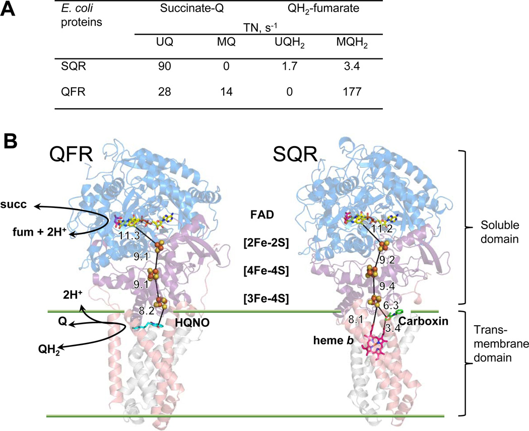
There are two homologous membrane-bound enzymes in Escherichia coli that catalyze reversible conversion between succinate/fumarate and quinone/quinol. Succinate:ubiquinone reductase (SQR) is a component of aerobic respiratory chains, whereas quinol:fumarate reductase (QFR) utilizes menaquinol to reduce fumarate in a final step of anaerobic respiration. Although, both protein complexes are capable of supporting bacterial growth on either minimal succinate or fumarate media, the enzymes are more proficient in their physiological directions. Here we evaluate factors that may underlie this catalytic bias. This article is part of a Special Issue entitled: Respiratory complex II: Role in cellular physiology and disease.
Copyright © 2013 Elsevier B.V. All rights reserved.
Figures

Similar articles
-
The quinone-binding and catalytic site of complex II.Biochim Biophys Acta. 2010 Dec;1797(12):1877-82. doi: 10.1016/j.bbabio.2010.02.015. Epub 2010 Feb 20. Biochim Biophys Acta. 2010. PMID: 20175986 Free PMC article. Review.
-
Comparison of catalytic activity and inhibitors of quinone reactions of succinate dehydrogenase (Succinate-ubiquinone oxidoreductase) and fumarate reductase (Menaquinol-fumarate oxidoreductase) from Escherichia coli.Arch Biochem Biophys. 1999 Sep 15;369(2):223-32. doi: 10.1006/abbi.1999.1359. Arch Biochem Biophys. 1999. PMID: 10486141
-
A threonine on the active site loop controls transition state formation in Escherichia coli respiratory complex II.J Biol Chem. 2008 May 30;283(22):15460-8. doi: 10.1074/jbc.M801372200. Epub 2008 Apr 2. J Biol Chem. 2008. PMID: 18385138 Free PMC article.
-
The di-heme family of respiratory complex II enzymes.Biochim Biophys Acta. 2013 May;1827(5):679-87. doi: 10.1016/j.bbabio.2013.02.012. Epub 2013 Mar 4. Biochim Biophys Acta. 2013. PMID: 23466335 Review.
-
Electroneutral and electrogenic catalysis by dihaem-containing succinate:quinone oxidoreductases.Biochem Soc Trans. 2008 Oct;36(Pt 5):996-1000. doi: 10.1042/BST0360996. Biochem Soc Trans. 2008. PMID: 18793177
Cited by
-
The assembly of succinate dehydrogenase: a key enzyme in bioenergetics.Cell Mol Life Sci. 2019 Oct;76(20):4023-4042. doi: 10.1007/s00018-019-03200-7. Epub 2019 Jun 24. Cell Mol Life Sci. 2019. PMID: 31236625 Free PMC article. Review.
-
Bioenergetic Inhibitors: Antibiotic Efficacy and Mechanisms of Action in Mycobacterium tuberculosis.Front Cell Infect Microbiol. 2021 Jan 11;10:611683. doi: 10.3389/fcimb.2020.611683. eCollection 2020. Front Cell Infect Microbiol. 2021. PMID: 33505923 Free PMC article. Review.
-
Mathematical Modeling of ROS Production and Diode-like Behavior in the SDHA/SDHB Subcomplex of Succinate Dehydrogenases in Reverse Quinol-Fumarate Reductase Direction.Int J Mol Sci. 2022 Dec 9;23(24):15596. doi: 10.3390/ijms232415596. Int J Mol Sci. 2022. PMID: 36555239 Free PMC article.
-
Hysteresis and bistability in the succinate-CoQ reductase activity and reactive oxygen species production in the mitochondrial respiratory complex II.Redox Biol. 2020 Oct;37:101630. doi: 10.1016/j.redox.2020.101630. Epub 2020 Jul 5. Redox Biol. 2020. PMID: 32747163 Free PMC article.
-
Investigation of candidate genes involved in the rhodoquinone biosynthetic pathway in Rhodospirillum rubrum.PLoS One. 2019 May 21;14(5):e0217281. doi: 10.1371/journal.pone.0217281. eCollection 2019. PLoS One. 2019. PMID: 31112563 Free PMC article.
References
-
- Cecchini G. Function and structure of complex II of the respiratory chain. Annu Rev Biochem. 2003;72:77–109. - PubMed
-
- Ackrell BA, Johnson MK, Gunsalus RP, Cecchini Cary. Chemistry and Biochemistry of Flavoenzymes. London: CRC Press; 1992. Structure and function of succinate dehydrogenase and fumarate reductase; pp. 229–297.
-
- Iwata F, Shinjyo N, Amino H, Sakamoto K, Islam MK, Tsuji N, Kita K. Change of subunit composition of mitochondrial complex II (succinate-ubiquinone reductase/quinol-fumarate reductase) in Ascaris suum during the migration in the experimental host. Parasitol Int. 2008;57:54–61. - PubMed
-
- Guest JR. Partial replacement of succinate dehydrogenase function by phage- and plasmid-specified fumarate reductase in Escherichia coli. J Gen Microbiol. 1981;122:171–179. - PubMed
Publication types
MeSH terms
Substances
Grants and funding
LinkOut - more resources
Full Text Sources
Other Literature Sources
Molecular Biology Databases
