. 2013 Mar;45(3):269-78.
doi: 10.1038/ng.2541. Epub 2013 Feb 10.

RNF212 is a dosage-sensitive regulator of crossing-over during mammalian meiosis

Affiliations

RNF212 is a dosage-sensitive regulator of crossing-over during mammalian meiosis

April Reynolds et al. Nat Genet. 2013 Mar.

Abstract

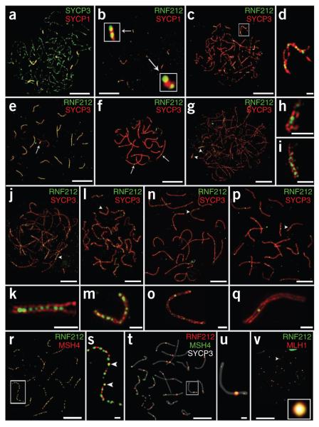
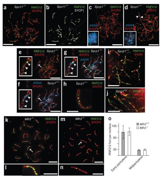
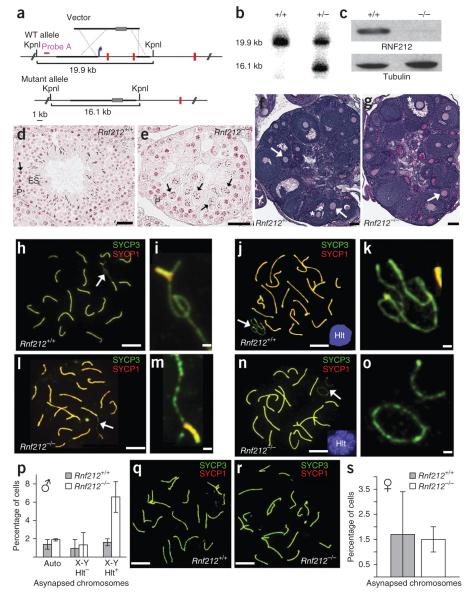
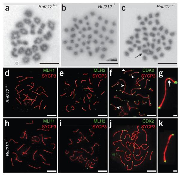
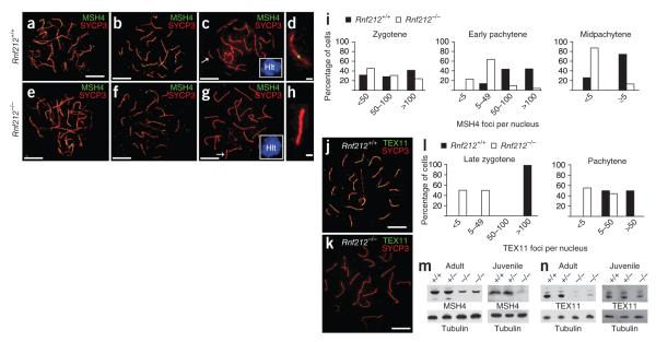
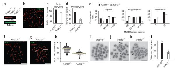
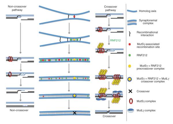
Crossing-over ensures accurate chromosome segregation during meiosis, and every pair of chromosomes obtains at least one crossover, even though the majority of recombination sites yield non-crossovers. A putative regulator of crossing-over is RNF212, which is associated with variation in crossover rates in humans. We show that mouse RNF212 is essential for crossing-over, functioning to couple chromosome synapsis to the formation of crossover-specific recombination complexes. Selective localization of RNF212 to a subset of recombination sites is shown to be a key early step in the crossover designation process. RNF212 acts at these sites to stabilize meiosis-specific recombination factors, including the MutSγ complex (MSH4-MSH5). We infer that selective stabilization of key recombination proteins is a fundamental feature of meiotic crossover control. Haploinsufficiency indicates that RNF212 is a limiting factor for crossover control and raises the possibility that human alleles may alter the amount or stability of RNF212 and be risk factors for aneuploid conditions.


Figures

Figure 1
Figure 1
Dynamic localization of RNF212 to synaptonemal complexes and crossover sites in mouse spermatocytes. (a,b) Nucleus at very early zygonema immunolabeled for SYCP1 and SYCP3 (a) and showing colocalization of SYCP1 and RNF212 (b). Insets show magnified views of two short stretches of synaptonemal complex with overlapping RNF212 foci. (c–f) Representative prophase nuclei immunolabeled for RNF212 and SYCP3. (c,d) Early zygotene nucleus (c) with magnified view of a chromosome pair (d), highlighting the exclusion of RNF212 from unsynapsed regions. (e) Early pachytene nucleus. The X-Y chromosome pair is highlighted by an arrow. (f) Midpachytene nucleus. Two RNF212 foci are highlighted by arrows. (g–q) SIM images of selected prophase nuclei immunolabeled for RNF212 and SYCP3. (g–i) Very early zygonema nucleus (g) with magnified views of synapsed regions indicated by arrowheads: left arrowhead (h), right arrowhead (i). (j,k) Midzygonema nucleus (j), with magnified view of the highlighted region (k). (l,m) Early pachynema nucleus (l), with magnified view of the indicated chromosome (m). (n,o) Early pachynema to midpachynema nucleus (n), with magnified view of the indicated chromosome (o). (p,q) Midpachynema nucleus (p), with magnified view of the chromosome indicated by the arrowhead (q). (r,s) Early pachytene nucleus costained for RNF212 and MSH4 (r), with magnified views of the two chromosome pairs highlighted by the white box (s). Arrowheads highlight RNF212 and MSH4 foci that are fully colocalized. (t,u) Midpachytene nucleus immunolabeled for RNF212, MSH4 and SYCP3 (t), with magnified view of the chromosome pair highlighted by the white box (u). Note the single site of RNF212-MSH4 colocalization. (v) Midpachytene nucleus immunolabeled for RNF212 and MLH1. The inset shows a single RNF212-MLH1 focus. Scale bars, 10 μm in a–c,e,f,r,t,v; 5 μm in g,j,l,n,p and 1 μm in d,h,i,k,m,o,q,s,u.
Figure 2
Figure 2
Genetic requirements for RNF212 localization. (a,b) Spermatocyte nucleus from a Spo11−/− mouse with immunolabeling for SYCP3 and SYCP1 (a) and RNF212 and SYCP1 (b). (c–g) Spermatocyte nuclei from a Sycp1−/− mutant mouse immunolabeled for RNF212, SYCP3 and γH2AX. (c) Zygotene-like nucleus with pannuclear γH2AX staining. (d) Pachytene-like nucleus with diminishing γH2AX staining. Insets in c and d show SYCP3 and γH2AX channels. Arrowheads in d highlight RNF212 foci localized to coaligned homolog axes. (e–g) Late-pachytene/early diplotene nucleus stained for RNF212 and SYCP3 (e) and γH2AX and SYCP3, showing residual γH2AX foci (f). (g) Merged staining of this nucleus for RNF212, SYCP3 and γH2AX. Insets in e–g show a single homolog pair, with RNF212-γH2AX foci highlighted by arrowheads. (h) Selected homolog pair from a Sycp1−/− spermatocyte showing RNF212 foci associated with one of the two SYCP3-staining axes. (i) Representative homolog pairs from a wild-type early pachytene spermatocyte immunolabeled for RNF212 and γH2AX. (j) Independent nucleus immunolabeled as in i. (k–n) Spermatocyte nuclei from an Mlh3−/− knockout mouse, immunolabeled for RNF212 and SYCP3. (k,l) Early pachynema nucleus (k), with magnified view of the chromosome indicated by an arrow (l). (m,n) Midpachynema nucleus (m), with magnified view of the indicated chromosome (n). (o) Quantification of RNF212 foci (± s.d.) in early pachytene and midpachytene spermatocytes from wild-type and Mlh3−/− mice. For wild-type cells, 16 early pachytene and 16 midpachytene nuclei were analyzed. For Mlh3−/− cells, 39 early pachytene and 10 midpachytene nuclei were analyzed. Scale bars, 10 μm in a–g,k,m and 5 μm in h–j,i,n.
Figure 3
Figure 3
Gonad morphology and homolog synapsis in Rnf212−/− knockout mice. (a) Rnf212 targeting scheme. Thick lines represent homology arms used for targeting. Gray rectangles represent the neomycin-resistance cassette. The blue arrow indicates the position of the promoter, and red vertical lines represent Rnf212 exons (not to scale). WT, wild type. (b) Southern analysis of KpnI-digested genomic DNA from wild-type and heterozygous embryonic stem cells hybridized with the probe shown in a. (c) Protein blot analysis of RNF212 in protein extracts from whole testes. (d,e) Representative seminiferous tubules stained with hematoxylin and eosin from testis sections from wild-type (d) and Rnf212−/− (e) mice. P, pachytene-stage cells; ES, elongated spermatids. Arrows indicate metaphase I cells. In Rnf212−/− tubules, cells progress to metaphase I (stage XII), but normal chromosome congression is not observed, and postmeiotic spermatogenic cells (spermatids and spermatozoa) are absent. (f,g) Representative ovary sections stained with periodic acid Schiff and hematoxylin from wild-type (f) and Rnf212−/− (g) mice. Asterisks indicate antral follicles with defined antral space. Arrows highlight secondary follicles surrounded by more than one layer of cuboidal granulosa cells. (h–o) Spread spermatocyte nuclei immunostained for SYCP3 and SYCP1. (h,i) Early pachynema nucleus from wild-type mouse (h), with magnified view of the sex chromosomes indicated by the arrow (i). (j,k) Midpachynema nucleus from wild-type cells (j). The inset shows H1t staining. (k) Magnified view of the indicated sex chromosomes in j. (l,m) Early pachynema nucleus from Rnf212−/− cells (l), with magnified view of the sex chromosomes indicated by an arrow (m). (n,o) Midpachynema nucleus from Rnf212−/− cells (n), with magnified view of the sex chromosomes (o). (p) Quantification of synapsis defects in pachytene spermatocytes. Numbers of nuclei analyzed in wild-type and Rnf212−/− cells, respectively: 293 and 521 for autosomal asynapsis (auto) and X-Y asynapsis in H1t-negative pachytene cells; 686 and 622 for X-Y asynapsis in H1t-positive pachytene cells. (q,r) Pachytene-stage fetal oocytes from wild-type (q) and Rnf212−/− mutant (r) animals immunostained for SYCP3 and SYCP1. (s) Quantification of synapsis defects in pachytene oocytes (191 and 272 wild-type and Rnf212−/− nuclei, respectively). Error bars in p and s, s.e.m. Scale bars, 100 μm in d–g, 10 μm in h,j,l,n,q,r and 1 μm in i,k,m,o.
Figure 4
Figure 4
RNF212 is required for chiasma formation and assembly of crossover-specific recombination complexes. (a–c) Chromosome spreads of cells in diakinesis/metaphase I stained with Giemsa from wild-type (a) and Rnf212−/− (b,c) mice. (b) Nucleus with no chiasmata. (c) Nucleus with a single chiasmate bivalent (highlighted by an arrow). (d–k) Pachytene spermatocytes. (d–g) Wild-type cells immunostained for MLH1 and SYCP3 (d), MLH1 and SYCP3 (e) or CDK2 and SYCP3 (f). (g) Magnified view of a chromosome from f. (h–k) Rnf212−/− cells immunostained for MLH1 and SYCP3 (h), MLH3 and SYCP3 (i) or CDK2 and SYCP3 (j). (k) Magnified view of a chromosome from j. The arrowheads and arrow (f,g) highlight interstitial CDK2 foci. Scale bars, 10 μm in a–f,h–j and 1 μm in g,k.
Figure 5
Figure 5
RNF212 stabilizes two ZMM factors, Mutsγ and TEX11. (a–h) Spermatocyte nuclei immunostained for MSH4 and SYCP3 at successive prophase stages. (a–d) Wild-type cells stained at zygonema (a), early pachynema (b) and midpachynema (c). (d) Magnified chromosome from c. (e–h) Rnf212−/− cells stained at zygonema (e), early pachynema (f) and midpachynema (g). (h) Magnified chromosome from g. Insets in c and g show H1t staining. (i) Quantification of MSH4 foci at successive prophase stages. Statistical comparisons: zygotene, P = 0.1 (G test; n = 83 Rnf212+/+ and 51 Rnf212−/− nuclei); early pachytene, P = 1 × 10⁻³² (G test; n = 131 Rnf212+/+ and 103 Rnf212−/− nuclei); midpachytene, P = 1 × 10–19 χ-square test; n = 121 Rnf212+/+ and 103 Rnf212−/− nuclei). (j,k) Early pachytene spermatocyte nuclei immunolabeled for TEX11 and SYCP3 from wild-type (j) and Rnf212−/− (k) animals. (l) Quantification of TEX11 foci. In late zygonema, TEX11 foci average 122 ± 11 (s.d.; n = 5) in wild-type nuclei and 21 ± 22 (s.d.; n = 10) in Rnf212−/− mutants (P = 0.0027, Mann-Whitney test). In pachynema, TEX11 foci average 56 ± 27 (s.d., n = 10) in wild-type nuclei and 10 ± 11 (s.d., n = 9) in Rnf212−/− mutants (P = 0.0004, Mann-Whitney test). (m,n) Protein blot analysis of MSH4 (m) and TEX11 (n) in protein extracts from whole testes. In adult testes, the levels of MSH4 and TEX11 are approximately 40% and 17% of wild-type levels, respectively; in juvenile testes (18 d postpartum), protein levels are ~20% and ~63% of wild-type levels. Scale bars, 10 μm in a–c,e–g,j,k and 1 μm in d,h.
Figure 6
Figure 6
Rnf212 is haploinsufficient. (a) Quantitative protein blot analysis of RNF212 in testis cell extracts from wild-type, Rnf212+/− and Rnf212−/− mice. (b) Midpachytene Rnf212+/− spermatocyte nucleus immunostained for RNF212 and SYCP3. White lines highlight synaptonemal complexes that lack RNF212 foci. (c,d) Numbers of RNF212 foci per nucleus ± s.e.m. in wild-type and Rnf212+/− spermatocytes at early pachynema (c) and midpachynema (d). (e) Numbers of MSH4 foci per nucleus in wild-type and Rnf212+/− spermatocytes at successive prophase substages. Statistical comparisons: zygotene, P = 0.04 (G test; n = 83 Rnf212+/+ and 68 Rnf212+/− nuclei); early pachytene, P = 9 × 10⁻¹³ (G test; n = 131 Rnf212+/+ and 270 Rnf212−/− nuclei); midpachytene, P = 0.01 χ-square test; n = 121 Rnf212+/+ and 126 Rnf212−/− nuclei). (f,g) Midpachytene spermatocyte nuclei from wild-type (f) and Rnf212+/− (g) mice immunolabeled for MLH1 and SYCP3. White lines in g highlight synaptonemal complexes that lack MLH1 foci. (h) Numbers of MLH1 foci per nucleus in midpachytene-stage spermatocytes. Each symbol represents a single nucleus. Yellow lines indicate the average numbers of foci. (i–k) Chromosome spreads of spermatocytes in diakinesis/metaphase I from Rnf212+/+ (i) and Rnf212+/− (j,k) mice. Independent examples from Rnf212−/− spermatocytes are shown in (j) and (k). Arrows in j and k highlight achiasmate univalent chromosomes. (l) Numbers of chiasmata per nucleus (± s.e.m.) in wild-type and Rnf212+/− diakinesis/metaphase I spermatocytes. Scale bars, 10 μm.
Figure 7
Figure 7
Summary and model of RNF212 function. Schematics showing the cytological development of recombination complexes and parallel molecular pathways of crossover and non-crossover recombination. Black and blue lines represent homologous DNA duplexes. Although four chromatids are present at this stage, for simplicity, only the two chromatids involved in recombination are shown. Both crossover and non-crossover pathways initiate from a common D-loop precursor. As synapsis ensues, binding of the MutSγ complex initially stabilizes most or all D-loops. In the absence of RNF212-mediated stabilization, MutSγ dissociates, and D-loops are unwound, resulting in non-crossover formation. At crossover sites, RNF212-dependent SUMOylation enhances the association of MutSγ, D-loops are stabilized, and formation of crossover-specific double Holliday junctions ensues. These crossover precursors become competent to assemble the crossover-specific resolution factor, MutLγ, and crossing-over occurs. Recombination sites that nucleate synaptonemal complex formation have a high probability of being the first sites where MutSγ and RNF212 colocalize. At such sites, a positive feedback loop locally enhances the binding of both MutSγ and RNF212. General binding of RNF212 to sites along the synaptonemal complex central element disfavors stabilization of MutSγ at other recombination sites.

Similar articles

Cited by

References

    1. Hunter N. Meiotic recombination. In: Aguilera A, Rothstein R, editors. Molecular Genetics of Recombination. Springer-Verlag; Heidelberg, Germany: 2006. pp. 381–442.
    1. Zickler D, Kleckner N. Meiotic chromosomes: integrating structure and function. Annu. Rev. Genet. 1999;33:603–754. - PubMed
    1. Sakuno T, Tanaka K, Hauf S, Watanabe Y. Repositioning of aurora B promoted by chiasmata ensures sister chromatid mono-orientation in meiosis I. Dev. Cell. 2011;21:534–545. - PubMed
    1. Hirose Y, et al. Chiasmata promote monopolar attachment of sister chromatids and their co-segregation toward the proper pole during meiosis I. PLoS Genet. 2011;7:e1001329. - PMC - PubMed
    1. Hassold T, Hall H, Hunt P. The origin of human aneuploidy: where we have been, where we are going. Hum. Mol. Genet. 2007;16(Spec. No. 2):R203–R208. - PubMed

Publication types