

The role of ultrasonography in the study of medical nephropathy
- PMID: 23396246
- PMCID: PMC3552768
- DOI: 10.1016/j.jus.2007.09.001
The role of ultrasonography in the study of medical nephropathy
Abstract
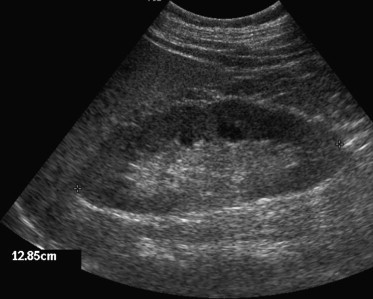
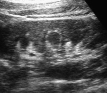
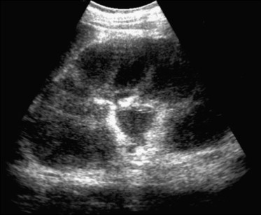
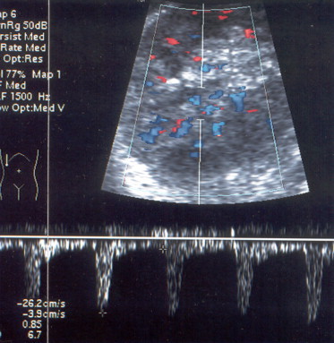
Diagnostic techniques in nephrology include clinical history, physical examination, laboratory tests, scintigraphy, diagnostic imaging techniques as well as renal biopsy. In kidney diseases, ultrasonography is used as a first-line imaging technique, and its role in medical nephropathy is to exclude urological pathologies, to differentiate between acute and chronic renal failure, to follow-up on the course of a disease, to guide needle biopsy, etc. Ultrasound images are useful at characterizing the pelvis, assessing renal dimensions and parenchymal echogenicity, sampling color-power Doppler signals and evaluating their characteristics and distribution as well as measuring parenchymal resistive index. Taken together, these data can provide useful clues to the diagnosis and help to reduce the number of possible differential diagnoses.
SommarioLa diagnostica in nefrologica comprende la storia clinica, l'esame fisico, gli esami di laboratorio, gli esami scintigrafici, la diagnostica per immagini e la biopsia renale. Nelle malattie renali l'ecografia rappresenta la tecnica per immagini di prima scelta e il suo ruolo nelle nefropatie mediche è quello di escludere una patologia urologica, differenziare fra un'insufficienza renale acuta e cronica, permettere il follow-up della malattia, guidare l'agobiopsia renale ecc. Le immagini ultrasonografiche permettono di caratterizzare la pelvi, di valutare le dimensioni renali e l'ecogenicità parenchimale, di campionare i segnali color–power Doppler e di valutarne caratteristiche e distribuzione, nonché di misurare gli indici di resistenza intraparenchimali. L'insieme di questi dati permette di ottenere importanti informazioni diagnostiche in molti casi, mentre in altri permette di ridurre le possibili diagnosi differenziali.
Keywords: Nephropathy; Resistive index; Ultrasonography.
Figures
Similar articles
-
Resistive Index of Intrarenal Artery in Evaluation of Diabetic Nephropathy.Bangladesh Med Res Counc Bull. 2015 Dec;41(3):125-130. doi: 10.3329/bmrcb.v41i3.29888. Bangladesh Med Res Counc Bull. 2015. PMID: 29870167
-
ARFI-based tissue elasticity quantification and kidney graft dysfunction: first clinical experiences.Clin Hemorheol Microcirc. 2011;49(1-4):527-35. doi: 10.3233/CH-2011-1503. Clin Hemorheol Microcirc. 2011. PMID: 22214724 Clinical Trial.
-
[Role of color Doppler US in the evaluation of renal transplant].Radiol Med. 2001 Apr;101(4):243-50. Radiol Med. 2001. PMID: 11398053 Italian.
-
Gray Scale Ultrasound, Color Doppler Ultrasound, and Contrast-Enhanced Ultrasound in Renal Parenchymal Diseases.Ultrasound Q. 2018 Dec;34(4):250-267. doi: 10.1097/RUQ.0000000000000383. Ultrasound Q. 2018. PMID: 30169495 Review.
-
Imaging in Chronic Kidney Disease.Contrib Nephrol. 2016;188:69-80. doi: 10.1159/000445469. Epub 2016 May 12. Contrib Nephrol. 2016. PMID: 27170301 Review.
Cited by
-
Role of Ultrasound in the Diagnosis of Chronic Kidney Disease and its Correlation with Serum Creatinine Level.Cureus. 2019 Mar 12;11(3):e4241. doi: 10.7759/cureus.4241. Cureus. 2019. PMID: 31131164 Free PMC article.
-
Diagnostic performance of renal ultrasonography in detecting chronic kidney disease of various severity.Asian Biomed (Res Rev News). 2020 Oct 31;14(5):195-202. doi: 10.1515/abm-2020-0028. eCollection 2020 Oct. Asian Biomed (Res Rev News). 2020. PMID: 37551269 Free PMC article.
-
Role of intrarenal resistive index and ElastPQ® renal shear modulus in early diagnosis and follow-up of diabetic nephropathy: A prospective study.Ultrasound. 2020 Nov;28(4):246-254. doi: 10.1177/1742271X20942249. Epub 2020 Jul 14. Ultrasound. 2020. PMID: 36959891 Free PMC article.
-
Cardiovascular risk and renal injury profile in subjects with type 2 diabetes and non-albuminuric diabetic kidney disease.Cardiovasc Diabetol. 2023 Dec 13;22(1):344. doi: 10.1186/s12933-023-02065-2. Cardiovasc Diabetol. 2023. PMID: 38093293 Free PMC article.
-
Sonographic assessment of kidneys in patients with hypertension co-existed with diabetes mellitus and ischemic heart disease.J Family Med Prim Care. 2020 May 31;9(5):2411-2415. doi: 10.4103/jfmpc.jfmpc_50_20. eCollection 2020 May. J Family Med Prim Care. 2020. PMID: 32754511 Free PMC article.
References
-
- Webb J.A. The role of ultrasonography in the diagnosis of intrinsic renal disease. Clin Radiol. 1994;49:589–591. - PubMed
-
- Brandt T.D., Neiman H.L., Dragowski M.J., Bulawa W., Claykamp G. Ultrasound assessment of normal renal dimensions. J Ultrasound Med. 1982;1:49–52. - PubMed
-
- Emamian S.A., Nielsen M.B., Pedersen J.F. Tenth percentiles of kidney length in adult volunteers. AJR Am J Roentgenol. 1994;163:748. - PubMed
-
- Webb J.A., Reznek R.H., White F.E., Cattell W.R., Fry I.K., Baker L.R. Can ultrasound and computed tomography replace high-dose urography in patients with impaired renal function? Q J Med. 1984;53:411–425. - PubMed
-
- Han B.K., Babcock D.S. Sonographic measurement and appearance of normal kidneys in children. AJR Am J Roentgenol. 1985;145:611–616. - PubMed
LinkOut - more resources
Full Text Sources